Applied anatomy of the hip and buttock
The hip joint
The hip joint is a ball-and-socket joint, formed by the femoral head and the acetabulum (Fig. 1, see Standring, Fig. 80.15). The articular surfaces are spherical with a marked congruity; this limits the range of movement but contributes to the considerable stability of the joint. In the anatomical position, the anterior/superior part of the femoral head is not covered by the acetabulum. This is because the axes of the femoral head and of the acetabulum are not in line with each other. The axis of the femoral head points superiorly, medially and anteriorly, while the axis of the acetabulum is directed inferiorly, laterally and anteriorly.
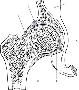
Fig 1 The hip joint: 1, ilium; 2, femur; 3, ligamentum teres; 4, labrum acetabulare; 5, transverse acetabular ligament; 6, joint capsule.
The cup-shaped acetabulum is a little below the middle third of the inguinal ligament. The acetabular articular surface is an incomplete cartilaginous ring, thickest and broadest above, where the pressure of body weight falls in the erect posture, narrowest in the pubic region. The rough lower part of the cup, the acetabular notch, is not covered by cartilage. The centre of the cup, the acetabular fossa, is also devoid of cartilage but contains fibroelastic fat. The acetabular labrum, a fibrocartilaginous ring attached to the acetabular rim, deepens the cup and enlarges the contact area with the femoral head (see Standring, Fig. 81.5). The part of the labrum that bridges the acetabular notch does not have cartilage cells and is called the transverse acetabular ligament. If forms a foramen through which vessels and nerves may enter the joint. The acetabular labrum is triangular in section. The base is attached to the acetabular rim and the apex is free.
The femoral head is ovoid or spheroid but not completely congruent with the reciprocal acetabulum. It is covered by articular cartilage except for a rough pit for the ligamentum teres, a flattened fibrous band, embedded in adipose tissue and lined by the synovial membrane (Figs 2, 3). The ligament connects the central part of the femoral head with the acetabular notch and its transverse acetabular ligament. The ligament is extra-articular and contains a tiny branch of the obturator artery partly responsible for the vascular supply of the femoral head.
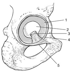
Fig 2 The acetabulum: 1, articular cartilage; 2, acetabular fossa; 3, ligamentum teres; 4, labrum acetabulare; 5, transverse acetabular ligament.
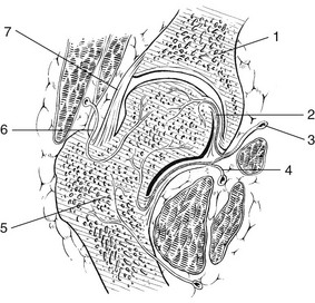
Fig 3 Coronal section through the hip: 1, ilium; 2, ligamentum teres; 3, branch of obturator artery; 4, branch of medial circumflex artery; 5, femur; 6, branch of lateral circumflex artery; 7, joint capsule strengthened by the iliofemoral ligament (lateral part).
The femoral head and neck also receive arterial supply from the capsular vessels, arising from the medial and lateral circumflex arteries (see Fig. 3).
Capsule and ligaments
The capsule is a cylindrical sleeve, running from the acetabular rim to the base of the femoral neck. It is supported by powerful ligaments.
Anteriorly these are two ligaments: the fan-shaped iliofemoral ligament of Bertin situated craniolaterally; and the pubofemoral ligament, in a more caudomedial orientation. Together they resemble the letter Z (Fig. 4). Posteriorly the capsule is strengthened by the ischiofemoral ligament (see Standring, Fig. 81.3). These three ligaments are coiled round the femoral neck. Extension ‘winds up’ and tautens the ligaments, thus stabilizing the joint passively (Fig. 5a); flexion slackens them (Fig. 5b). Lateral rotation tightens the iliofemoral ligament and also the pubofemoral ligament. Medial rotation tightens the ischiofemoral ligament. Abduction tightens the pubofemoral and the ischiofemoral ligaments. Adduction tightens the lateral part of the iliofemoral ligament.
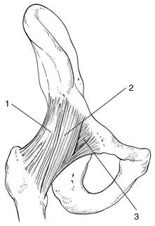
Fig 4 Anterior ligaments: 1, iliofemoral, lateral part; 2, iliofemoral, medial part; 3, pubofemoral.
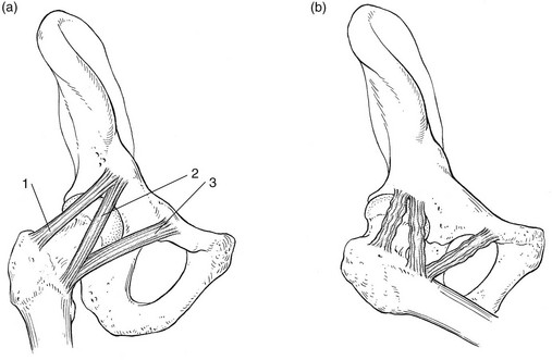
Fig 5 Lateral view of the ligaments in extension (a) and flexion (b): 1, iliofemoral, lateral part; 2, iliofemoral, medial part; 3, pubofemoral.
The ligamentum teres plays only a minor role in the control of hip movements. Adduction from a semi-flexed position is the only movement where this ligament is under tension.
Muscles
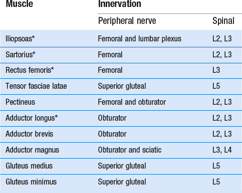
The hip joint is surrounded by a large number of muscles. According to their function these are divided into six groups: (1) flexors, (2) extensors, (3) abductors, (4) adductors, (5) lateral rotators, (6) medial rotators. In this chapter the anatomical and kinesiological aspects of particular importance in orthopaedic medicine are discussed.
Flexor muscles
The flexor muscles of the hip joint (Table 1) are anterior to the axis of flexion and extension.
The iliopsoas is the most powerful of the flexors (Fig. 6). It originates at the lumbar vertebrae and the corresponding intervertebral discs of the last thoracic and all the lumbar vertebrae, the superior two-thirds of the bony iliac fossa and the iliolumbar and ventral sacroiliac ligaments. The insertion is to the lesser trochanter. Although its main function is flexion, it is also a weak adductor and lateral rotator. The distal part of the muscle is palpable just deep to the inguinal ligament, where it lies bordered by the sartorius muscle laterally and the femoral artery medially (Fig. 7).
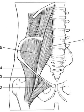
Fig 6 The iliopsoas and associated structures: 1, psoas; 2, pectineus; 3, iliopsoas tendon; 4, inguinal ligament; 5, iliacus.
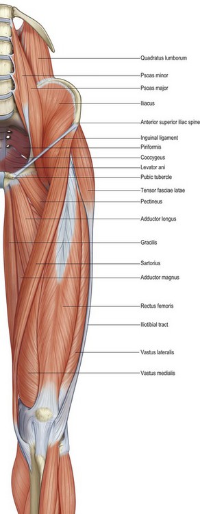
Fig 7 Anterior view of the hip muscles. From Standring, Gray’s Anatomy, 40th edn. Churchill Livingstone/Elsevier, Philadelphia, 2009 with permission.
The sartorius is mainly a flexor of the hip, originating at the anterior superior iliac spine and inserting at the proximal part of the medial surface of the tibia (see Fig. 7). Consequently the muscle acts on two joints, with the accessory function of lateral rotation and abduction of the hip as well as flexion and medial rotation of the knee. At the surface, the muscle divides the anterior aspect of the thigh into a medial and a lateral femoral triangle. During active flexion, abduction and lateral rotation at the hip and 90° flexion at the knee, the muscle becomes prominent and is easily palpable.
The rectus femoris combines movements of flexion at the hip and extension at the knee. Its origin is at the anterior inferior iliac spine, a groove above the acetabulum and the fibrous capsule of the hip joint and inserts into the common quadriceps tendon at the proximal border of the patella (see Fig. 7). The origin can be palpated only in a sitting position because of tension in the overlying structures. The tendon and muscle belly are bordered medially by the sartorius muscle, and laterally by the tensor fasciae latae and the vastus lateralis, the largest part of the quadriceps.
The tensor fasciae latae (see Fig. 7) originates at the outer surface of the anterior superior iliac spine, and inserts into the proximal part of the iliotibial tract – a strong band which thickens the fascia lata at its lateral aspect. Thus the course of the tensor is dorsal and distal. Acting through the iliotibial tract the muscle extends and rotates the knee laterally. It may also assist in flexion, abduction and medial rotation of the hip. In the erect posture, it helps to steady the pelvis on the head of the femur (Fig. 8). The muscle can be palpated easily during resisted flexion and abduction of the hip with the knee extended.
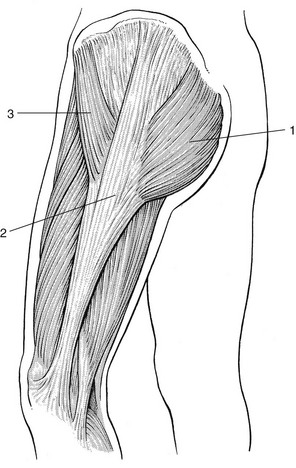
Fig 8 Lateral view of the hip muscles: 1, gluteus maximus; 2, iliotibial tract; 3, tensor fasciae latae.
A number of other muscles are also active during flexion of the hip joint but only via their accessory function. These are the pectineus, adductor longus and brevis, and the most anterior fibres of the adductor magnus and the glutei (medius and minimus).
Extensor muscles
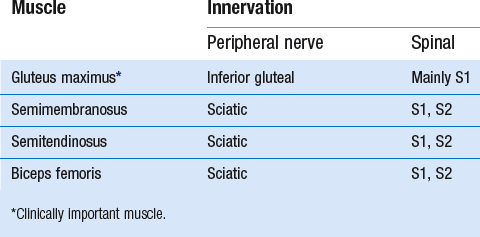
These (Table 2) are posterior to the axis of flexion and extension of the hip.
Gluteus maximus
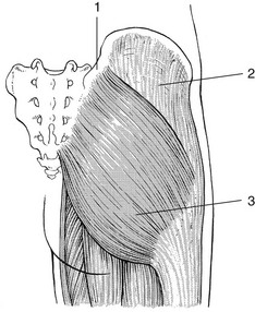
The most important extensor is the gluteus maximus (Fig. 9) which takes origin from the posterior gluteal line and crest of the ilium, the lower part of the sacrum, the coccyx and the sacrotuberous ligament, and runs in a lateral and caudal direction. Three-quarters of the muscle inserts at the proximal part of the iliotibial tract (see Fig. 8) and the other part into the gluteal tuberosity of the femur. It is a strong extensor. The lower fibres also have a function in lateral rotation and adduction and the upper fibres assist in powerful abduction. In standing, the muscle is inactive and remains so during the forward bending at the hip joint. However, in conjunction with the hamstrings, it is active in raising the trunk after stooping.
Hamstrings: semimembranosus, semitendinosus and biceps femoris
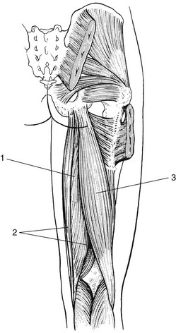
These muscles (Fig. 10) are also important extensors of the hip. Because the muscles cross two joints, their efficiency at the hip depends on the (extended) position of the knee. Their origins are from the upper area of the ischial tuberosity.
The semimembranosus inserts at the posterior aspect of the medial condyle of the tibia. An additional attachment is to the posterior capsule of the knee joint. Throughout its extent the muscle is partly overlapped by the semitendinosus and is therefore only palpable on each side of the latter.
The semitendinosus inserts at the proximal part of the medial surface of the tibia, behind the attachments of the sartorius and gracilis. Both muscles have accessory functions in medial rotation and adduction of the thigh, and in flexion and medial rotation of the knee joint.
The tendon of the biceps femoris splits round the fibular collateral ligament and inserts into the lateral side of the head of the fibula. The accessory function of this muscle is lateral rotation and adduction of the thigh, and flexion and lateral rotation of the knee.
The collective origin of these muscles at the tuberosity of the ischium is best palpated in the side-lying position or supine lying, with the hip flexed to 90°. This position moves the gluteus maximus upwards so permitting direct palpation of the tuberosity. Palpation of the muscle bellies is performed in prone lying, with the knee flexed to less than 90° and during slight contraction of the muscles. Resisted medial rotation of the lower leg makes semitendinosus and semimembranosus prominent. Resisted lateral rotation of the lower leg tenses the biceps and also makes the tendon visible and palpable at the lateral and distal part of the thigh.
Abductor muscles
The main abductor muscle is the gluteus medius (Fig. 11). It originates from the external surface of the ilium, just below the iliac crest. Insertion is on the lateral surface of the greater trochanter of the femur. The muscle is partially covered by two other muscles: anteriorly the tensor fasciae latae, posteriorly the gluteus maximus.
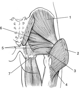
Fig 11 The gluteus medius muscle – the main abductor of the hip joint: 1, gluteus medius; 2, gluteus maximus (reflected back); 3, quadratus femoris; 4, gluteal bursa; 5, gemelli; 6, piriformis; 7, ischial bursa.
The gluteus medius stabilizes the pelvis in the transverse direction. Standing on one limb, strong action of the gluteus medius, powerfully assisted by gluteus minimus and tensor fasciae latae, keeps the pelvis horizontal. This stabilization of the pelvis is essential for normal walking. In mild or moderate weakness of these muscles, the characteristic sign of Duchenne–Trendelenburg syndrome is demonstrated, i.e. the patient is unable to keep the pelvis horizontal, which is tilted to the opposite side. In gross weakness an excessive movement of the trunk towards the affected side compensates the paralysed hip abductors (‘abduction lurch’).
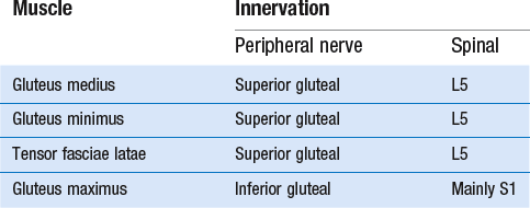
The other important abductors are the gluteus minimus, the tensor fasciae latae and the upper part of the gluteus maximus (Table 3).
The lateral aspect of the hip is covered by a wide muscular fan (see Fig. 8) made up of two muscle bellies: anteriorly the tensor fasciae latae, posteriorly the superficial fibres of the gluteus maximus. Both insert into the anterior and posterior borders of the iliotibial tract at its proximal aspect. Because of the triangular shape and the anatomical and functional similarity to the deltoid muscle of the shoulder joint, these muscles are sometimes known as the ‘deltoid of the hip’.
The iliotibial tract is a long and strong band, which is part of the fascia lata and attached to the anterior aspect of the lateral tibial condyle. This structural arrangement permits the muscles to influence the stability of the extended knee joint and thus help to maintain the erect posture.
The iliotibial tract overrides the greater trochanter, where it is vulnerable to strain.
Adductor muscles
The adductors lie medial to the central axis of the hip joint. Although the adductor magnus is the most powerful it is clinically not important. The adductor longus (see Fig. 7) is more easily strained. Its origin is at the anterior aspect of the pubis at the junction of the pubic crest and symphysis, and it inserts at the middle third of the linea aspera of the femur. During resisted adduction the adductor longus is the most prominent muscle of the adductor group and forms the medial border of the femoral triangle. Its accessory function is flexion of the hip.
Other adductors are the pectineus, adductor magnus, quadratus femoris, external obturator and the greatest part of the gluteus maximus. Another adductor is the gracilis, the most superficial of the adductor group. It arises broadly from the inferior ramus of the pubis. The muscle belly lies just dorsal to the adductor longus (see Fig. 7). The tendon of this biarticular muscle passes across the medial condyle of the femur, posterior to the tendon of the sartorius, and is attached to the upper part of the medial surface of the tibia. Because of this course it is also a flexor and medial rotator of the knee.
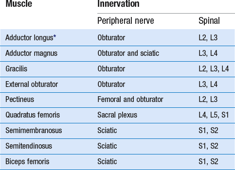
Finally, the semitendinosus, semimembranosus and biceps femoris also assist in adduction of the hip (Table 4).
There is a strong functional relationship between the abdominal muscles and the adductors of the hip joint. ‘Adductor tendinitis’ and ‘rectus abdominus tendinitis’ are often seen simultaneously.
Lateral rotator muscles
When the resisted lateral rotation test is painful, the quadratus femoris should be sought first because the lesion is usually in this muscle (see Fig. 11). This flat quadrilateral muscle arises from the upper part of the lateral border of the tuberosity of the ischium and inserts just distally from the intertrochanteric crest of the femur.
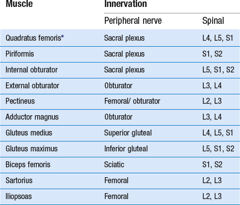
Other lateral rotators of the hip (Table 5) that cross the joint posteriorly, such as the piriformis, the obturator muscles, pectineus, the posterior fibres of the adductor magnus, the gluteus maximus and the posterior fibres of gluteus medius, are clinically unimportant (see Standring, Fig. 80.21).
The long head of biceps also laterally rotates the thigh when the hip is extended.
Anteriorly, the sartorius and iliopsoas have only an accessory function during lateral rotation.
Medial rotator muscles
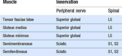
The medial rotators (Table 6) that cross the hip anteriorly are the tensor fasciae latae and the anterior fibres of gluteus medius and minimus. Posteriorly, semimembranosus and semitendinosus have an accessory function in medial rotation when the hip is extended.
Bursae
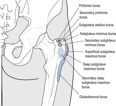
The gluteal bursae are situated deeply, just above and behind the greater trochanter underneath the gluteus medius and maximus (see Fig. 11).
The trochanteric bursa (or subgluteus maximus bursa) is situated more superficially, laterally between the greater trochanter and the iliotibial tract (Fig. 12).
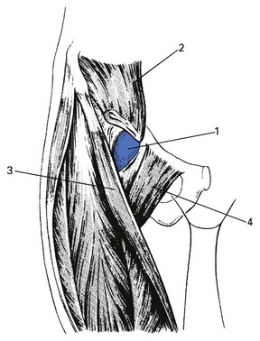
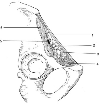
The psoas bursa, also called the bursa iliopectinea, lies deep to the iliopsoas muscle at the floor of the femoral triangle and just in front of the hip joint, with which it may communicate (Fig. 13).
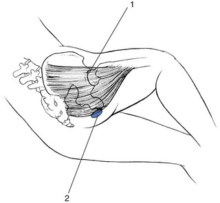
The ischial bursa lies distally at the tuberosity just covered by the edge of the gluteus maximus (see Fig. 11). In a flexed, i.e. sitting, position the muscle is pulled up slightly so that in bursitis pain results because of compression of the inflamed bursa between the seat and the tuberosity.
Nerves
The femoral nerve (see Fig. 15) arises mainly from the second and third lumbar spinal nerves. It passes down between the psoas major and iliacus muscles, then behind the inguinal ligament to enter the thigh. At this proximal level, the nerve lies just lateral to the femoral artery (see Standring, Fig. 80.28). Muscular branches supply the iliacus, pectineus, sartorius and quadriceps muscles. The skin on the front of the thigh is supplied by several cutaneous branches.
The lateral cutaneous nerve of the thigh arises from the second and third lumbar spinal nerves. It passes behind or through the inguinal ligament about 1 cm medial to the anterior superior iliac spine. At the proximal part of the sartorius it divides into two branches to supply the anterolateral part of the thigh as far as the knee.
The obturator nerve arises mainly from the third and fourth lumbar spinal nerves. It enters the thigh through the obturator foramen. Some cutaneous branches are given to the skin on the medial side of the thigh, whereas another branch supplies the capsule of the hip joint. Muscular branches are distributed to the pectineus, adductor longus, gracilis, adductor brevis, external obturator and adductor magnus.
The sciatic nerve (see Fig. 17), the largest nerve in the body, arises from the fourth and fifth lumbar and first and second sacral spinal nerves. It passes out of the pelvis through the greater sciatic foramen below the piriformis muscle. On its medial side it is accompanied by the inferior gluteal artery and the posterior cutaneous nerve of the thigh. The nerve descends just medial to the midpoint of a line joining the greater trochanter of the femur and the tuberosity of the ischium. Muscular branches are distributed to the semimembranosus, semitendinosus and biceps femoris muscles. It also supplies the capsule of the hip joint through articular branches.
Blood vessels
The abdominal aorta bifurcates at the level of the fourth lumbar vertebrae into right and left common iliac arteries (Fig. 14).
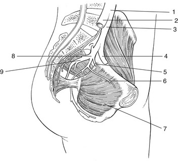
Fig 14 Arteries: 1, aorta; 2, right common iliac artery; 3, left common iliac artery; 4, external iliac artery; 5, superior gluteal artery; 6, obturator artery; 7, internal obturator muscle; 8, internal iliac artery; 9, inferior gluteal artery.
At the level of the sacroiliac joint, these vessels divide into external and internal iliac arteries. Passing behind the inguinal ligament just midway between symphysis pubis and anterior superior iliac spine, the external artery becomes the femoral artery. The internal iliac arteries divide into three: the superior and inferior gluteal arteries supplying the buttock and the obturator artery which supplies the hip joint and the adductor muscles (Fig. 15).
Topographical anatomy
Anterior side
The anterior superior iliac spines (ASIS) are easily palpable except in obese patients. They form the point of origin of the sartorius muscle anteriorly and the tensor fasciae latae laterally. The spines are not level when there is leg length difference. The same occurs in pelvic torsion. However, the level of the posterior superior iliac spine (PSIS) on the same side is then just reversed, i.e. in a posterior rotation of the right innominate bone the ASIS on that side is higher whereas the PSIS is lower compared with the other side.
The anterior inferior iliac spines, lying just beneath the superior ones, are best palpated in a sitting position, with the hip flexed to 90°. These are the points of origin of the rectus femoris muscles.
Towards the midline, the pubic tubercles provide attachment for the medial end of the inguinal ligament. They are palpable as bony prominences lying at the same level as the superior aspect of the greater trochanters. The rectus abdominus inserts with a medial and lateral head just cranial to these tubercles at the crest of the pubic bone.
For topographical reasons, it is also important to have an understanding of the anatomy of the femoral triangle (see Fig. 7), which is defined superiorly by the inguinal ligament, medially by the adductor longus and laterally by the sartorius. The floor of the triangle is formed by portions of the iliopsoas on the lateral side and the pectineus on the medial side. The femoral artery lies superficial and medial to the iliopsoas muscle and is easily found by palpation of the pulse as it emerges from behind the inguinal ligament (see Fig. 15). Deep to the iliopsoas lies the psoas bursa and still deeper the hip joint.
The femoral head, however, is not palpable because of the presence of the overlying muscles. The femoral nerve and vein (lateral and medial to the artery, respectively) are not palpable.
Several lymph nodes are situated medially in the femoral triangle. They can be palpated only when they are enlarged.
Lateral side
Palpation of the greater trochanter is relatively easy at its posterior edge, where the bone is not covered by muscles. The greater trochanter is an important landmark and, in the standing position, both greater trochanters should be level and at the same distance from the iliac crest and the anterior superior iliac spines. Their upper aspects should also be on the same level as the pubic tubercles.
Posterior side
The ischial tuberosity is covered by the gluteus maximus and adipose tissue (Fig. 16).
If the hip is flexed, the gluteus maximus moves upwards and the ischial tuberosity becomes easily palpable. It provides attachment for the hamstring muscles posteriorly and the quadratus femoris laterally. At its distal aspect, the bone is covered by a bursa (see Fig. 11).
Midway between the greater trochanter and the ischial tuberosity the sciatic nerve passes to the leg (Fig. 17). With the hip in a flexed position this nerve may be palpable underneath the adipose tissue.
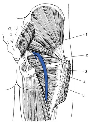
Fig 17 The sciatic nerve: 1, piriformis; 2, gemellus superior; 3, sciatic nerve; 4, gluteus maximus (resected); 5, ischial tuberosity.
The posterior superior iliac spines (see Fig. 9) are easily palpable, where they lie directly underneath the visible dimples just above and medial to the buttocks.
Joint movements
The hip has three degrees of freedom: flexion–extension, adduction–abduction, and medial and lateral rotation.
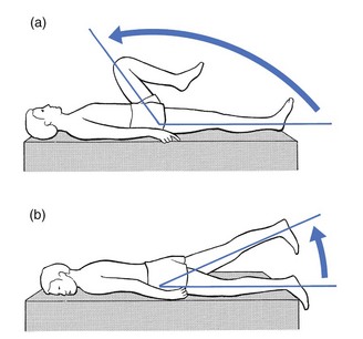
Flexion is forward movement in the sagittal plane (Fig. 18a). The range depends on the position of the knee. With the knee flexed and the movement performed passively, the anterior aspect of the thigh comes in close contact with the abdomen, so that the range exceeds 140°. With the knee extended, flexion is limited by the tension of the hamstrings. These muscles are biarticular and therefore restrict hip flexion when the knee is extended – the constant-length phenomenon.
Extension backward movement in the sagittal plane (Fig. 18b) is considerably restricted by the tension of the iliofemoral ligament. Passively an average range of 30° can be reached.
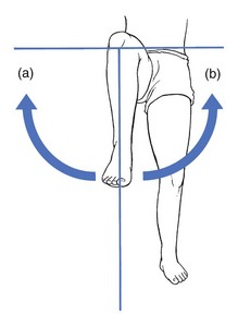
Adduction and abduction are movements in the frontal plane (Fig. 19). The average range of adduction is 30°, and abduction 45°. In the latter, the constant-length phenomenon again plays a vital part. Here the biarticular gracilis restricts the range of abduction when the knee joint is fully extended.
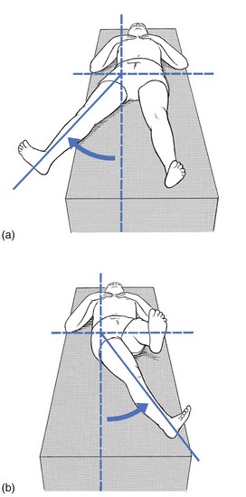
Rotational movements of the hip are measured in the supine position, with the thigh in 90° of flexion (Fig. 20), or lying face down, the hip at 0° and the knee flexed to 90°. The normal range of medial rotation is about 45°, whereas lateral rotation reaches about 60°. With the thigh flexed, lateral rotation can be increased, because of relaxation of the iliofemoral and pubofemoral ligaments.
Cyriax, J. Textbook of Orthopaedic Medicine, 8th ed. London: Baillière Tindall; 1982.
Hoppenfeld, S. Physical Examination of the Spine and Extremities. New York: Appleton-Century-Crofts; 1976.
Kapandji, IA. The Physiology of the Joints. Vol 2, Lower Limb, 2nd ed. London: Churchill Livingstone; 1970.
Kendall, HO, Kendall, FP, Wadsworth, GE. Muscle Testing and Function, 2nd ed. Baltimore: Williams & Wilkins; 1971.
Martens, M, Hansen, C, Mulier, J, Adductor tendinitis and musculus rectus abdominus tendopathy. Am J Sports Med 1987; 4:156–158. ![]()
Warwick, R, Williams, PL. Gray’s Anatomy, 35th ed. London: Longman; 1973.

