Anatomy of the female pelvis

Published on 09/03/2015 by admin

Filed under Obstetrics & Gynecology

Last modified 22/04/2025

Print this page

rate 1 star rate 2 star rate 3 star rate 4 star rate 5 star
Your rating: none, Average: 0 (0 votes)

This article have been viewed 7642 times

Anatomy of the female pelvis

Caroline de Costa

Knowledge of the major features of the female pelvis is essential to the understanding of the processes of reproduction and childbearing and to the effect that various pathological processes may have on the pelvic organs and on the health of the woman.

The structure and function of the genital organs vary considerably with the age of the individual and her hormonal status, as will be apparent in chapter 16, which covers the changes that take place in puberty and the menopause. This chapter aims to outline the major structures comprising the female pelvis, predominantly in the sexually mature female.
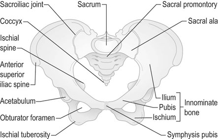
The bony pelvis

The bony pelvis consists of the paired innominate bones (each consisting of ilium, ischium and pubis) and the sacrum and coccyx (Fig. 1.1).

The innominate bones are joined anteriorly at the symphysis pubis and each articulates posteriorly with the sacrum in the sacroiliac joints. All three joints are fixed in the non-pregnant state, but during pregnancy there is relaxation of the joints to allow some mobility during labour and birth. The sacrum articulates with the fifth lumbar vertebra superiorly and the coccyx inferiorly.

The bony pelvis is divided into the false pelvis and the true pelvis by the pelvic brim. The true pelvis is divided into three sections: the pelvic inlet (bounded anteriorly by the superior surface of the pubic bones and posteriorly by the promontory and alae of the sacrum); the mid-pelvis (at the level of the ischial spines); and the pelvic outlet (bounded anteriorly by the lower border of the symphysis, laterally by the ischial tuberosities and posteriorly by the tip of the sacrum).

The external genitalia

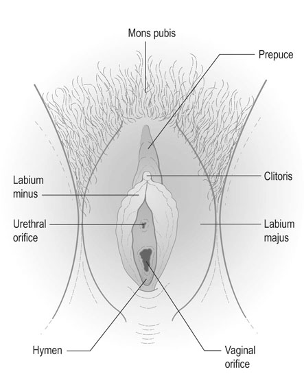
The term vulva is generally used to describe the female external genitalia, and includes the mons pubis, the labia majora, the labia minora, the clitoris, the external urinary meatus, the vestibule of the vagina, the vaginal orifice and the hymen (Fig. 1.2).

The mons pubis, sometimes known as the mons veneris, is composed of a fibrofatty pad of tissue that lies above the pubic symphysis and, in the mature female, is covered with dense pubic hair. The upper border of this hair is usually straight or convex upwards and differs from the normal male distribution. Pubic hair generally begins to appear between the ages of 11 and 12 years.

The labia majora consist of two longitudinal cutaneous folds that extend downwards and posteriorly from the mons pubis anteriorly to the perineum posteriorly. The labia are composed of an outer surface covered by hair and sweat glands and an inner smooth layer containing sebaceous follicles. The labia majora enclose the pudendal cleft into which the urethra and vagina open.

Posterior to the vaginal orifice, the labia merge to form the posterior commissure and the area between this structure and the anterior verge of the anus constitutes the obstetric perineum.

The labia majora are homologous with the male scrotum.

The labia minora are enclosed by the labia majora and are cutaneous folds that enclose the clitoris anteriorly and fuse posteriorly behind the vaginal orifice to form the posterior fourchette or posterior margin of the vaginal introitus. Anteriorly, the labia minora divide to enclose the clitoris, the anterior fold forming the prepuce and the posterior fold the frenulum. They are richly vascularized and innervated and are erectile. They do not contain hair but are rich in sebaceous glands.

The clitoris is the female homologue of the penis and is situated between the anterior ends of the labia minora. The body of the clitoris consists of two corpora cavernosa of erectile tissue enclosed in a fibrous sheath. Posteriorly, these two corpora divide to lie along the inferior rami of the pubic bones. The free end of the clitoris contains the glans, composed of erectile tissue covered by skin and richly supplied with sensory nerve endings and hence very sensitive. The clitoris plays an important role in sexual stimulation and function.

The vestibule consists of a shallow depression lying between the labia minora. The external urethral orifice opens into the vestibule anteriorly and the vaginal orifice posteriorly. The ducts from the two Bartholin’s glands drain into the vestibule at the posterior margin of the vaginal introitus and the secretions from these glands have an important lubricating role during sexual intercourse.

Skene’s ducts lie alongside the lower 1 cm of the urethra and also drain into the vestibule. Although they have some lubricating function, it is minor compared to the function of Bartholin’s glands.

The bulb of the vestibule consists of two erectile bodies that lie on either side of the vaginal orifice and are in contact with the surface of the urogenital diaphragm. The bulb of the vestibule is covered by a thin layer of muscle known as the bulbocavernosus muscle.

The external urethral orifice lies 1.5–2 cm below the base of the clitoris and is often covered by the labia minora, which also function to direct the urinary stream. In addition to Skene’s ducts, there are often a number of paraurethral glands without associated ducts and these sometimes form the basis of paraurethral cysts.

The vaginal orifice opens into the lower part of the vestibule and, prior to the onset of sexual activity, is partly covered by the hymenal membrane. The hymen is a thin fold of skin attached around the circumference of the vaginal orifice. There are various types of opening within the hymen and the membrane varies in consistency. Once the hymen has been penetrated, the remnants are represented by the carunculae myrtiformes, which are nodules of fibrocutaneous material at the edge of the vaginal introitus.

Bartholin’s glands are a pair of racemose glands located at either side of the vaginal introitus and measuring 0.5–1.0 cm in diameter. The ducts are approximately 2 cm in length and open between the labia minora and the vaginal orifice. Their function is to secrete mucus during sexual arousal. Cyst formation is relatively common but is the result of occlusion of the duct, with fluid accumulation in the duct and not in the gland.

Although it does not strictly lie within the description of the vulva, the perineum as described in relation to obstetric function is defined as the area that lies between the posterior fourchette anteriorly and the anus posteriorly; it lies over the perineal body, which occupies the area between the anal canal and the lower one-third of the posterior vaginal wall.

The internal genital organs

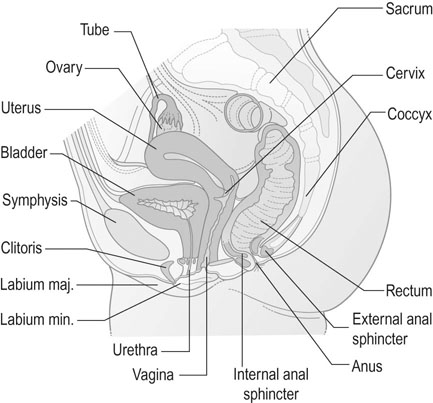
The internal genitalia include the vagina, the uterus, the Fallopian tubes and the ovaries. Situated in the pelvic cavity, these structures lie in close proximity to the urethra and urinary bladder anteriorly and the rectum, anal canal and pelvic colon posteriorly (Fig. 1.3).

The vagina

The vagina is a muscular tube some 6–7.5 cm long in the mature female. It is lined by non-cornified squamous epithelium and is more capacious at the vault than at the introitus. In cross-section, the vagina is H-shaped and it is capable of considerable distension, particularly during parturition when it adapts to accommodate the passage of the fetal head. Anteriorly, it is intimately related to the trigone of the urinary bladder and the urethra. Posteriorly, the lower part of the vagina is separated from the anal canal by the perineal body. In the middle third, it lies in apposition to the ampulla of the rectum and in the upper segment it is covered by the peritoneum of the rectovaginal pouch (pouch of Douglas).

The uterine cervix protrudes into the vaginal vault. Four zones are described in the vaginal vault: the anterior fornix; the posterior fornix; and the two lateral fornices. The lateral fornices lie under the base of the broad ligament in close proximity to the point where the uterine artery crosses the ureter.

The pH of the vagina in the sexually mature non-pregnant female is between 4.0 and 5.0. This has an important antibacterial function that reduces the risk of pelvic infection. The functions of the vagina are copulation, parturition and the drainage of menstrual loss.

The uterus

The uterus is a hollow, muscular, pear-shaped organ situated in the pelvic cavity between the bladder anteriorly and the rectum and pouch of Douglas posteriorly. The size of the uterus depends on the hormonal status of the female. In the sexually mature female, the uterus is approximately 7.5 cm long and 5 cm across at its widest point. The uterus normally lies in a position of anteversion such that the uterine fundus is anterior to the uterine cervix. In about 10% of women, the uterus lies in a position of retroversion in the pouch of Douglas. The uterus may also be curved anteriorly in its longitudinal axis, a feature that is described as anteflexion, or posteriorly, when it is described as retroflexion.

It consists of a body or corpus, an isthmus and a cervix.

The corpus uteri consists of a mass of smooth muscle cells, the myometrium, arranged in three layers. The external layers contain smooth muscle cells that pass transversely across the uterine fundus into the lateral angles of the uterus, where their fibres merge with the outer layers of the smooth muscle of the Fallopian tubes and the ovarian and round ligaments. The muscle fibres in the middle layer are arranged in a circular manner and the inner layer contains a mixture of longitudinal, circular and oblique muscle fibres.

The cavity of the uterus is triangular in shape and is flattened anteroposteriorly so that the total volume of the cavity in the non-pregnant state is approximately 2 mL. It is lined by endometrium that consists on the surface of mucus-secreting columnar epithelium. The nature of the endometrium depends on the phase of the menstrual cycle. Following menstruation, the endometrium in the proliferative phase is only 1–2 mm thick. By the second half (secretory phase) of the cycle the endometrium has grown to a thickness of up to 1 cm.

The endometrial cavity is in contact with the vaginal cavity inferiorly via the cervical canal and superiorly with the peritoneal cavity through the Fallopian tubes.

The cervix is a barrel-shaped structure extending from the external cervical os, which opens into the vagina at the apex of the vaginal portion of the cervix, to the internal cervical os in its supravaginal portion. The internal os opens into the uterine cavity through the isthmus of the uterus. In non-parous women the external os is round or oval, but it becomes transverse following vaginal birth and this can be noted in clinical examination when a speculum is passed, for example, when taking a Pap smear.

The cervical canal is fusiform in shape and is lined by ciliated columnar epithelium that is mucus-secreting. The transition between this epithelium and the stratified squamous epithelium of the vaginal ectocervix forms the squamocolumnar junction. The exact site of this junction is related to the hormonal status of the woman. Some of the cervical glands in the endocervical lining are extensively branched and mucus-secreting. If the opening to these glands becomes obstructed, small cysts may form, known as nabothian follicles.

The cervix consists of layers of circular bundles of smooth muscle cells and fibrous tissue. The outer longitudinal layer merges with the muscle layer of the vagina.

The isthmus of the uterus joins the cervix to the corpus uteri and in the non-pregnant uterus is a narrow, rather poorly defined, area some 2–3 mm in length. In pregnancy, it enlarges and contributes to the formation of the lower segment of the uterus, which is the normal site for the incision of caesarean section. In labour it becomes a part of the birth canal but does not contribute significantly to the expulsion of the fetus.

Supports and ligaments of the uterus

The uterus and the pelvic organs are supported by a number of ligaments and fascial thickenings of varying strength and importance. The pelvic organs also depend for support on the integrity of the pelvic floor: a particular feature in the human female is that, an upright posture having been adopted, the pelvic floor has to contain the downward pressure of the viscera and the pelvic organs.

The anterior ligament is a fascial condensation that, with the adjacent peritoneal uterovesical fold, extends from the anterior aspect of the cervix across the superior surface of the bladder to the peritoneal peritoneum of the anterior abdominal wall. It has a weak supporting role.

Posteriorly, the uterosacral ligaments play a major role in supporting the uterus and the vaginal vault. These ligaments and their peritoneal covering form the lateral boundaries of the rectouterine pouch (of Douglas). The ligaments contain a considerable amount of fibrous tissue and non-striped muscle and extend from the cervix onto the anterior surface of the sacrum.

Laterally, the broad ligaments are reflected folds of peritoneum that extend from the lateral margins of the uterus to the lateral pelvic walls. They cover the Fallopian tubes and the round ligaments, the blood vessels and nerves that supply the uterus, tubes and ovaries, and the mesovarium and ovarian ligaments that suspend the ovaries from the posterior surface of the broad ligament. Like the anterior ligaments, the broad ligaments play only a weak supportive role for the uterus.

The round ligaments are two fibromuscular ligaments that extend from the anterior surface of the uterus. In the non-pregnant state, they are a few millimetres thick and are covered by the peritoneum of the broad ligaments. They arise from the anterolateral surface of the uterus just below the entrance of the tubes and extend diagonally and laterally for 10–12 cm to the lateral pelvic walls, where they enter the abdominal inguinal canal, and blend into the upper part of the labia majora. These ligaments have a weak supporting role for the uterus but do play a role in maintaining its anteverted position. In pregnancy, they become much thickened and strengthened, and during contractions may pull the uterus anteriorly and align the long axis of the fetus in such a way as to improve the direction of entry of the presenting part into the pelvic cavity.

The cardinal ligaments (transverse cervical ligaments) form the strongest supports for the uterus and vaginal vault and are dense fascial thickenings that extend from the cervix to the fascia over the obturator fossa on each pelvic side wall. Medially, they merge with the mass of fibrous tissue and smooth muscle that encloses the cervix and the vaginal vault and is known as the parametrium. The uterosacral ligaments merge with the parametrium. Close to the cervix, the parametrium contains the uterine arteries, nerve plexuses and the ureter passing through the ureteric canal to reach the urinary bladder. Lower down, the muscular activity of the pelvic floor muscles and the integrity of the perineal body play a vital role in preventing the development of uterine prolapse (see Chapter 21).

The Fallopian tubes

The Fallopian tubes or uterine tubes are the oviducts. They extend from the superior angle of the uterus, where the tubal canal at the tubal ostium opens into the lateral and uppermost part of the uterine cavity. The tubes are approximately 10–12 cm long and lie on the posterior surface of the broad ligament, extending laterally in a convoluted fashion so that, eventually, the tubes open into the peritoneal cavity in close proximity to the ovaries.

The tubes are enclosed in a mesosalpinx, a superior fold of the broad ligament, and this peritoneal fold, apart from the tube, also contains the blood vessels and nerve supply to the tubes and the ovaries. It also houses various embryological remnants such as the epoophoron, the paroophoron, Gartner’s duct and the hydatid of Morgagni. These embryological remnants are significant in that they may form para-ovarian cysts, which are difficult to differentiate from true ovarian cysts. They are generally benign.

The tube is divided into four sections:

The tubes are lined by a single layer of ciliated columnar epithelium which serves to assist the movement of the oocyte down the tube. The tubes are richly innervated and have an inherent rhythmicity that varies according to the stage of the menstrual cycle and whether or not the woman is pregnant.

The ovaries

The ovaries are paired almond-shaped organs that have both reproductive and endocrine functions.

They are approximately 2.5–5 cm in length and 1.5–3.0 cm in width. Each ovary lies on the posterior surface of the broad ligaments in a shallow depression known as the ovarian fossa in close proximity to the external iliac vessels and the ureter on the lateral pelvic walls. Each has a medial and a lateral surface, an anterior border, a posterior border that lies free in the peritoneal cavity, an upper or tubal pole and a lower or uterine pole.

The anterior border of the ovary is attached to the posterior layer of the broad ligament by a fold in the peritoneum known as the mesovarium. This fold contains the blood vessels and nerves supplying the ovary. The tubal pole of the ovary is attached to the pelvic brim by the suspensory ligament (infundibulopelvic fold) of the ovary. The lower pole is attached to the lateral border of the uterus by a musculofibrous condensation known as the ovarian ligament.

The surface of the ovary is covered by a cuboidal or low columnar type of germinal epithelium. This surface opens directly into the peritoneal cavity.

Beneath the germinal epithelium is a layer of dense connective tissue that effectively forms the capsule of the ovary; this is known as the tunica albuginea. Beneath this layer lies the cortex of the ovary, formed by stromal tissue and collections of epithelial cells that form the Graafian follicles at different stages of maturation and degeneration. These follicles can also be found in the highly vascular, central portion of the ovary: the medulla. The blood vessels and nerve supply enter the ovary through the medulla.

The blood supply to the pelvic organs

Internal iliac arteries

The major part of the blood supply to the pelvic organs is derived from the internal iliac arteries (sometimes known as the hypogastric arteries), which originate from the bifurcation of the common iliac vessels into the external iliac arteries and the internal iliac vessels (Fig. 1.4).

The internal iliac artery arises at the level of the lumbosacral articulation and passes over the pelvic brim, continuing downward on the posterolateral wall of the cavity of the true pelvis beneath the peritoneum until it crosses the psoas major and the piriformis muscles. It then reaches the lumbosacral trunk of the sacral plexus of nerves and, at the upper margin of the greater sciatic notch, it divides into anterior and posterior divisions. It then continues as the umbilical artery, which shortly after birth, becomes obliterated to form the lateral umbilical ligament. Thus, in fetal life, this is the major vascular network, which delivers blood via the internal iliac anterior division and its continuation as the umbilical artery to the placenta.

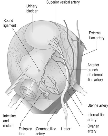
The branches of the two divisions of the internal iliac artery are as follows.

Anterior division

The anterior division provides the structure for the umbilical circulation as previously described. It also provides the superior, middle and inferior vesical arteries that provide the blood supply for the bladder. The superior and middle branches, having passed medially to the lateral and superior surfaces of the bladder, anastomose with branches from the contralateral vessels and with the branches of the uterine and vaginal arteries.

It also forms the middle haemorrhoidal artery.

The uterine artery becomes the major vascular structure arising from this division during pregnancy, when there is a major increase in uterine blood flow. It initially runs downward in the subperitoneal fat under the inferior attachment of the broad ligament towards the cervix.

The artery crosses over the ureter shortly before that structure enters the bladder approximately 1.5–2 cm from the lateral fornix of the vagina. At the point of contact with the vaginal fornix, it gives off a vaginal branch that runs downwards along the lateral vaginal wall. The main uterine artery then follows a tortuous course along the lateral wall of the uterus, giving off numerous branches into the substance of the uterus and finally diverging laterally into the broad ligament to anastomose with the ovarian artery, thus forming a continuous loop that provides the blood supply for the ovaries and the tubes as well as the uterine circulation.

There are also parietal branches of the anterior division of the internal iliac artery and these include the obturator artery, the internal pudendal artery and the inferior gluteal artery.

The ovarian vessels

The other important blood supply to the pelvic organs comes from the ovarian arteries. These arise from the front of the aorta between the origins of the renal and inferior mesenteric vessels. They descend behind the peritoneum on the surface of the corresponding psoas muscle until they reach the brim of the pelvis, where they cross into the corresponding infundibulopelvic fold and from there to the base of the mesovarium, and on to anastomose with the uterine vessels. Both the uterine and ovarian arteries are accompanied by a rich plexus of veins.

The pelvic lymphatic system

The lymphatic vessels follow the course of the blood vessels but have a specific nodal system that is of particular importance in relation to malignant disease of the pelvis (Fig. 1.5).

The lymphatic drainage from the lower part of the vagina, the vulva and perineum and anus passes to the superficial inguinal and adjacent superficial femoral nodes.

The superficial inguinal nodes lie in two groups with an upper group lying parallel with the inguinal ligament and a lower group situated along the upper part of the great saphenous vein.

Some of these nodes drain into the deep femoral nodes, which lie medial to the upper end of the femoral vein.

One of these nodes, known as the gland of Cloquet, occupies the femoral canal.

There are also pelvic parietal nodes grouped around the major pelvic vessels. These include the common iliac, external iliac and internal iliac nodes, which subsequently drain to the aortic chain of nodes.

The lymphatics of the cervix, the uterus and the upper portion of the vagina drain into the iliac nodes whereas the lymphatics of the fundus of the uterus, the Fallopian tubes and the ovaries follow the ovarian vessels to the aortic nodes. Some of the lymphatics from the uterine fundus follow the round ligament into the deep and superficial inguinal nodes.

Nerves of the pelvis

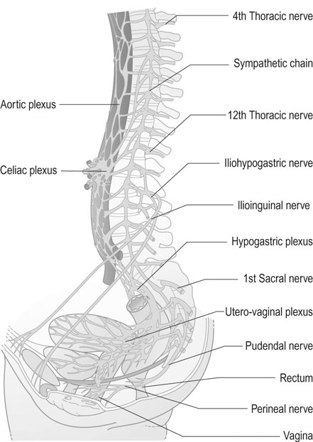
The nerve supply to the pelvis and the pelvic organs has both a somatic and an autonomic component. While the somatic innervation is both sensory and motor in function and relates predominantly to the external genitalia and the pelvic floor, the autonomic innervation provides the sympathetic and parasympathetic nerve supply to the pelvic organs (Fig. 1.6).

Somatic innervation

The somatic innervation to the vulva and pelvic floor is provided by the pudendal nerves that arise from the S2, S3 and S4 segments of the spinal cord. These nerves include both efferent and afferent components.

The pudendal nerves arise in the lumbosacral plexus and leave the pelvis under the sacrospinous ligament to enter Alcock’s canal and pass through the layers of the wall of the ischiorectal fossa to enter the perineum. Motor branches provide innervation of the external anal sphincter muscle, the superficial perineal muscles and the external urethral sphincter.

Sensory innervation is provided to the clitoris through the branch of the dorsal nerve of the clitoris. The sensory innervation of the skin of the labia and of the perineum is also derived from branches of the pudendal nerves. Additional cutaneous innervation of the mons and the labia is derived from the ilioinguinal nerves (L1) and the genitofemoral nerves (L1 and L2) and of the perineum through the posterior femoral cutaneous nerve from the sacral plexus (S1, S2 and S3).

Autonomic innervation

Sympathetic innervation arises from preganglionic fibres at the T10/T11 level and supplies the ovaries and tubes through sympathetic fibres that follow the ovarian vessels.

The body of the uterus and the cervix receive sympathetic innervation through the hypogastric plexus, which accompanies the branches of the iliac vessels, and also contain fibres that signal stretching.

The parasympathetic innervation to the uterus, bladder and anorectum arises from the S1, S2 and S3 segments; these fibres are important in the control of smooth muscle function of the bladder and the anal sphincter system.

Uterine pain is mediated through sympathetic afferent nerves passing up to T11/T12 and L1/L2; the pain is felt in the lower abdomen and the high lumbar spine.

Cervical pain is mediated through the parasympathetic afferent nerves passing backwards to S1, S2 and S3; perineal pain is felt at the site and is mediated through the pudendal nerves.

The pelvic floor

The pelvic floor provides a diaphragm across the outlet of the true pelvis that contains the pelvic organs and some of the organs of the abdominal cavity. The pelvic floor is naturally breached by the vagina, the urethra and the rectum. It plays an essential role in parturition and in urinary and faecal continence (Fig. 1.7). The principal supports of the pelvic floor are the constituent parts of the levator ani muscles. These are described in three sections:
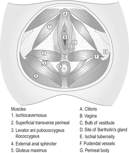
These muscles play an important role in defecation, coughing, vomiting and parturition.

The perineum

The perineum is the region defined as the inferior aperture of the pelvis and consists of all the pelvic structures that lie below the pelvic floor. The area is bounded anteriorly by the inferior margin of the pubic symphysis, the subpubic arch and the ischial tuberosities. Posteriorly, the boundaries are formed by the sacrotuberous ligaments and the coccyx.

The perineum is divided into anterior and posterior triangles by a line drawn between the two ischial tuberosities. The anterior portion is known as the urogenital triangle and includes part of the urethra; the urogenital diaphragm is a condensation of fascia below the level of the pelvic floor muscles and is traversed by the vagina. The posterior or anal triangle includes the anus, the anal sphincter and the perineal body. The two triangles have their bases on the deep transverse perineal muscles.

The ischiorectal fossa lies between the anal canal and the lateral wall of the fossa formed by the inferior ramus of the ischium covered by the obturator internus muscle and fascia. Posteriorly, the fossa is formed by the gluteus maximus muscle and the sacrotuberous ligament, and anteriorly by the posterior border of the urogenital diaphragm.

The pudendal nerve and internal pudendal vessels pass through the lateral aspect of the fossa enclosed in the fascial layer of Alcock’s canal.

image   Essential information

The internal genital organs