[level-membership-for-pulmolory-and-respiratory-category]
Anatomy and Physiology of the Nervous System
I Structure of the Nerve Fiber
A Each nerve fiber has three components: (Figure 16-1)
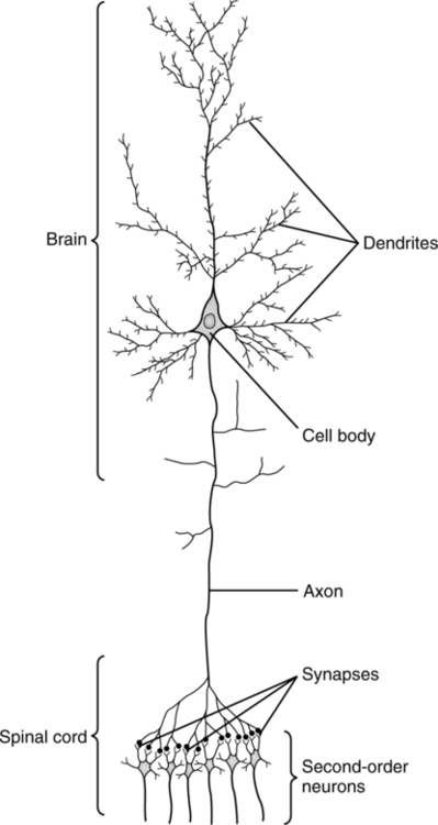
1. Cell body or soma: The primary metabolic area of the nerve, site of initial synthesis of transmitter substances.
2. Dendrite: The structures that carry impulses to the cell body. A single neuron may contain 10,000 dendrites.
3. Axon: The structure that conducts impulses away from the cell body. Usually only one axon leaves a cell body; however, it may branch extensively.
A Each neuron is classified by the direction of impulse transmission.
1. Sensory (afferent) neurons: Transmit nerve impulses to the spinal cord or brain.
2. Motor (efferent) neurons: Transmit nerve impulses from the brain or spinal cord to muscles or glands (effector organs).
3. Interneurons (internuncial neurons): Conduct impulses from sensory to motor neurons entirely within the central nervous system (CNS).
III Nerve Cell Membrane Potential (Figure 16-2)
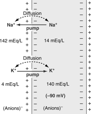
A In the resting state, the inner surface of the cell membrane is negative compared with the positive outer surface. This sets up an electric potential across the cell membrane (normal polarity).
1. Intracellular: The primary cation, potassium (K+), is readily diffusible across the cell membrane.
2. Extracellular: The primary cation, sodium (Na+), is poorly diffusible across the cell membrane.
3. High extracellular Na+ (142 mEq/L) and low extracellular K+ (5 mEq/L) levels, along with high intracellular K+ (140 mEq/L) and low intracellular Na+ (10 mEq/L) levels, are maintained in the resting state by the Na+ pump (active transport).
4. Active transport (Na+ pump): The movement of Na+ from the intracellular space to the extracellular space and the movement of K+ from the extracellular space to the intracellular space. Adenosine triphosphate (ATP) provides the energy for the uphill transport of these ions.
A For a nerve impulse to be transmitted, an alteration in the cell’s resting membrane potential must be activated.
B An action potential is a stimulus that is capable of significantly increasing the cell membrane’s permeability to sodium.
C The action potential is an all-or-nothing phenomenon (i.e., the stimulus must be strong enough to allow for a reversal in the membrane potential); Na+ moves into the cells causing the intracellular side of the membrane to become positive. If the stimulus is not of sufficient strength, an action potential does not occur.
V Nerve Impulse Propagation (Figure 16-3)
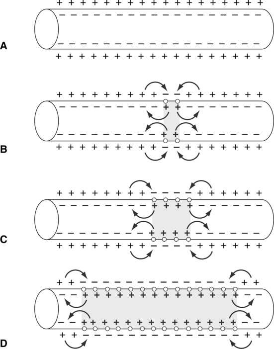
A The nerve impulse is a self-propagating wave of electric charge that travels along the surface of the neuron’s membrane (i.e., the movement of the action potential along the cell membrane changing its charge). The nerve impulse travels in one direction only, from dendrite to cell body to axon.
B Depolarization: Stage 1 of the action potential, in which an impulse travels along the nerve fiber.
1. Sodium ions rush to the inside of the cell membrane.
2. A positive intracellular membrane charge is set up with a negative extracellular membrane charge.
3. There is a complete reversal of the resting membrane potential.
C Repolarization: Stage 2 of the action potential, in which the membrane returns to the normal resting membrane potential.
A The nerve synapse (synaptic cleft) is the junction between one neuron and another neuron, muscle, or gland. It is an actual space between nerve fibers.
B Transmission of an impulse from the axon of one nerve to a dendrite, soma, or effector organ is a chemical process occurring across the synapse. The distance across the synapse is approximately 200 Å.
C Presynaptic terminals are located on the axon and contain vesicles that synthesize, store, and secrete a transmitter substance into the synapse. The transmitter substance stimulates the dendrite or soma of the next nerve, causing an action potential.
D Postsynaptic terminals are located on dendrites, somas, or effector organs. After being stimulated, these terminals secrete a substance into the synapse to metabolize the transmitter substance.
E Acetylcholine as the transmitter substance (Figure 16-4)
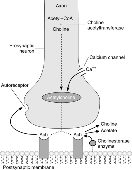
1. Acetylcholine is synthesized in the terminal endings of cholinergic nerve fibers. After synthesis the acetylcholine is transported into the presynaptic vesicles where it is stored and released. It is formed from the reaction of acetyl coenzyme A with choline in the presence of choline acetyltransferase.
2. Vesicles storing acetylcholine are formed in the cell body and migrate to the surface of the presynaptic terminal.
3. When an action potential reaches the presynaptic terminal, acetylcholine is released into the synapse.
4. The acetylcholine moves across the synapse and stimulates a receptor on the postsynaptic terminal.
5. After stimulation the acetylcholine is released back into the synapse, and the postsynaptic terminal releases cholinesterase (acetylcholinesterase and acetylcholine esterase are terms synonymous with cholinesterase), which metabolizes acetylcholine, forming choline and acetate ion.
6. The choline is reabsorbed into the presynaptic terminal and is available to form more acetylcholine.
F Norepinephrine as the transmitter substance (Figure 16-5)
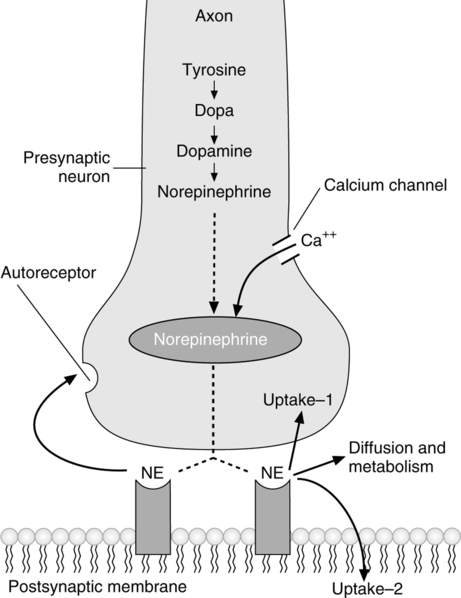
1. Norepinephrine is synthesized inside and outside the presynaptic vesicles, where it is stored and released.
2. The synthesis of norepinephrine proceeds as follows:
3. Norepinephrine is released via a mechanism identical to acetylcholine (see section VI-E).
4. After stimulation norepinephrine has three possible immediate fates:
a. Reabsorption into the presynaptic terminal (50% to 80% of secreted volume) for future secretion.
b. Metabolism in adjoining tissue by catechol-O-methyl-transferase (COMT) or in the nerve ending by monoamine oxidase (MAO).
c. Diffusion to surrounding tissues and eventually into the blood.
5. Complete metabolism of by-products formed in the synapse or norepinephrine entering the blood occurs in the liver via MAO and COMT.
6. In the adrenal medulla the formation of norepinephrine may proceed one step further to epinephrine by the process of methylation.
7. Duration of effect of norepinephrine in the synapse is only a few seconds. However, the norepinephrine and epinephrine released into the blood by the adrenal medulla remain active for up to several minutes.
VII Alteration of Nerve Impulse Transmission
A This junction is the site of transmission of impulses from nerves to skeletal muscle fibers.
B The axon of the nerve branches at its end to form a structure called the motor endplate, which invaginates into muscle fiber but does not penetrate the muscle membrane.
C The motor endplate’s terminal aspects are referred to as sole feet.
D Sole feet provide a large area of “contact” on the muscle surface. It is from the sole feet that the transmitter substance acetylcholine is secreted.
E After stimulation cholinesterase is secreted by the muscle fiber to metabolize the acetylcholine.
IX Reflex Arc or Reflex Action
A The reflex arc is a functional process of the nervous system. Most neuromuscular and neuroglandular mechanisms are controlled by a reflex arc.
B The reflex arc consists of a series of neurons in which impulses are transmitted from a receptor to the CNS and then to a motor neuron to elicit a response.
C Most simple reflexes consist of an impulse from a sensory neuron being transmitted (normally in the spinal cord) to a motor neuron.
D Complex reflexes may involve many internuncial neurons.
E Reflex actions are involuntary, specific, predictable, and adaptive.
X Organization of the Nervous System
A The nervous system is composed of the brain, spinal cord, ganglia (aggregations of nerve cell bodies), and nerves, which regulate and coordinate bodily activities.
B Divisions of the nervous system
1. The CNS is composed of the brain and spinal cord, which acts as a switchboard, receiving and sending impulses to all areas of the body.
2. The peripheral nervous system is composed of neurons that enter and leave the CNS. The peripheral nervous system has two main divisions:
1. The CNS is subdivided into the following structures:
2. The cerebral cortex is primarily responsible for:
a. All higher brain functions (e.g., memory, reasoning, sight, and hearing)
3. The diencephalon is located between the hemispheres of the cerebral cortex and the brain stem containing the following structures:
b. The subthalamus is responsible for the coordination of fine motor activity (extrapyramidal) along with the cerebellum.
4. The brain stem lies within the cranium and connects the diencephalon with the spinal cord. It contains the following structures:
(1) The medulla contains control centers for the following:
(2) The ascending reticular activating system (ARAS) travels through the medulla. The ARAS helps to modulate behavior.
b. The pons contains two centers that effect respiration (see Chapter 6):
c. The midbrain acts as a relay station, controlling vision and hearing.
d. The cerebellum coordinates fine motor activity along with the subthalamus.
D Somatic nervous system (Figure 16-6)
1. The somatic nervous system is responsible for all voluntary muscular activities.
2. Contains sensory and motor neurons.
3. Ganglia, a highly dense collection of nerve cell bodies, are located within the spinal cord.
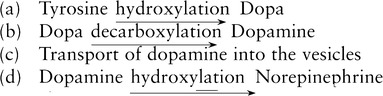
1. Contains only motor neurons (Figure 16-7).
2. Input to the ANS is via the somatic nervous system that is coordinated by the CNS.
3. Each nerve of the ANS has a ganglion, which is located outside of the spinal cord.
4. The ANS is responsible for all involuntary bodily activities.
6. The two divisions are somewhat antagonistic.
7. The ANS is activated and controlled primarily by the:
8. The ANS is activated secondarily by the:
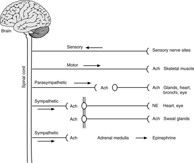
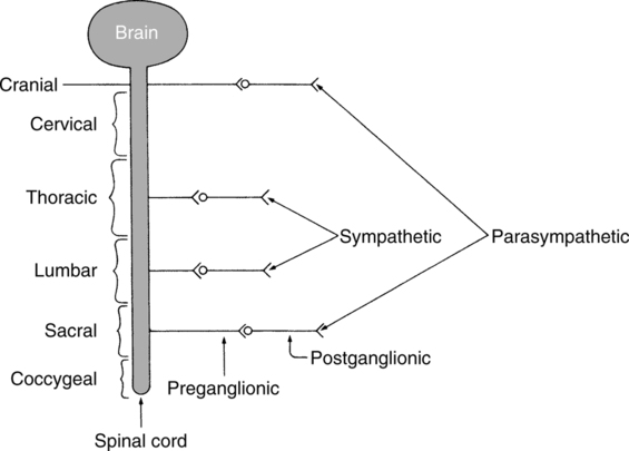
9. SNS: Nerves of the SNS originate from the spinal cord at levels T-1 (thoracic) through L-2 (lumbar) (see Figure 16-7; Figure 16-8).
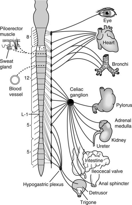
a. Nerve fibers that transmit SNS impulses are either:
b. The SNS fibers synapse primarily at ganglia located along the spinal cord. Here they form a chain of interconnecting ganglia referred to as the sympathetic chain.
c. Some SNS fibers synapse at peripheral ganglia (celiac ganglion and hypogastric plexus), bypassing the sympathetic chain.
10. PNS: Nerves of the PNS originate from sacral nerves 1 through 4 and cranial nerves III, VII, IX, and X. Eighty percent of all PNS impulses originate from cranial nerve X (vagus nerve) (see Figure 16-7; Figure 16-9).
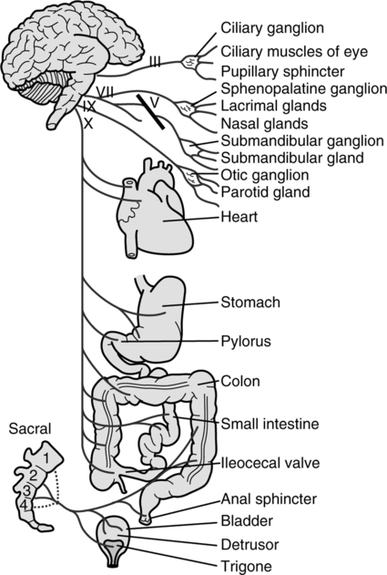
a. Nerve fibers that transmit PNS impulses are either:
b. These fibers synapse at the ganglia of the PNS, which are located close to the organs they innervate.
c. The transmitter substance for preganglionic and postganglionic fibers is acetylcholine (metabolized by cholinesterase) (Table 16-1).
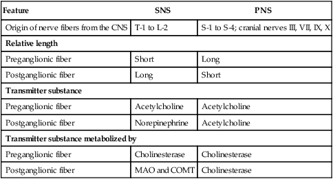
TABLE 16-1
Comparison of the Structure of the Sympathetic Nervous System and the Parasympathetic Nervous System
| Feature | SNS | PNS |
| Origin of nerve fibers from the CNS | T-1 to L-2 | S-1 to S-4; cranial nerves III, VII, IX, X |
| Relative length | ||
| Preganglionic fiber | Short | Long |
| Postganglionic fiber | Long | Short |
| Transmitter substance | ||
| Preganglionic fiber | Acetylcholine | Acetylcholine |
| Postganglionic fiber | Norepinephrine | Acetylcholine |
| Transmitter substance metabolized by | ||
| Preganglionic fiber | Cholinesterase | Cholinesterase |
| Postganglionic fiber | MAO and COMT | Cholinesterase |
F Adrenal medulla: Secretes epinephrine (75% by volume) and norepinephrine (25% by volume) into the bloodstream, resulting in stimulation of the SNS.
1. Innervation is via specific preganglionic fibers of the SNS.
2. Adrenal secretions assist in maintaining normal tone of the SNS.
G Effects of activation of the SNS and PNS:
1. Adrenergic effect: An effect activated or transmitted by norepinephrine or epinephrine.
2. Adrenergic receptor: A receptor stimulated by norepinephrine or epinephrine.
3. Cholinergic effect: An effect activated or transmitted by acetylcholine.
4. Cholinergic receptor: A receptor stimulated by acetylcholine.
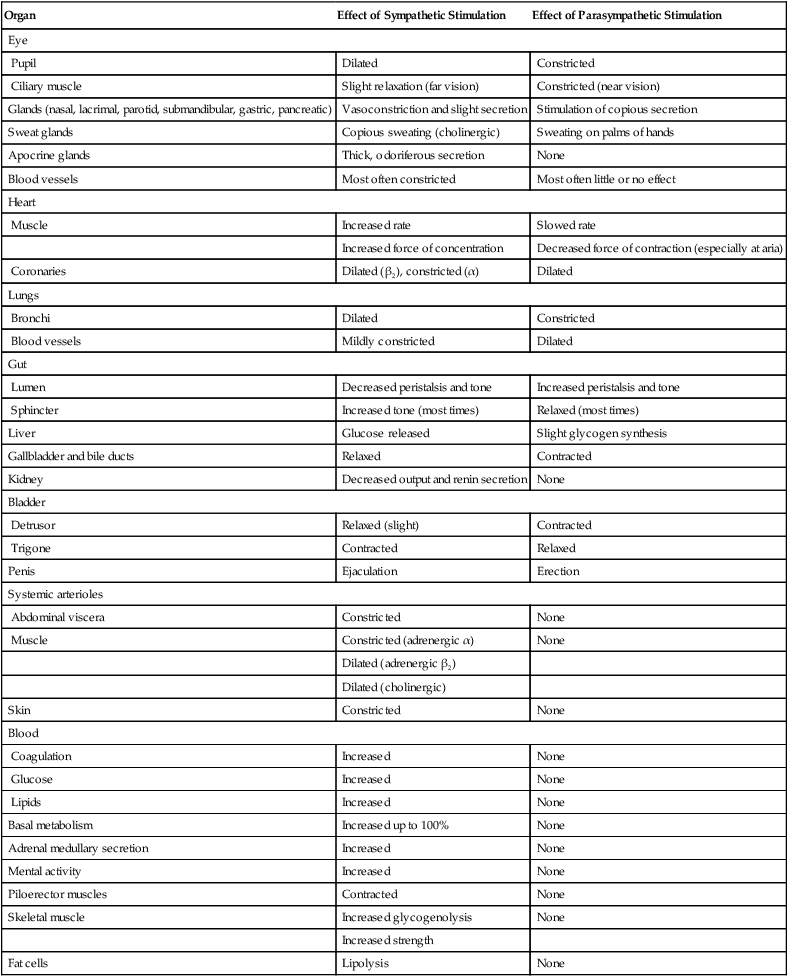
5. Table 16-2 lists the effects of the SNS and the PNS on various organs.
TABLE 16-2
Autonomic Effects on Various Organs of the Body
| Organ | Effect of Sympathetic Stimulation | Effect of Parasympathetic Stimulation |
| Eye | ||
| Pupil | Dilated | Constricted |
| Ciliary muscle | Slight relaxation (far vision) | Constricted (near vision) |
| Glands (nasal, lacrimal, parotid, submandibular, gastric, pancreatic) | Vasoconstriction and slight secretion | Stimulation of copious secretion |
| Sweat glands | Copious sweating (cholinergic) | Sweating on palms of hands |
| Apocrine glands | Thick, odoriferous secretion | None |
| Blood vessels | Most often constricted | Most often little or no effect |
| Heart | ||
| Muscle | Increased rate | Slowed rate |
| Increased force of concentration | Decreased force of contraction (especially at aria) | |
| Coronaries | Dilated (β2), constricted (α) | Dilated |
| Lungs | ||
| Bronchi | Dilated | Constricted |
| Blood vessels | Mildly constricted | Dilated |
| Gut | ||
| Lumen | Decreased peristalsis and tone | Increased peristalsis and tone |
| Sphincter | Increased tone (most times) | Relaxed (most times) |
| Liver | Glucose released | Slight glycogen synthesis |
| Gallbladder and bile ducts | Relaxed | Contracted |
| Kidney | Decreased output and renin secretion | None |
| Bladder | ||
| Detrusor | Relaxed (slight) | Contracted |
| Trigone | Contracted | Relaxed |
| Penis | Ejaculation | Erection |
| Systemic arterioles | ||
| Abdominal viscera | Constricted | None |
| Muscle | Constricted (adrenergic α) | None |
| Dilated (adrenergic β2) | ||
| Dilated (cholinergic) | ||
| Skin | Constricted | None |
| Blood | ||
| Coagulation | Increased | None |
| Glucose | Increased | None |
| Lipids | Increased | None |
| Basal metabolism | Increased up to 100% | None |
| Adrenal medullary secretion | Increased | None |
| Mental activity | Increased | None |
| Piloerector muscles | Contracted | None |
| Skeletal muscle | Increased glycogenolysis | None |
| Increased strength | ||
| Fat cells | Lipolysis | None |
From Guyton AC, Hall JE: Textbook of Medical Physiology, ed 10. Philadelphia, WB Saunders, 2000.
1. There are two major types of adrenergic receptors: alpha and beta. β receptors are divided into β1 and β2 receptors.
2. Table 16-3 lists the responses elicited by activation of these receptors.
TABLE 16-3
Adrenergic Receptors and Responses
| Alpha | Beta1 | Beta2 |
| Vasoconstriction | Increased heart rate | Vasodilation |
| Iris dilation | Increased myocardial strength | Intestinal relaxation |
| Intestinal relaxation | Lipolysis | Uterus relaxation |
| Intestinal sphincter contraction | Bronchodilation | |
| Calorigenesis | ||
| Pilomotor contraction | Glycogenolysis | |
| Bladder sphincter contraction | Bladder wall relaxation |
From Guyton A, Hall J: Textbook of Medical Physiology, ed 10, Saunders, New York, 2001.
3. Norepinephrine mainly excites α receptors but will excite β receptors but to a lesser extent.
4. Epinephrine excites both types of receptors to an equal extent.
1. There are two major types of cholinergic receptors: muscarinic and nicotinic.
2. Muscarinic receptors are those receptors affected by muscarine, a poison produced by toadstools.
3. Nicotinic receptors are those receptors affected by nicotine.
4. Muscarinic receptors are on effector cells stimulated by the postganglionic fibers of the PNS and the postganglionic cholinergic fibers of the SNS.
5. Nicotinic receptors are found at the synapsis between preganglionic and postganglionic fibers of SNS and PNS and at many nonautonomic nerve endings, specifically the neuromuscular junction.
[/level-membership-for-pulmolory-and-respiratory-category][not-level-membership-for-pulmolory-and-respiratory-category]
Anatomy and Physiology of the Nervous System
I Structure of the Nerve Fiber
A Each nerve fiber has three components: (Figure 16-1)
1. Cell body or soma: The primary metabolic area of the nerve, site of initial synthesis of transmitter substances.
2. Dendrite: The structures that carry impulses to the cell body. A single neuron may contain 10,000 dendrites.
3. Axon: The structure that conducts impulses away from the cell body. Usually only one axon leaves a cell body; however, it may branch extensively.
A Each neuron is classified by the direction of impulse transmission.
1. Sensory (afferent) neurons: Transmit nerve impulses to the spinal cord or brain.
2. Motor (efferent) neurons: Transmit nerve impulses from the brain or spinal cord to muscles or glands (effector organs).
3. Interneurons (internuncial neurons): Conduct impulses from sensory to motor neurons entirely within the central nervous system (CNS).
III Nerve Cell Membrane Potential (Figure 16-2)
A In the resting state, the inner surface of the cell membrane is negative compared with the positive outer surface. This sets up an electric potential across the cell membrane (normal polarity).
1. Intracellular: The primary cation, potassium (K+), is readily diffusible across the cell membrane.
2. Extracellular: The primary cation, sodium (Na+), is poorly diffusible across the cell membrane.
3. High extracellular Na+ (142 mEq/L) and low extracellular K+ (5 mEq/L) levels, along with high intracellular K+ (140 mEq/L) and low intracellular Na+ (10 mEq/L) levels, are maintained in the resting state by the Na+ pump (active transport).
4. Active transport (Na+ pump): The movement of Na+ from the intracellular space to the extracellular space and the movement of K+ from the extracellular space to the intracellular space. Adenosine triphosphate (ATP) provides the energy for the uphill transport of these ions.
A For a nerve impulse to be transmitted, an alteration in the cell’s resting membrane potential must be activated.
B An action potential is a stimulus that is capable of significantly increasing the cell membrane’s permeability to sodium.
C The action potential is an all-or-nothing phenomenon (i.e., the stimulus must be strong enough to allow for a reversal in the membrane potential); Na+ moves into the cells causing the intracellular side of the membrane to become positive. If the stimulus is not of sufficient strength, an action potential does not occur.
V Nerve Impulse Propagation (Figure 16-3)
A The nerve impulse is a self-propagating wave of electric charge that travels along the surface of the neuron’s membrane (i.e., the movement of the action potential along the cell membrane changing its charge). The nerve impulse travels in one direction only, from dendrite to cell body to axon.
B Depolarization: Stage 1 of the action potential, in which an impulse travels along the nerve fiber.
1. Sodium ions rush to the inside of the cell membrane.
2. A positive intracellular membrane charge is set up with a negative extracellular membrane charge.
3. There is a complete reversal of the resting membrane potential.
C Repolarization: Stage 2 of the action potential, in which the membrane returns to the normal resting membrane potential.
A The nerve synapse (synaptic cleft) is the junction between one neuron and another neuron, muscle, or gland. It is an actual space between nerve fibers.
B Transmission of an impulse from the axon of one nerve to a dendrite, soma, or effector organ is a chemical process occurring across the synapse. The distance across the synapse is approximately 200 Å.
C Presynaptic terminals are located on the axon and contain vesicles that synthesize, store, and secrete a transmitter substance into the synapse. The transmitter substance stimulates the dendrite or soma of the next nerve, causing an action potential.
D Postsynaptic terminals are located on dendrites, somas, or effector organs. After being stimulated, these terminals secrete a substance into the synapse to metabolize the transmitter substance.
E Acetylcholine as the transmitter substance (Figure 16-4)
1. Acetylcholine is synthesized in the terminal endings of cholinergic nerve fibers. After synthesis the acetylcholine is transported into the presynaptic vesicles where it is stored and released. It is formed from the reaction of acetyl coenzyme A with choline in the presence of choline acetyltransferase.
2. Vesicles storing acetylcholine are formed in the cell body and migrate to the surface of the presynaptic terminal.
3. When an action potential reaches the presynaptic terminal, acetylcholine is released into the synapse.
4. The acetylcholine moves across the synapse and stimulates a receptor on the postsynaptic terminal.
5. After stimulation the acetylcholine is released back into the synapse, and the postsynaptic terminal releases cholinesterase (acetylcholinesterase and acetylcholine esterase are terms synonymous with cholinesterase), which metabolizes acetylcholine, forming choline and acetate ion.
6. The choline is reabsorbed into the presynaptic terminal and is available to form more acetylcholine.
F Norepinephrine as the transmitter substance (Figure 16-5)
1. Norepinephrine is synthesized inside and outside the presynaptic vesicles, where it is stored and released.
2. The synthesis of norepinephrine proceeds as follows:
3. Norepinephrine is released via a mechanism identical to acetylcholine (see section VI-E).
4. After stimulation norepinephrine has three possible immediate fates:
a. Reabsorption into the presynaptic terminal (50% to 80% of secreted volume) for future secretion.
b. Metabolism in adjoining tissue by catechol-O-methyl-transferase (COMT) or in the nerve ending by monoamine oxidase (MAO).
c. Diffusion to surrounding tissues and eventually into the blood.
5. Complete metabolism of by-products formed in the synapse or norepinephrine entering the blood occurs in the liver via MAO and COMT.
6. In the adrenal medulla the formation of norepinephrine may proceed one step further to epinephrine by the process of methylation.
7. Duration of effect of norepinephrine in the synapse is only a few seconds. However, the norepinephrine and epinephrine released into the blood by the adrenal medulla remain active for up to several minutes.
VII Alteration of Nerve Impulse Transmission
A This junction is the site of transmission of impulses from nerves to skeletal muscle fibers.
B The axon of the nerve branches at its end to form a structure called the motor endplate, which invaginates into muscle fiber but does not penetrate the muscle membrane.
C The motor endplate’s terminal aspects are referred to as sole feet.
D Sole feet provide a large area of “contact” on the muscle surface. It is from the sole feet that the transmitter substance acetylcholine is secreted.
E After stimulation cholinesterase is secreted by the muscle fiber to metabolize the acetylcholine.
IX Reflex Arc or Reflex Action
A The reflex arc is a functional process of the nervous system. Most neuromuscular and neuroglandular mechanisms are controlled by a reflex arc.
[/not-level-membership-for-pulmolory-and-respiratory-category]
