[level-membership-for-radiology-category]
Chapter 20 Anatomy and physiology
 The respiratory system functions as a series of air passages through which air enters the lungs and travels to the alveoli where gaseous exchange occurs.


SKELETAL SYSTEM
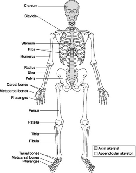
The skeletal system is made up of bones and joints that work together with muscles and ligaments to provide a framework for the body (Fig. 20.1).
BONES
General structure and appearance
Bone has an outer covering called the periosteum, which is a tough outer membrane made up of fibrous tissue and containing blood vessels. Thecortex is made up of compact tissue and lies directly below the periosteum. The inner layer of is made of spongy or cancellous bone, which is softer, compared to the tough outer cortex. Lastly, the innermost part of the bone is formed from ‘fatty’ yellow bone marrow, which contains a few white blood cells, and red marrow containing red blood cells.
Functions of bone
| Shape | Description | Location in the body |
|---|---|---|
| Long bones | Length is greater than width |
JOINTS
A joint forms at a point where two bones and cartilages meet or where adjacent bones and cartilages are joined. Although bone gives the body protective structure and muscles provide the ability to move, it is actually the joints that provide the mechanism by which movement takes place. Radiographic contrast can sometimes be injected into a joint space (e.g. in the glenoid cavity of the shoulder) to visualise any underlying pathology. This procedure is known as an arthrogram.
Classification
A joint can be classified according to the range of movement it provides or by its articular surface structure. All joints in the body can be classified as shown in Table 20.2; however, certain areas of the body may have a combination of two joints; for example the temporomandibular joint (TMJ) comprises gliding and pivot joints.
| Type of joint | Structure | Description of movement, with examples |
|---|---|---|
| Fibrous |
CARDIOVASCULAR SYSTEM
This system forms the transport network for the body.
THE HEART
The heart is a muscle that acts as a pump and provides the energy and force to keep blood circulating throughout the body. Blood is circulated via a closed transport system; that is, oxygenated blood leaves the heart via arteries, passes through a tiny network of capillaries where transfer of oxygen and nutrients take place, and then deoxygenated blood returns to the heart via the veins.
Size, shape and location
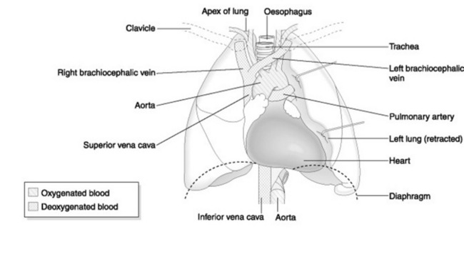
The heart is conical in shape and, under normal circumstances, about the size of its owner’s clenched fist. It is located anteriorly in the centre of the thorax, with about two-thirds of its bulk lying towards the left of the sternal margin. The tip of the ‘cone’ is called the apex and the flat portion is called the base. The base, normally found at the level of T5–T8, faces forwards and downwards to the left, ending in the apex. The apex lies at the level of the fifth intercostal space on the left midclavicular line (Fig. 20.2).
Occasionally patients may present with dextrocardia, a condition where the heart and great vessels originate with the same structure but opposite in direction (i.e. the apex lies towards the right side of the thorax rather than the left). This is a normal variant and is usually discovered as an incidental finding when the patient presents for a chest examination for an unrelated symptom.
Structure
The heart wall is made up of three layers of tissue (Fig. 20.3):
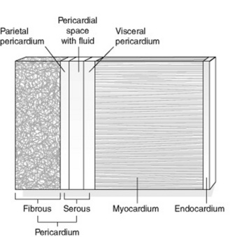
The pericardium consists of two layers of tissue. The outer layer is a tough fibrous layer that serves to protect the heart wall and secure its position within the thorax. The inner layer is a serous layer. This serous pericardium is furtherdivided into an outer parietal layer, which forms the inner lining of the fibrous pericardium, and an inner visceral layer that forms the outer covering of the heart (also known as epicardium). Between these layers is a potential space, called the pericardial cavity. This contains serous fluid, which allows for flexibility in the movement of the heart during contraction and relaxation phases (heartbeats), thus reducing friction during these movements.
Chambers of the heart
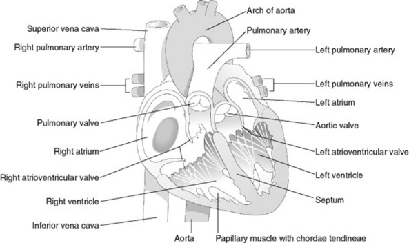
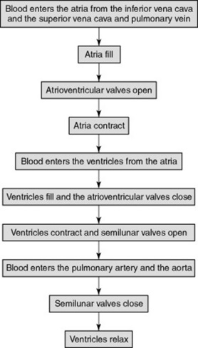
The heart is divided into left and right halves by a muscular septum. Each half has an upper atrium and a lower ventricle (Fig. 20.4). The atria are separated from each other by an interatrial septum and the ventricles are separated from each other by the interventricular septum. The atria are linked to the ventricles by atrioventricular valves. In a normal heart, blood flows from atria to ventricles and not the reverse. Figure 20.5 shows the sequence of events during the cardiac cycle.
Pumping action of the heart
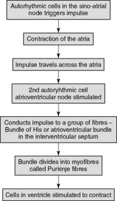
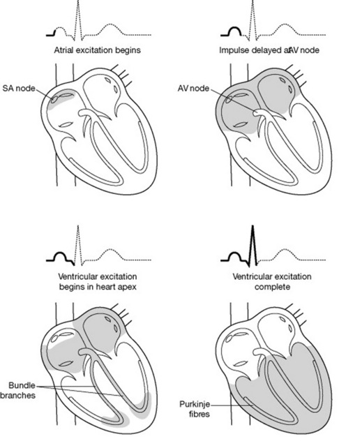
The heart’s own inherent autorhythmic cells act as a pacemaker to initiate and maintain the beating and pumping actions of the heart. These cells are also responsible for conducting these impulses throughout the cardiac muscle, thus creating an action in the path in which it travels (Fig. 20.6). Because the ventricles are responsible for sending blood out of the heart, the pressure in the ventricles is greater than the pressure inthe atria. The normal rhythm of a heart beat can be seen on an ECG (electrocardiograph) trace (see Fig. 20.7).
BLOOD VESSELS
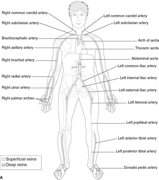
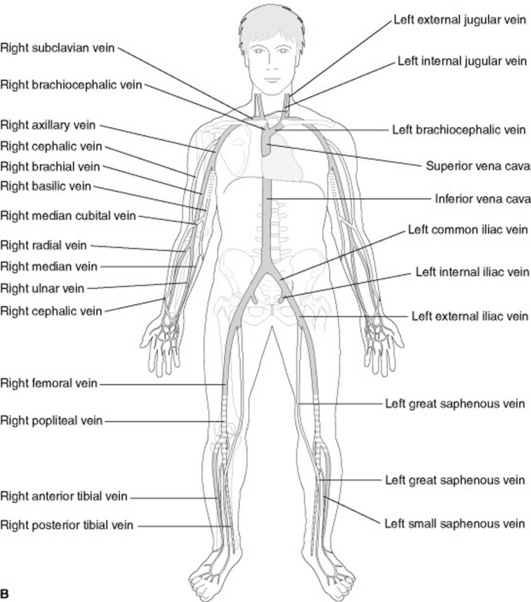
The aorta and other arteries carry oxygenated blood to the body (Fig. 20.8) and have thicker walls to withstand the high pressure at which blood is pumped into them. The closer the artery is to the heart, the thicker the walls of the artery. Arteries and veins can be demonstrated radiographically by using contrast media (e.g. in angiography and venography).
BLOOD
Blood is the ‘life’ of the body. It is a viscous tissue made up of liquid plasma and a combination of formed elements (blood cells). The plasma helpsthe body to maintain its normal state of hydration by responding to change in the internal environment via osmosis.
These blood cells have different functions (Table 20.3). Together they serve to:
| Blood cell | Function |
|---|---|
| Erythrocytes (red blood cells) | Contain haemoglobin (iron-containing pigment attached to a globular protein) for absorption and transport of oxygen from the lungs to cells and tissues |
| Leucocytes (white blood cells) | Provides immunity for the body by protecting against harmful invasion by disease-causing microorganisms – removes these, and debris from dead or damaged cells, from the blood. |
| Thrombocytes (blood platelets) | Responsible for clotting of the blood |
LYMPH AND LYMPH VESSELS
RESPIRATORY SYSTEM
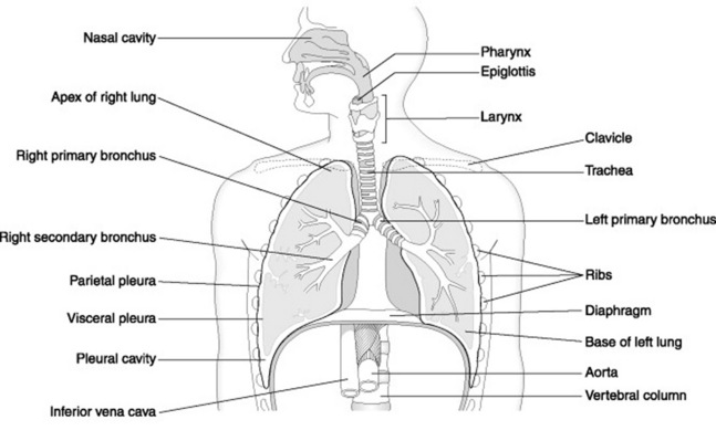
The thorax is probably the most frequently imaged body part in radiology departments today because a single chest X-ray is sometimes sufficient to make a diagnosis and determine the overall health of a patient. The mechanism of breathing (inspiration and expiration) occurs within this system. It functions as a series of passages through which air travels from the outside (atmosphere) to the inside(lungs). In addition, this system contributes to wider ranging functions of voice production, coughing and sneezing.
LARYNX
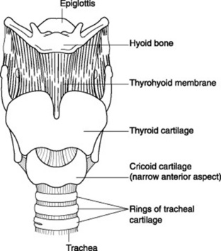
The larynx, or voice box, is composed of cartilages, the most important of which are the thyroid, cricoid and the epiglottis (Fig. 20.9). The thyroidcartilage is situated at the level of approximately C5 (fifth cervical vertebra) but may extend roughly to about the level of T1 (first thoracic vertebra). It is made up of two segments of cartilages that form an angle anteriorly and is therefore prominently visible.
TRACHEA
This is the continuation of the respiratory tract that is composed of C-shaped cartilaginous rings and extends from the larynx (around the level of C5) down to the upper lungs where it bifurcates into a left and a right main bronchus (Fig. 20.10). These incomplete cartilaginous rings help support the trachea. The trachea’s inner surface is lined with ciliated columnar epithelial tissue that traps dust and other particles that have passed down from the nasal cavity. If the particles are large, a coughing reflex is initiated to expel the foreign body.
BRONCHI AND BRONCHIOLES
Each bronchus then branches further into smaller branches or secondary bronchioles and these further subdivide into much smaller branches of terminal bronchioles. These continue to branch and end in respiratory bronchioles. These respiratory bronchioles, in turn, then branch into alveolar ducts, which form the pathway to thealveolar air sacs. Gaseous exchange takes place in the alveolar air sacs or alveoli, which form the functional unit of the lung. The alveoli are thin walled and lined with special excretory cells that produce a substance called surfactant. Surfactant serves to lubricate the lining of the alveoli to keep the walls inflated and thus reduce surface tension during the phases of gaseous exchange, when expansion and contraction of the alveoli occur.
INSPIRATION AND EXPIRATION
Look out for bronchiole ‘tree’ appearances on chest radiographs and compare lung appearance on images taken on good inspiration with that of those taken on poor inspiration (Fig. 20.11). To evaluate a good inspiratory radiograph, approximately six anterior ribs or ten posterior ribs should be visible on a posteroanterior (PA) chest radiograph.
DIGESTIVE SYSTEM
The digestive system functions to allow food to be taken into the mouth, swallowed (ingested), digested, absorbed and eliminated.
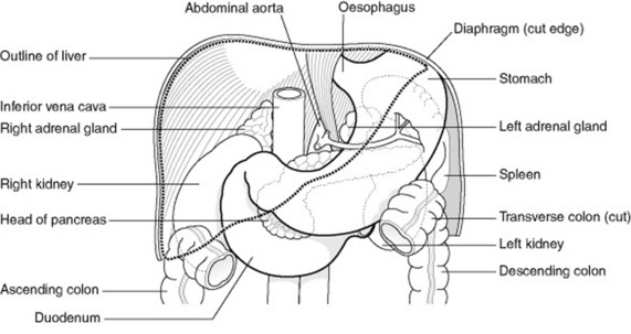
Various components make up this vast and varied tract and they will be discussed in the manner in which they represent anatomically. At this point the tract can be divided into two regions: that part of the digestive tract that lies above the diaphragm (upper tract) and then the part that lies below the diaphragm (lower tract or gastrointestinal tract) (Fig. 20.12). In addition, there are accessory organs associated with digestion; that is, the liver, gall bladder and pancreas.
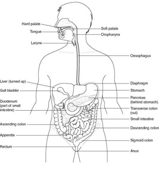
UPPER DIGESTIVE TRACT
This is made up of the following components:
Mouth/buccal cavity
This is the oral cavity that is found at the very beginning of the digestive tract, through which food enters. The mouth is lined by mucus secreting stratified epithelial cells. Its anterior and lateral boundaries are the lips and cheeks, respectively.These help to keep the food in the mouth and have a role in speech.
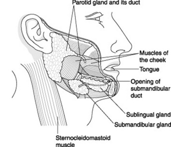
Salivary glands
Each gland (Fig. 20.13) is surrounded by a fibrous outer capsule and is lined with mucous-secreting cells. The secretions are called saliva, which is made up of water, mineral salts, a digestive enzyme called salivary amylase, mucous, lysosomes (to remove/repair/replace injured cells), immunoglobulins (protection against microorganisms) and certain blood clotting factors. Secretions are controlled by the autonomic nervous system. The presence of food in the mouth triggers a reflex secretion of saliva. In addition, sight, smell or even the thought of food may have the same effect.
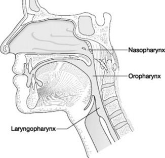
Pharynx
This is a funnel-shaped structure that contributes to both the digestive and respiratory tract (Fig. 20.14). Once food leaves the mouth it enters the pharynx. As discussed in the respiratory system, the pharynx continues inferiorly as the oesophagus. The epiglottis acts as a gate allowing food to enter the oesophagus by closing off the entrance to the trachea. The digestive function of the pharynx is to aid swallowing together with the effort of the muscles associated with the tongue, hyoid and soft palate.
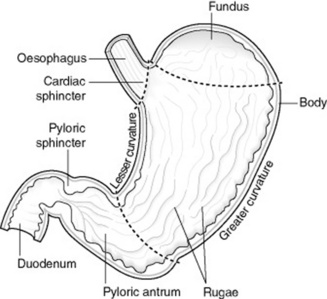
Oesophagus
At its lower end the oesophagus has a band of smooth muscle that acts as a sphincter, which functions to let food pass into the stomach as well as preventing gastric contents from being refluxed back into the oesophagus. If the sphincter does not function properly, it could allow the acidicgastric contents to move up the oesophagus causing a burning sensation, commonly referred to as heartburn.
LOWER DIGESTIVE TRACT (GASTROINTESTINAL TRACT)
This is made up of the following components:
Stomach
The stomach is a sac-like structure that can be located under the left hemi-diaphragm and functions to store food and aid in mechanical and chemical digestion (Fig. 20.15). Food enters the stomach from the oesophagus via the cardiac sphincter.
Small intestine
This is a narrow tube that is about 6 m long and divided into three parts:
Jejunum and ileum
These parts form the second and third portions of the small intestine and continue from the duodenum to end at the caecum. Most of the nutrients from digested food are absorbed in this part of the small intestine. The mucosal lining has little protrusions called villi; these contain a rich blood and lymph supply, which helps the absorption of useful substances from the food. Between the villi are intestinal glands which produce enzymes and intestinal juice for further digestion as food moves along in the small intestine by peristalsis (Table 20.4).
| Part of digestive tract | Enzyme |
|---|---|
| Mouth or buccal cavity salivary glands | Salivary amylase |
| Stomach | Gastric enzymes – pepsin, gastric amylase and gastric lipase |
| Small intestine | Intestinal enzymes – sucrase, lipase, maltase and lactase, and receives bile and pancreatic digestive secretions |
| Pancreas | Pancreatic enzymes – trypsinogens, pancreatic amylase and lipase |
Large intestine or colon
This part of the tract extends from the terminal ileum to the anus. It is made up of various segments:
ACCESSORY DIGESTIVE ORGANS
The accessory digestive organs are depicted in Figure 20.16.
Pancreas
The pancreas lies posterior to the stomach in the retroperitoneal space and acts as both an endocrine (see Endocrine system, p. 283) and exocrine gland. Its exocrine function is to produce digestive juices (pancreatic juices) and enzymes that are transported via the pancreatic duct to empty into the duodenum to help in digestion.
UROGENITAL SYSTEM
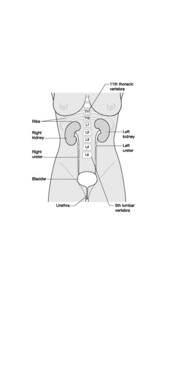
The urinary system (Fig. 20.17) comprises the following:
KIDNEYS
The position of the kidneys in the abdomen is important for accurate radiographic technique required during intravenous urogram procedures (IVUs) (Fig. 20.18).
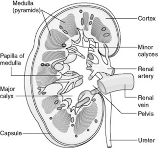
Internal anatomy
There are three distinct areas on the internal surface of the kidney: a cortex, a medulla and a renal pelvis (Fig. 20.19). The cortex forms the outermost part of the internal structure of the kidney, whilst the middle portion is called the medulla and contains cone-shaped tissue called medullary pyramids. On a longitudinal section these pyramids appear to have stripes or striations. This is because they are composed of urine-collecting tubules arranged in a parallel manner. The pyramids have ‘finger like’ projections called papillae, which empty into the minor calyces. The renal pelvis is found medially and is the dilated portion of the ureter, which has two divisions known as the major and minor calyces. The minor calyces act as cups to collect the urine from the collectingtubules of the pyramids and propels it, with the help of its smooth muscle containing layer, towards the pelvis. The pelvis acts as a reservoir to collect the urine and then transport it to the bladder for excretion.
Filtration, formation and flow of urine
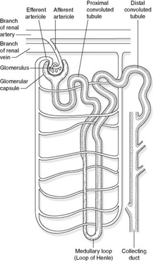
Kidneys function to regulate the water, nutrient and electrolyte balance of the body. The component responsible for this activity is a microscopic, blood-filtering and functional unit called the nephron (Fig. 20.20). The nephron is associated with many renal tubules. These structures filter the blood and form urine. Collecting ducts then collect the urine, which is emptied into the minor calyx of the renal pelvis.
Functions of the kidney
URETERS
Urine is transported into the ureters via the renal pelvis. The ureters transport the urine from the kidneys to the bladder. The ureters do not lie vertically in structure; instead they travel down along the posterior peritoneal wall and curve medially before entering the posterior bladder wall in an oblique manner. This morphology helps prevent the backflow of urine from the bladder into the ureters.
URETHRA
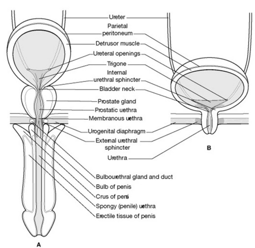
The urethra is a muscular tube that transports the urine from the bladder to the exterior. The urethra has sphincters that control the flow of urine and prevents urine leakage. The location and structure of the urethra differs in males and females (Fig. 20.21).
NERVOUS SYSTEM
NERVOUS TISSUE
Axons and dendrites make up the white matter of the nervous system. Dendrites function to receive an impulse and direct it towards the cell body of the neuron. The axon functions to conduct this impulse away from the cell along its path of conduction. There are two types of nerve:
CENTRAL NERVOUS SYSTEM
Brain
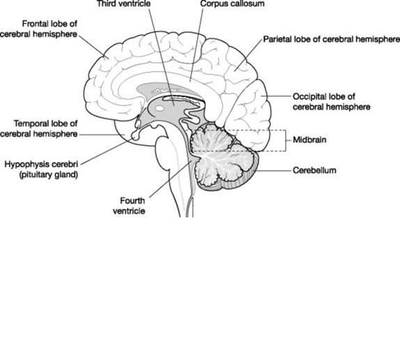
The brain is a large mass of white and grey matter that would slump and sag if it was not supported by the cranium and meninges (Fig. 20.22). The cranium is the skull vault that surrounds the brain and protects it from injury.
Meninges
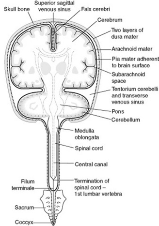
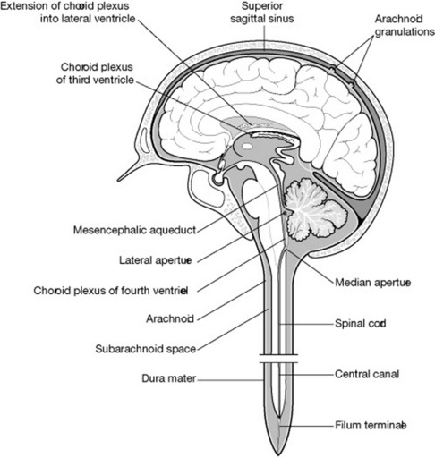
Directly surrounding the brain are the meninges (Fig. 20.23). These layers of tissue protect thebrain and, together with the CSF, act as shock absorbers to prevent injury. There are three meningeal layers:
Cerebrum
The two hemispheres form the left and the right lobes. The lobes are made up of grey matter on the outer cortex and the inner layers are made up ofwhite matter. The cerebrum consists of nerve tracts for communication between the lobes. The lobes are connected to each other by a dense bundle of nerve fibres called the corpus callosum. This relays impulses between the left and right lobes.
Brain stem
The brain stem consists of long tracts of ascending and descending pathways that conduct impulses from the body to the cerebrum and from the cerebrum to the rest of the body. It houses animportant structure called the reticular formation that controls the vital life-sustaining role of maintaining respiration and cardiac activity. It is also responsible for regulating consciousness and levels of ‘awareness’. The reticular fibres desensitise the repetitive sounds and create levels of awareness for ‘important’ sounds.
Ventricles and cerebrospinal fluid (CSF)
The ventricles are cavities within the brain that are filled with CSF (Fig. 20.24). These cavities are lined with cuboidal epithelial cells (called ependymal cells) that make contact with the pia mater at various points along its surface to form blood vessel network structures known as choroid plexuses.
PERIPHERAL NERVOUS SYSTEM (PNS)
It is divided into the somatic nervous system and the visceral nervous system.
Visceral nervous system
This is also composed of sensory and motor pathways. The sensory pathway receives impulses from sensory receptors of visceral systems(e.g. cardiovascular, digestive and urinary). These impulses are perceived on an unconscious level; however, some impulses are conveyed on a conscious level – e.g. pain, taste and feeling of fullness of the bladder.
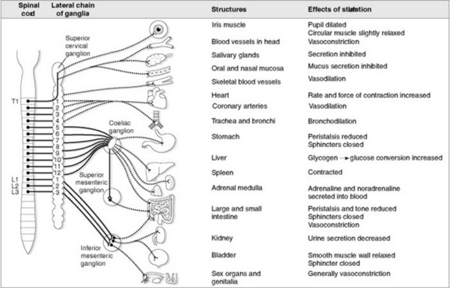
The motor pathway of the visceral nervous system is known as the autonomic nervous system.
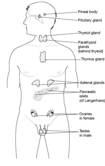
ENDOCRINE SYSTEM
It has been mentioned before that the endocrine system together with the nervous system controls body activities and maintains homeostasis. Whereas the action of the nervous system is rapid, with immediate but shorter-lived responses, the response of the endocrine system is slower but the effects are longer lasting. The nervous system influences bodily activities by means of chemical messengers known as neurotransmitters. The endocrine system influences metabolic activities of body cells by means of chemical messengers better known as hormones. Organs or glands that can be classified into exocrine and endocrine structures secrete these chemical messengers.
HORMONES
Hormones are derived from two different sources.
Examples of hormones, their gland or organ of origin and functions are listed in Table 20.5. Hormone stimulation, release and inhibition are controlled via a feedback mechanism. This method of control is used by the endocrine system to regulate the amount of secretion required. Feedback may be positive or negative.
| Organ/gland | Hormone | Functions |
|---|---|---|
| Hypothalamus | Has nerve cells that control the pituitary by producing neurochemicals that either stimulate or suppress the secretions from the pituitary. Acts as a primary link between the nervous system (brain) and endocrine system (mainly the pituitary) | |
| Antidiuretic hormone (ADH) | Regulates the body’s fluid balance | |
| Oxytocin | Stimulates contraction of uterine muscles during childbirth; ejection of milk during lactation | |
| Pituitary gland (hypophysis) | The most important endocrine gland because its responses control the action of some of the other endocrine glands | |
| Anterior pituitary or adenohypophysis | Growth hormone (GH) | Bone, muscle and body tissue growth |
| Prolactin (PRL) | Activates milk production in the mammary glands during pregnancy | |
| Thyroid stimulating hormone (TSH) | Stimulates the thyroid gland to produce thyroid hormones | |
| Adrenocorticotropic hormone (ACTH) | Stimulates the adrenal glands to produce hormones | |
| Follicle stimulating hormone (FSH) – also known as gonadotropin | Regulates the functions of the ovaries and testes, including the production and secretion of oestrogen and progesterone | |
| Posterior pituitary or neurohypophysis | Does not produce its own hormones | Receives and stores the neurohormones ADH and oxytocin (see below) from the hypothalamus |
| Thyroid gland | Thyroxine triiodothryonine | Affect almost every cell in the body by regulating the metabolic rates of carbohydrates, proteins and fat; increase heat production as energy for use by the body; stimulate oxygen take-up by the body (except the brain), important role in skeletal muscle growth and development of the nervous system in children. |
| Calcitonin | Decreases the level of calcium in the blood when the levels are high | |
| Parathyroid gland | Parathyroid hormone (PTH) | Increases the level of calcium in the blood when the levels are low |
| Adrenal glands | ||
| Adrenal cortex | Secretion of corticosteroids: | |
| Glucocorticoids | Regulate glucose levels and metabolism of food; acts as an anti-inflammatory agent for immunity; affects growth and development and provides resistance to physical and emotional stress | |
| Mineralocorticoids | Regulate water and ion balance in the body together with the enzyme renin, produced by the kidneys (see below), and angiotensin (produced in the liver) | |
| Gonadocorticoids | These have a small effect on onset of puberty and sexual development | |
| Adrenal medulla | During stress these hormones produce the ‘fright, fight or flight’ response in the body by causing the body to act quickly; e.g. increases heart rate and blood pressure; dilating blood vessels, etc. | |
| Epinephrine (adrenalin) | Given as an intravenous injection during allergic reactions; e.g. reactions to iodine during radiographic procedures, and has the greater effect on the heart and metabolism | |
| Norepinephrine (noradrenalin) | Acts as neurotransmitter as part of the nervous system and has the greater effect on the blood vessels | |
| Pancreas (islets of Langerhans) | Insulin | Lowers blood nutrient levels when they are high, especially glucose |
| Glucagon | Increase blood glucose levels when they are low by converting glycogen to glucose | |
| Levels of both insulin and glucogon are regulated by somatostatin that is produced by the hypothalamus | ||
| Gonads | Testosterone | Stimulates the production of sperm; maintains growth of sexual organs and development of sexual behaviour; stimulates growth changes associated with puberty; e.g. facial and pubic hair, deepening of voice, etc. |
| Oestrogen | Regulates menstrual cycle, development of mammary glands and female sexual characteristics | |
| Others | ||
| Kidneys | Renin | Influences blood pressure; sodium–water balance and volume |
| Erythropoietin | Stimulates bone marrow to produce red blood cells | |
| Pineal gland | Melatonin | Helps to regulate the sleep–wake cycle |
| Thymus | Thymosin | Helps in the development of T lymphocytes (also known as T cells) for immunity |
| Heart | Atrial natriuretic peptide (ANP) | Helps in the regulation of blood pressure |
| Digestive tract | Secretin | Neutralises gastric acid |
| Cholecystokinin | Activates contraction of the gall bladder to secrete bile | |
| Placenta | Oestrogen, progesterone and chorionic gonadotropin | Occurs during pregnancy to help maintain pregnancy |
| Prostate and throughout the body | Prostaglandins | Regulate blood pressure, digestive secretions; aid immunity by providing an anti-inflammatory response |
| Blood cells (basophils) | Histamine | Provides a response during an allergic or inflammatory process (e.g. causes bronchoconstriction); is reversed by the action of antihistamines |
Control of hormone secretion is regulated by the hypothalamus via the feedback mechanism (Fig. 20.26).
ENDOCRINE ORGANS AND GLANDS
As already mentioned, organs and glands can be classified as being either exocrine or endocrine.
[/level-membership-for-radiology-category][not-level-membership-for-radiology-category]
Chapter 20 Anatomy and physiology
The respiratory system functions as a series of air passages through which air enters the lungs and travels to the alveoli where gaseous exchange occurs.SKELETAL SYSTEM
The skeletal system is made up of bones and joints that work together with muscles and ligaments to provide a framework for the body (Fig. 20.1).
BONES
General structure and appearance
Bone has an outer covering called the periosteum, which is a tough outer membrane made up of fibrous tissue and containing blood vessels. Thecortex is made up of compact tissue and lies directly below the periosteum. The inner layer of is made of spongy or cancellous bone, which is softer, compared to the tough outer cortex. Lastly, the innermost part of the bone is formed from ‘fatty’ yellow bone marrow, which contains a few white blood cells, and red marrow containing red blood cells.
Functions of bone
| Shape | Description | Location in the body |
|---|---|---|
| Long bones | Length is greater than width |
JOINTS
A joint forms at a point where two bones and cartilages meet or where adjacent bones and cartilages are joined. Although bone gives the body protective structure and muscles provide the ability to move, it is actually the joints that provide the mechanism by which movement takes place. Radiographic contrast can sometimes be injected into a joint space (e.g. in the glenoid cavity of the shoulder) to visualise any underlying pathology. This procedure is known as an arthrogram.
Classification
A joint can be classified according to the range of movement it provides or by its articular surface structure. All joints in the body can be classified as shown in Table 20.2; however, certain areas of the body may have a combination of two joints; for example the temporomandibular joint (TMJ) comprises gliding and pivot joints.
| Type of joint | Structure | Description of movement, with examples |
|---|---|---|
| Fibrous |
CARDIOVASCULAR SYSTEM
This system forms the transport network for the body.
THE HEART
The heart is a muscle that acts as a pump and provides the energy and force to keep blood circulating throughout the body. Blood is circulated via a closed transport system; that is, oxygenated blood leaves the heart via arteries, passes through a tiny network of capillaries where transfer of oxygen and nutrients take place, and then deoxygenated blood returns to the heart via the veins.
Size, shape and location
The heart is conical in shape and, under normal circumstances, about the size of its owner’s clenched fist. It is located anteriorly in the centre of the thorax, with about two-thirds of its bulk lying towards the left of the sternal margin. The tip of the ‘cone’ is called the apex and the flat portion is called the base. The base, normally found at the level of T5–T8, faces forwards and downwards to the left, ending in the apex. The apex lies at the level of the fifth intercostal space on the left midclavicular line (Fig. 20.2).
Occasionally patients may present with dextrocardia, a condition where the heart and great vessels originate with the same structure but opposite in direction (i.e. the apex lies towards the right side of the thorax rather than the left). This is a normal variant and is usually discovered as an incidental finding when the patient presents for a chest examination for an unrelated symptom.
Structure
The heart wall is made up of three layers of tissue (Fig. 20.3):
The pericardium consists of two layers of tissue. The outer layer is a tough fibrous layer that serves to protect the heart wall and secure its position within the thorax. The inner layer is a serous layer. This serous pericardium is furtherdivided into an outer parietal layer, which forms the inner lining of the fibrous pericardium, and an inner visceral layer that forms the outer covering of the heart (also known as epicardium). Between these layers is a potential space, called the pericardial cavity. This contains serous fluid, which allows for flexibility in the movement of the heart during contraction and relaxation phases (heartbeats), thus reducing friction during these movements.
Chambers of the heart
The heart is divided into left and right halves by a muscular septum. Each half has an upper atrium and a lower ventricle (Fig. 20.4). The atria are separated from each other by an interatrial septum and the ventricles are separated from each other by the interventricular septum. The atria are linked to the ventricles by atrioventricular valves. In a normal heart, blood flows from atria to ventricles and not the reverse. Figure 20.5 shows the sequence of events during the cardiac cycle.
Pumping action of the heart
The heart’s own inherent autorhythmic cells act as a pacemaker to initiate and maintain the beating and pumping actions of the heart. These cells are also responsible for conducting these impulses throughout the cardiac muscle, thus creating an action in the path in which it travels (Fig. 20.6). Because the ventricles are responsible for sending blood out of the heart, the pressure in the ventricles is greater than the pressure inthe atria. The normal rhythm of a heart beat can be seen on an ECG (electrocardiograph) trace (see Fig. 20.7).
BLOOD VESSELS
The aorta and other arteries carry oxygenated blood to the body (Fig. 20.8) and have thicker walls to withstand the high pressure at which blood is pumped into them. The closer the artery is to the heart, the thicker the walls of the artery. Arteries and veins can be demonstrated radiographically by using contrast media (e.g. in angiography and venography).
BLOOD
Blood is the ‘life’ of the body. It is a viscous tissue made up of liquid plasma and a combination of formed elements (blood cells). The plasma helpsthe body to maintain its normal state of hydration by responding to change in the internal environment via osmosis.
These blood cells have different functions (Table 20.3). Together they serve to:
| Blood cell | Function |
|---|---|
| Erythrocytes (red blood cells) | Contain haemoglobin (iron-containing pigment attached to a globular protein) for absorption and transport of oxygen from the lungs to cells and tissues |
| Leucocytes (white blood cells) | Provides immunity for the body by protecting against harmful invasion by disease-causing microorganisms – removes these, and debris from dead or damaged cells, from the blood. |
| Thrombocytes (blood platelets) | Responsible for clotting of the blood |
LYMPH AND LYMPH VESSELS
RESPIRATORY SYSTEM
The thorax is probably the most frequently imaged body part in radiology departments today because a single chest X-ray is sometimes sufficient to make a diagnosis and determine the overall health of a patient. The mechanism of breathing (inspiration and expiration) occurs within this system. It functions as a series of passages through which air travels from the outside (atmosphere) to the inside(lungs). In addition, this system contributes to wider ranging functions of voice production, coughing and sneezing.
