[level-membership-for-neurosurgery-category]
Chapter 1 Anatomy and Function in the Upper Extremity
Note: Unless otherwise indicated, arrows in figures indicate the direction of patient motion.
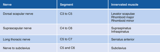
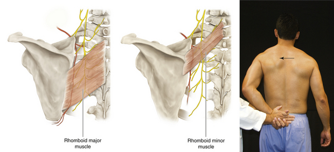
Figure 1-2 Rhomboid Major and Minor Muscles
• Muscle attachments: Spinous processes of T2 to T5 (rhomboid major) and ligamentum nuchae and spinous processes of C7 and T1 (rhomboid minor) to medial border of scapula
• Innervation: Dorsal scapular nerve (C4 and C5)
• Function: Adduction; rotation of scapula
• Physical examination: The patient places a hand on his or her back and pushes backward against resistance. (Arrow: muscle bellies can be seen.)
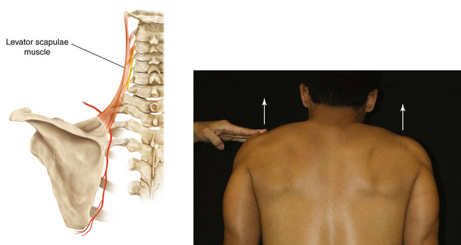
Figure 1-3 Levator Scapulae Muscle
• Muscle attachments: Transverse processes of C1 through C4 to medial border of scapula
• Innervation: Dorsal scapular (C5) and cervical (C3 and C4) nerves
• Function: Raises scapula and inclines neck to corresponding side if scapula is fixed
• Physical examination: The patient tries to shrug the shoulders (arrows) against resistance.
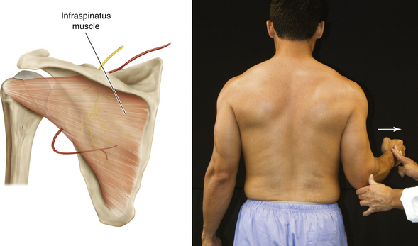
Figure 1-5 Infraspinatus Muscle
• Muscle attachments: Infraspinatus fossa of scapula to middle facet on greater tubercle of humerus
• Innervation: Suprascapular nerve (C5 and C6)
• Function: External rotation of head of humerus at the shoulder joint
• Physical examination: The patient externally rotates (arrow) the upper arm at the shoulder against resistance.
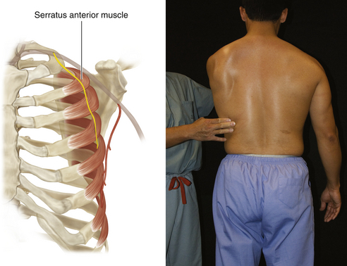
Figure 1-6 Serratus Anterior Muscle
• Muscle attachments: Anterior surfaces of first eight or nine ribs to medial border of anterior surface of scapula
• Innervation: Long thoracic nerve (C5 to C7)
• Function: Abduction of scapula
• Physical examination: Patient pushes against resistance (e.g., the examiner’s hand or a wall). (If the serratus anterior is paralyzed, winging of the scapula can be observed.)
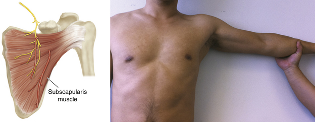
Figure 1-8 Subscapularis Muscle
• Muscle attachments: Subscapular fossa to lesser tubercle of humerus
• Innervation: Upper and lower subscapular nerves (C5 to C7)
• Function: Internal rotation of humerus
• Physical examination: The patient internally rotates the upper arm against resistance. (The main mover is the pectoralis major.)
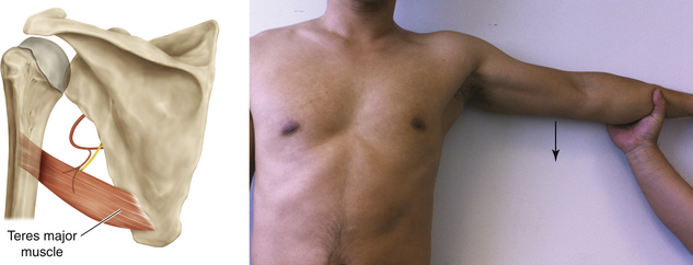
Figure 1-9 Teres Major Muscle
• Muscle attachments: Dorsal surface of inferior angle of scapula to intertubercular groove of humerus
• Innervation: Lower subscapular nerve (C6 and C7)
• Function: Internal rotation and adduction of humerus at shoulder joint
• Physical examination: The patient tries to adduct (arrow) the elevated upper arm against resistance.
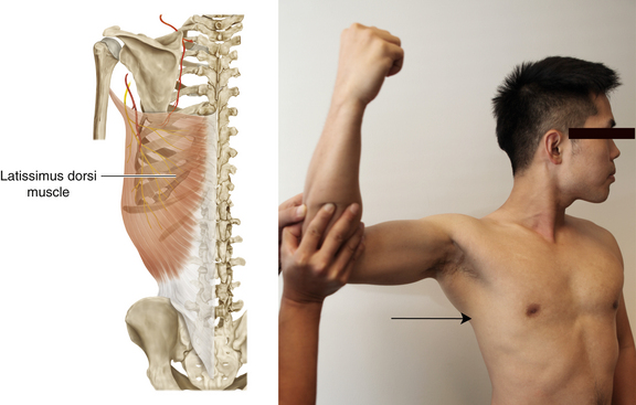
Figure 1-10 Latissimus Dorsi Muscle
• Muscle attachments: Spinous processes of T6 to T12, thoracolumbar fascia, iliac crest, and inferior three or four ribs to floor of intertubercular groove of humerus
• Innervation: Thoracodorsal nerve (C6 to C8)
• Function: Internal rotation and adduction of humerus at shoulder joint
• Physical examination: The upper arm is horizontal, and the patient is asked to adduct it against resistance. Also, in a relaxed, upright position, the patient is asked to cough; muscle bellies can be felt to contract when the patient coughs.
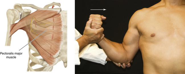
Figure 1-11 Pectoralis Major Muscle
• Muscle attachments: Anterior surface of medial half of clavicle (clavicular head) and anterior surface of sternum, superior six costal cartilages, and aponeurosis of external oblique muscle (sternocostal head) to lateral lip of intertubercular groove of humerus
• Innervation: Medial and lateral pectoral nerves—clavicular head supplied by C5, C6 (lateral pectoral nerve) and sternocostal head supplied by C6, C7, and C8 (medial and lateral pectoral nerves)
• Function: Internal rotation and adduction of humerus at shoulder joint
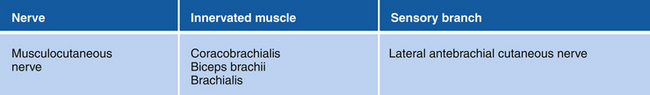
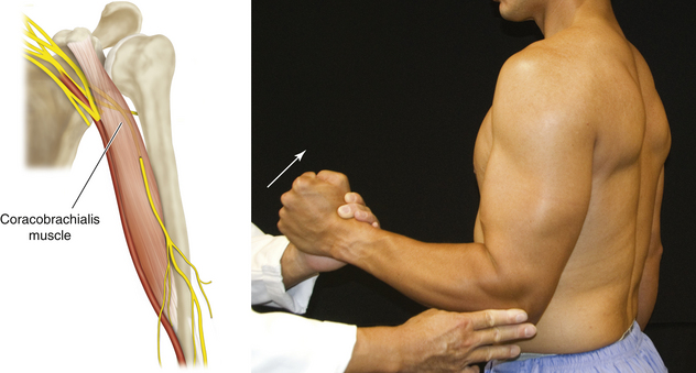
Figure 1-16 Coracobrachialis Muscle
• Muscle attachments: Coracoid process to medial surface of humerus
• Innervation: Musculocutaneous nerve (C5 to C7)
• Function: Internal rotation, flexion, and adduction of shoulder joint
• Physical examination: With the arm flexed and laterally rotated at the shoulder joint, the elbow completely flexed, and the forearm supinated, the patient flexes (arrow) the shoulder joint against resistance.
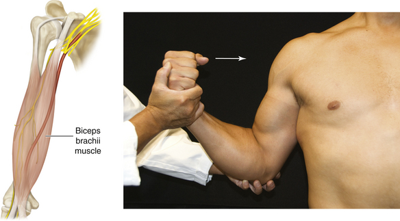
Figure 1-17 Biceps Brachii Muscle
• Muscle attachments: Coracoid process (short head) and supraglenoid tubercle of scapula (long head) to tuberosity of radius
• Innervation: Musculocutaneous nerve (C5 and C6)
• Function: Flexion at elbow joint and supination at forearm
• Physical examination: When the patient flexes (arrow) the supinated forearm against resistance, the muscle can be seen.

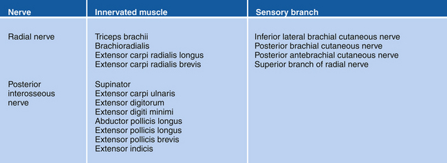
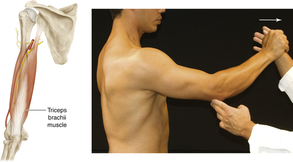
Figure 1-25 Triceps Brachii Muscle
• Muscle attachments: Infraglenoid tubercle of scapula (long head), posterior surface of humerus (lateral head), and posterior surface of humerus (medial head) to proximal end of olecranon of ulna and fascia of forearm
• Innervation: Radial nerve (C6 to C8)
• Function: Extension of elbow joint
• Physical examination: The patient extends the elbow (arrow) against resistance.
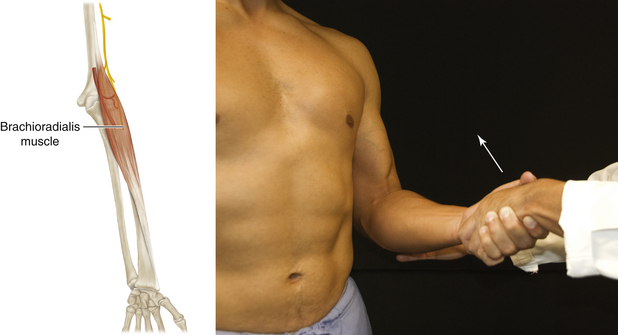
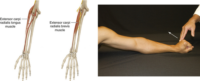
Figure 1-27 Extensor Carpi Radialis Longus and Brevis Muscles
• Muscle attachments: Distal third of lateral supracondylar ridge and lateral epicondyle of humerus to the radial side of dorsal surface of base second and third metacarpals
• Innervation: Posterior interosseous nerve of radial nerve (C7 and C8)
• Function: Extension and abduction of wrist joint
• Physical examination: The patient extends and abducts the wrist joint (arrow) against resistance.
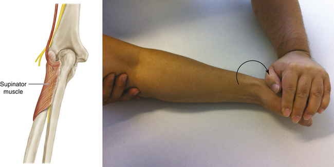
Figure 1-28 Supinator Muscle
• Muscle attachments: Lateral epicondyle of humerus, radial collateral and annular ligaments, supinator fossa, crest of ulna to radius
• Innervation: Posterior interosseous nerve, branch of radial nerve (C5 and C6)
• Function: Supination of forearm
• Physical examination: The patient supinates the forearm (arrow) against resistance, with the forearm extended at the elbow.

Figure 1-29 Extensor Carpi Ulnaris Muscle
• Muscle attachments: Lateral epicondyle of humerus and ulna to fifth metacarpal bone
• Innervation: Posterior interosseous nerve of radial nerve (C7 and C8)
• Function: Extension and adduction of wrist joint
• Physical examination: The muscle can be tested and seen when the patient extends and adducts the wrist (arrow) against resistance.
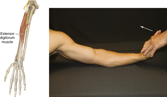
Figure 1-30 Extensor Digitorum Muscle
• Muscle attachments: Lateral epicondyle of humerus to dorsal aspects of bases of middle and distal phalanges of medial four digits via “extensor hoods”
• Innervation: Posterior interosseous nerve of radial nerve (C7 and C8)
• Function: Extension of medial four digits; assists in extension of wrist joint
• Physical examination: While the patient’s hand is firmly supported by the examiner’s hand, extension at the metacarpophalangeal joints (arrow) is maintained against resistance.
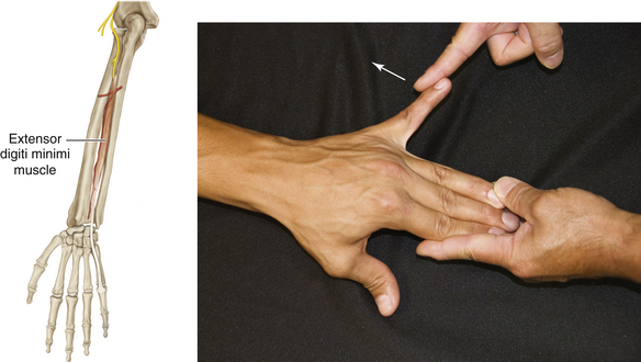
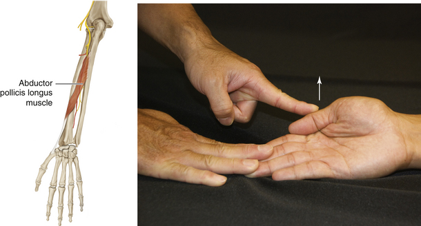
Figure 1-32 Abductor Pollicis Longus Muscle
• Muscle attachments: Ulna, radius, and interosseous membrane to first metacarpal
• Innervation: Posterior interosseous nerve of radial nerve (C7 and C8)
• Function: Abduction of carpometacarpal joint of the thumb.
• Physical examination: The patient abducts thumb at the carpometacarpal joint in a right angle plane to the palm (arrow).
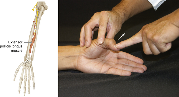
Figure 1-33 Extensor Pollicis Longus Muscle
• Muscle attachments: Ulna and interosseous membrane to distal phalanx of thumb
• Innervation: Posterior interosseous nerve, branch of radial nerve (C7 and C8)
• Function: Extension of terminal phalanx of thumb
• Physical examination: The patient extends the thumb at the interphalangeal joint (arrow) against resistance.
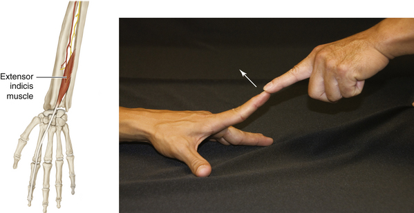
Figure 1-34 Extensor Indicis Muscle
• Muscle attachments: Ulna and interosseous membrane to second digit
• Innervation: Posterior interosseous nerve, branch of radial nerve (C7 and C8)
• Function: Extension of metacarpophalangeal and interphalangeal joint of index finger
• Physical examination: The patient extends the second (index) finger at the interphalangeal joint (arrow) against resistance.
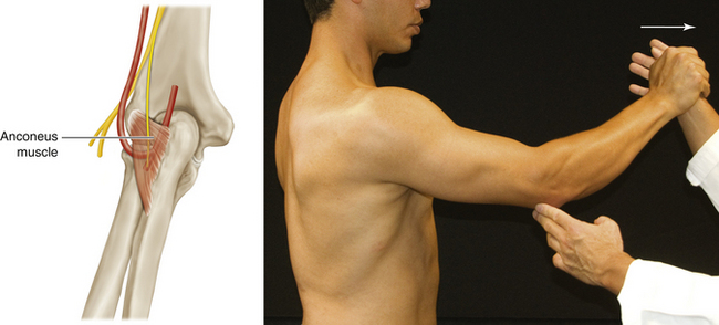
Figure 1-35 Anconeus Muscle
• Muscle attachments: Lateral epicondyle of humerus to olecranon and ulna
• Innervation: Radial nerve (C7 to T1)
• Function: Minor stabilizer of the elbow joint
• Physical examination: With the arm slightly abducted and the forearm slightly flexed, the patient extends the forearm (arrow) against resistance.

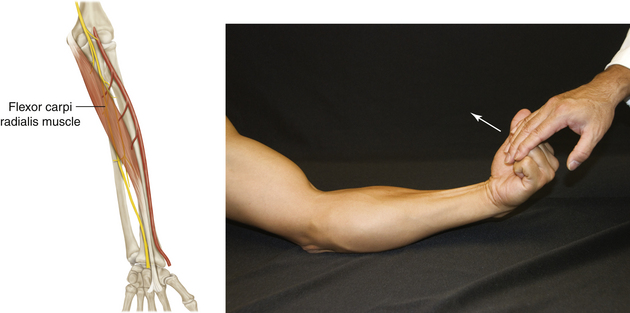
Figure 1-39 Flexor Carpi Radialis Muscle
• Muscle attachments: Medial epicondyle of humerus to second metacarpal
• Innervation: Median nerve (C6 and C7)
• Function: Flexion of wrist joint; assists in pronation and abduction of wrist joint
• Physical examination: The tendon of this muscle can be observed when the patient flexes and abducts the wrist (arrow) against resistance.
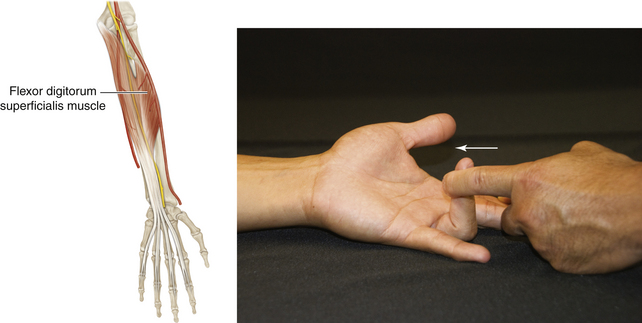
Figure 1-41 Flexor Digitorum Superficialis Muscle
• Muscle attachments: Epicondyle of humerus, ulnar collateral ligament, coronoid process (humeroulnar head), and radius (radial head) to middle phalanges of medial four digits
• Innervation: Median nerve (C7 to T1)
• Function: Flexion of middle and proximal phalanges of medial four digits; flexion of wrist joint
• Physical examination: With the proximal phalanx fixed, the patient flexes the proximal interphalangeal joint (arrow) against resistance.
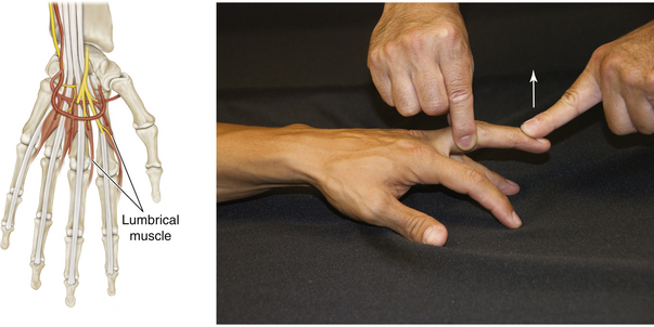
Figure 1-42 First and Second Lumbrical Muscles
• Muscle attachments: Lateral two tendons of flexor digitorum profundus to lateral sides of extensor expansions of digits 2 to 5
• Innervation: Median nerve (C8 and T1)
• Function: Flexion of second and third fingers at the metacarpophalangeal joint; extension of second and third fingers at the interphalangeal joint
• Physical examination: With the metacarpophalangeal joint hyperextended and fixed, the patient extends the finger at the proximal interphalangeal joint (arrow) against resistance.
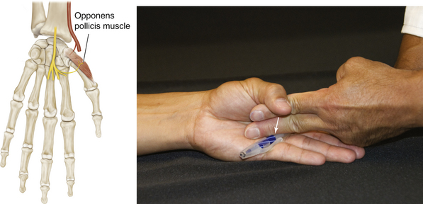
Figure 1-43 Opponens Pollicis Muscle
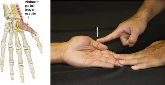
Figure 1-44 Abductor Pollicis Brevis Muscle
• Muscle attachments: Flexor retinaculum and tubercles of scaphoid and trapezium bones to proximal phalanx of thumb
• Innervation: Recurrent branch of median nerve (C8 and T1)
• Function: Abduction of the thumb at right angles to plane of palm
• Physical examination: The patient abducts the thumb at the carpometacarpal joint (arrow), in a right angle plane to the palm.
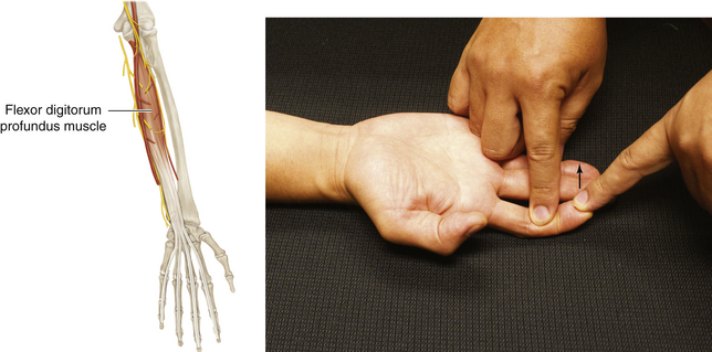
Figure 1-45 Flexor Digitorum Profundus Muscle
• Muscle attachments: Anterior and medial surfaces of proximal ulna to anterior surface of distal phalanges
• Function: Flexion of terminal phalanges of medial four digits after superficialis flexes third phalanges; flexion of wrist
• Physical examination: The patient flexes the distal interphalangeal joint (arrow) against resistance while the middle phalanx is fixed.
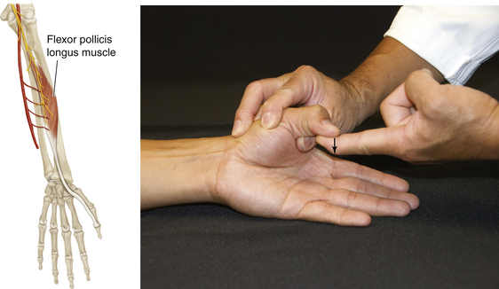
Figure 1-46 Flexor Pollicis Longus Muscle
• Muscle attachments: Anterior surface of radius and interosseous membrane to distal phalanx of thumb
• Innervation: Anterior interosseous nerve of median nerve (C8 and T1)
• Physical examination: The patient flexes the distal phalanx of the thumb (arrow) against resistance while the proximal phalanx is fixed.
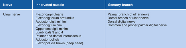
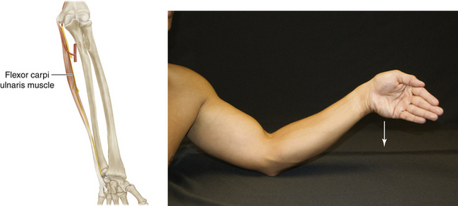
Figure 1-50 Flexor Carpi Ulnaris Muscle
• Muscle attachments: Medial epicondyle of humerus (humeral head) and olecranon and posterior border of ulna (ulnar head) to pisiform bone, hook of hamate bone, and fifth metacarpal bone
• Innervation: Ulnar nerve (C7 and C8)
• Function: Flexion and adduction of wrist joint
• Physical examination: The patient flexes and adducts the hand at the wrist (arrow) against resistance.
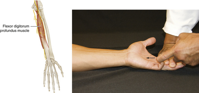
Figure 1-51 Flexor Digitorum Profundus Muscle
• Muscle attachments: Ulna to distal phalanges
• Function: Flexion of terminal phalanges of medial four digits after superficialis flexes third phalanges; flexion of wrist
• Physical examination: The patient flexes the distal interphalangeal joint (arrow) against resistance while the middle phalanx is fixed.
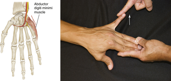
Figure 1-52 Abductor Digiti Minimi Muscle
• Muscle attachments: Pisiform bone to proximal phalanx of digit 5
• Innervation: Ulnar nerve (C8 and T1)
• Function: Flexion and abduction of metacarpophalangeal joint of fifth finger
• Physical examination: While the patient’s hand and fingers are flat upon the table, the patient abducts the little finger (arrow) against resistance.
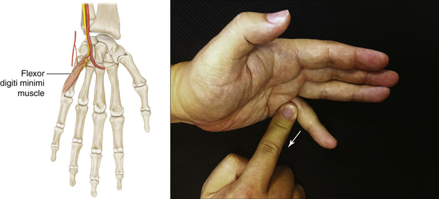
Figure 1-53 Flexor Digiti Minimi Muscle
• Muscle attachments: Hook of hamate bone and flexor retinaculum to proximal phalanx and metacarpal bone of digit 5
• Innervation: Ulnar nerve (C8 and T1)
• Function: Flexion and abduction of metacarpophalangeal joint of fifth finger
• Physical examination: With the interphalangeal joints held extended, the patient flexes the fifth finger at the metacarpophalangeal joint (arrow) against resistance.
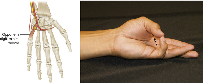
Figure 1-54 Opponens Digiti Minimi Muscle
• Muscle attachments: Hook of hamate bone and flexor retinaculum to fifth metacarpal bone
• Innervation: Ulnar nerve (C8 and T1)
• Function: Draws metacarpal joint of fifth digit in the palmar direction
• Physical examination: The patient touches the tip of the little finger with the thumb (arrow) against resistance.
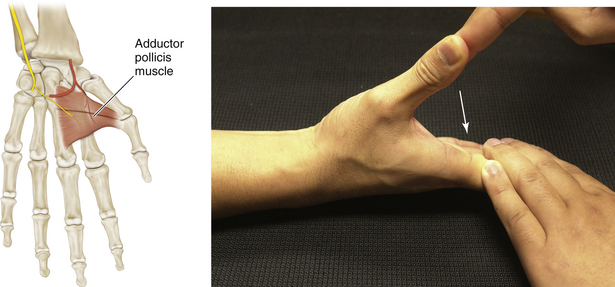
Figure 1-55 Adductor Pollicis Muscle
• Muscle attachments: Second and third metacarpals (oblique head) and third metacarpal (transverse head) to proximal phalanx of thumb
• Innervation: Ulnar nerve (C8 and T1)
• Function: Adduction of thumb
• Physical examination: The patient adducts (arrow) the thumb at right angles to the palm against the resistance of the examiner’s finger.
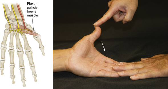
Figure 1-56 Flexor Pollicis Brevis Muscle
• Muscle attachments: Flexor retinaculum and tubercles of scaphoid and trapezium bones to proximal phalanx of thumb
• Function: Flexion and opposition of carpometacarpal joint of the thumb; flexion of metacarpophalangeal joint of the thumb
• Physical examination: The patient flexes the proximal phalanx of the thumb (arrow) against resistance.
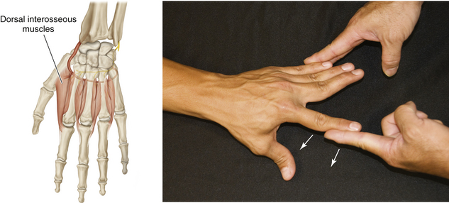
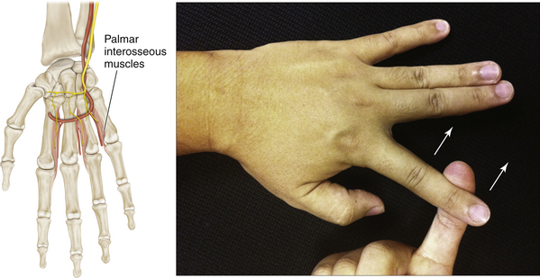
Figure 1-58 Palmar Interosseous Muscles
• Muscle attachments: Palmar surfaces of second, fourth, and fifth metacarpal bones to extensor expansions of digits and bases of proximal phalanges of digits 2, 4, and 5
• Innervation: Ulnar nerve (C8 and T1)
• Function: Flexion of second to fourth finger metacarpophalangeal joints; extension and adduction of second to fourth finger proximal and distal interphalangeal joints
• Physical examination: The patient adducts the second finger toward the middle finger (arrow).
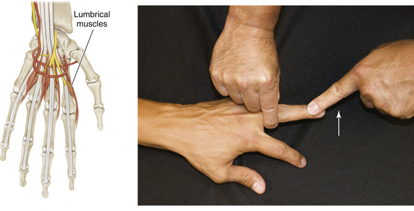
Figure 1-59 Third and Fourth Lumbrical Muscles
• Muscle attachments: Medial three tendons of flexor digitorum profundus to lateral sides of digits 2 to 5
• Innervation: Deep branch of ulnar nerve (C8 and T1)
• Function: Flexion of the fourth and fifth fingers at the metacarpophalangeal joint; extension of the fourth and fifth fingers at the interphalangeal joint
• Physical examination: The patient extends the finger at the proximal interphalangeal joint (arrow) against resistance, with the metacarpophalangeal joint hyperextended and fixed.
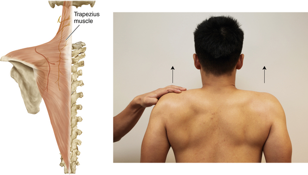
Figure 1-61 Trapezius Muscle
• Muscle attachments: Medial third of superior nuchal line, external occipital protuberance, ligamentum nuchae, and spinous processes of C7 through T12 to lateral third of clavicle, acromion, and spine of scapula
• Innervation: Spinal accessory nerve (CN XI)
• Function: Draws scapula upward; draws scapula medially at the transverse part; draws scapula downward. With serratus anterior, rotates scapula in shoulder abduction.
• Physical examination: The patient tries to elevate the shoulders (arrows) against resistance.
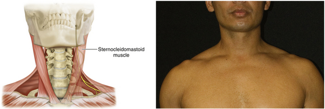
Figure 1-62 Sternocleidomastoid Muscle
• Muscle attachments: Mastoid process of temporal bone and lateral half of superior nuchal line to manubrium of sternum (sternal head) and medial third of clavicle (clavicular head)
• Innervation: Spinal accessory nerve (CN XI)
• Function: Protrusion and rotation of the head
• Physical examination: The examiner places a hand against the patient’s forehead. The patient attempts to protrude his or her head against resistance. The examiner places a hand on the side of the patient’s chin. The patient attempts to rotate his or her head against resistance.
[/level-membership-for-neurosurgery-category][not-level-membership-for-neurosurgery-category]
Chapter 1 Anatomy and Function in the Upper Extremity
Note: Unless otherwise indicated, arrows in figures indicate the direction of patient motion.
Figure 1-2 Rhomboid Major and Minor Muscles
• Muscle attachments: Spinous processes of T2 to T5 (rhomboid major) and ligamentum nuchae and spinous processes of C7 and T1 (rhomboid minor) to medial border of scapula
• Innervation: Dorsal scapular nerve (C4 and C5)
• Function: Adduction; rotation of scapula
• Physical examination: The patient places a hand on his or her back and pushes backward against resistance. (Arrow: muscle bellies can be seen.)
Figure 1-3 Levator Scapulae Muscle
• Muscle attachments: Transverse processes of C1 through C4 to medial border of scapula
• Innervation: Dorsal scapular (C5) and cervical (C3 and C4) nerves
• Function: Raises scapula and inclines neck to corresponding side if scapula is fixed
• Physical examination: The patient tries to shrug the shoulders (arrows) against resistance.
Figure 1-5 Infraspinatus Muscle
• Muscle attachments: Infraspinatus fossa of scapula to middle facet on greater tubercle of humerus
• Innervation: Suprascapular nerve (C5 and C6)
• Function: External rotation of head of humerus at the shoulder joint
• Physical examination: The patient externally rotates (arrow) the upper arm at the shoulder against resistance.
Figure 1-6 Serratus Anterior Muscle
• Muscle attachments: Anterior surfaces of first eight or nine ribs to medial border of anterior surface of scapula
• Innervation: Long thoracic nerve (C5 to C7)
• Function: Abduction of scapula
• Physical examination: Patient pushes against resistance (e.g., the examiner’s hand or a wall). (If the serratus anterior is paralyzed, winging of the scapula can be observed.)
Figure 1-8 Subscapularis Muscle
• Muscle attachments: Subscapular fossa to lesser tubercle of humerus
• Innervation: Upper and lower subscapular nerves (C5 to C7)
• Function: Internal rotation of humerus
• Physical examination: The patient internally rotates the upper arm against resistance. (The main mover is the pectoralis major.)
Figure 1-9 Teres Major Muscle
• Muscle attachments: Dorsal surface of inferior angle of scapula to intertubercular groove of humerus
• Innervation: Lower subscapular nerve (C6 and C7)
• Function: Internal rotation and adduction of humerus at shoulder joint
• Physical examination: The patient tries to adduct (arrow) the elevated upper arm against resistance.
Figure 1-10 Latissimus Dorsi Muscle
• Muscle attachments: Spinous processes of T6 to T12, thoracolumbar fascia, iliac crest, and inferior three or four ribs to floor of intertubercular groove of humerus
• Innervation: Thoracodorsal nerve (C6 to C8)
• Function: Internal rotation and adduction of humerus at shoulder joint
• Physical examination: The upper arm is horizontal, and the patient is asked to adduct it against resistance. Also, in a relaxed, upright position, the patient is asked to cough; muscle bellies can be felt to contract when the patient coughs.
Figure 1-11 Pectoralis Major Muscle
• Muscle attachments: Anterior surface of medial half of clavicle (clavicular head) and anterior surface of sternum, superior six costal cartilages, and aponeurosis of external oblique muscle (sternocostal head) to lateral lip of intertubercular groove of humerus
• Innervation: Medial and lateral pectoral nerves—clavicular head supplied by C5, C6 (lateral pectoral nerve) and sternocostal head supplied by C6, C7, and C8 (medial and lateral pectoral nerves)
• Function: Internal rotation and adduction of humerus at shoulder joint
Figure 1-16 Coracobrachialis Muscle
• Muscle attachments: Coracoid process to medial surface of humerus
• Innervation: Musculocutaneous nerve (C5 to C7)
• Function: Internal rotation, flexion, and adduction of shoulder joint
• Physical examination: With the arm flexed and laterally rotated at the shoulder joint, the elbow completely flexed, and the forearm supinated, the patient flexes (arrow) the shoulder joint against resistance.
Figure 1-17 Biceps Brachii Muscle
• Muscle attachments: Coracoid process (short head) and supraglenoid tubercle of scapula (long head) to tuberosity of radius
• Innervation: Musculocutaneous nerve (C5 and C6)
• Function: Flexion at elbow joint and supination at forearm
• Physical examination: When the patient flexes (arrow) the supinated forearm against resistance, the muscle can be seen.
[/not-level-membership-for-neurosurgery-category]
