Anaphylaxis

Published on 06/06/2015 by admin

Filed under Pediatrics

Last modified 22/04/2025

Print this page

rate 1 star rate 2 star rate 3 star rate 4 star rate 5 star
Your rating: none, Average: 0 (0 votes)

This article have been viewed 2088 times

18 Anaphylaxis

Anaphylaxis is an acute, rapidly progressive, potentially life-threatening systemic allergic reaction. Traditionally, anaphylactic reactions were caused by an immunoglobulin E– (IgE-) mediated mechanism, and “anaphylactoid” reactions were caused by non-IgE mediated mechanisms. More recently, definitions have changed. Anaphylactic reactions are caused by any immune-mediated mechanism (IgE, IgG, or immune complex), and nonallergic anaphylaxis refers to nonimmune-mediated reactions. Regardless of the mechanism, the reactions are indistinguishable. Clinically, patients typically present with varying degrees of dermatologic (e.g., hives), respiratory (e.g., wheezing), gastrointestinal (e.g., vomiting), and circulatory (e.g., hypotension) manifestations. Reactions can range from mild to severe and may be fatal. The most common causes of anaphylaxis in the pediatric population include foods, drugs, venom, and latex. The true incidence of anaphylactic reactions is unknown because of underdiagnosis and underreporting. In the United States, the potential risk of anaphylaxis approximates 1% of the general population. Currently, it is approximated that the death rate of anaphylaxis is 1 to 3 per million people per year.

Etiology and Pathogenesis

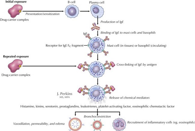
IgE-mediated anaphylaxis, a type I hypersensitivity reaction, is the most understood form of anaphylaxis (Figure 18-1). A person is exposed to an antigen, and upon reexposure, cross-linkage of IgE occurs followed by an immediate release of potent mediators from tissue mast cells and peripheral basophils. These mediators include histamine, leukotrienes, nitric oxide, and neutral proteases, which all lead to vasodilatation, increased vascular permeability, bronchoconstriction, and additional inflammation. At times, the reaction occurs with the first known exposure.

Other mechanisms include direct stimulation of mast cells and basophils, as is observed with morphine and exercise- and cold-induced anaphylaxis. Blood products and radiocontrast media may lead to activation of complement and subsequent reactions. Anaphylaxis to aspirin and nonsteroidal antiinflammatory drugs (NSAIDs) may result from the interference of the arachidonic acid pathway. Other agents may act through one or more of the above mechanisms.

The most common IgE-mediated reactions occur with food, drugs, venom, and latex. The leading cause of anaphylaxis in children is food. In the United States, the most common foods implicated in anaphylactic reactions include milk, eggs, soy, wheat, peanuts, tree nuts, and fish (although almost any food can cause a reaction). Children often develop tolerance and outgrow reactions to milk, egg, soy, and wheat; this is less likely to occur with peanuts, tree nuts, and fish. Drug allergy is another common cause of IgE-mediated anaphylaxis, with penicillin and other β-lactam antibiotics being the most commonly implicated agents. Other medications, such as aspirin and NSAIDs, may also lead to reactions. Fire ants and hymenoptera (e.g., honey bees, yellow jackets, hornets, and wasps) are common causes of anaphylaxis in both children and adults. Children with spina bifida and health care workers are at higher risk for latex allergy. Although latex allergy had been on the rise, current use of latex precautions, latex-free gloves, and health care provider awareness have stabilized the incidence of latex reactions. There is also an entity known as exercise-induced anaphylaxis. Three groups of patients present with anaphylaxis after exercising: some of whom have known specific food triggers, others in whom any food ingestion may lead to symptoms, and a third group in which there is no known food trigger. Those with a food trigger have symptoms when they exercise within 4 hours of a meal. Other causes of anaphylaxis include immunizations, radio contrast material, blood products, allergy immunotherapy, and those that remain unknown (idiopathic).

Clinical Presentation

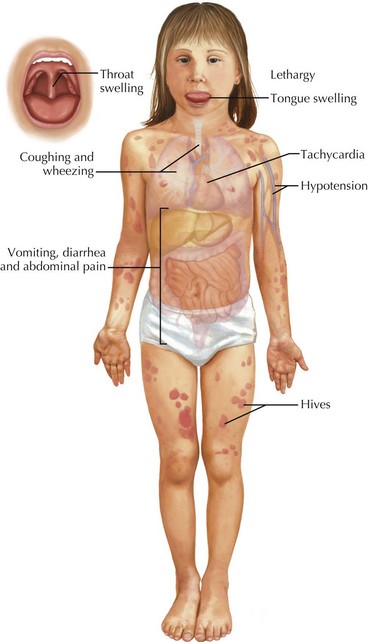
Patients with anaphylaxis may have different clinical manifestations (Table 18-1). Anaphylaxis is often underdiagnosed or misdiagnosed because of clinicians’ failure to recognize symptoms. There has been a recent attempt to standardize the diagnostic criteria to help clinicians to better recognize anaphylaxis (Box 18-1 and Figure 18-2). Approximately 90% of children with allergic reactions have skin manifestations, which include hives, angioedema (see Chapter 20, Figures 20-1 and 20-2), pruritus, or flushing. Although the remainder may not have skin involvement, they are still having a reaction, and that reaction may be more severe than those that occur with skin findings present. Tongue and throat swelling, dysphagia, and choking are manifestations of upper airway edema. Lower respiratory tract symptoms, such as coughing and wheezing, are the next most common symptoms. Vomiting, diarrhea, and abdominal pain are often seen, especially in food-induced anaphylaxis. Cardiovascular manifestations include tachycardia, hypotension, shock, and (rarely) bradycardia. Children may also be lethargic, and some have described a “feeling of impending doom.”

Table 18-1 Signs and Symptoms of Anaphylactic Reactions

Systems Signs and Symptoms
Cutaneous

Respiratory Upper Respiratory Lower Respiratory Cardiovascular Gastrointestinal Neurologic

Symptoms can develop within minutes of exposure, although most occur within 30 minutes to 1 hour. Most anaphylactic reactions are uniphasic in which the patient has a reaction, is treated, and improves. A biphasic response may also occur in which a patient becomes asymptomatic after the initial reaction and then develops a second reaction that may be the same or more severe than the initial reaction. Protracted anaphylaxis has also been described in which patients have symptoms that persist for days. Both biphasic and protracted reactions seem to occur less frequently in the pediatric population.

Differential Diagnosis

Given the involvement of multiple organ systems in anaphylaxis, many other life-threatening diagnoses can present similarly (Table 18-2). Vocal cord dysfunction presents with inspiratory stridor and coughing. Wheezing and stridor are also signs of an airway foreign body, croup, bronchiolitis, or an asthma exacerbation. Patients complaining of oral pruritus after food ingestion may have an oral allergy. This is typically self-limited and does not progress to anaphylaxis. Circulatory failure in the form of hypotension, tachycardia, and poor peripheral perfusion may be indicative of a systemic inflammatory response or shock (see Chapter 2). Vasovagal reactions usually present with history of syncope (see Chapter 48). Although urticaria can be an early sign of anaphylaxis, it also is an isolated local cutaneous reaction or may be associated with underlying infection (see Chapter 20). Flushing may occur after ingestion of particular food products, including additives such as monosodium glutamate (MSG) and sulfites, which are found in smoked foods and preservatives. “Red man syndrome” occurs with the use of vancomycin, presents with flushing, and typically resolves with termination of the infusion. Other diagnoses that present with multisystemic involvement include panic attacks, capillary leak syndrome, hereditary angioedema, and systemic mastocytosis. A good history will help distinguish these diagnoses from anaphylactic reactions.

Table 18-2 Differential Diagnosis by Symptoms

System Diagnosis
Circulatory

Cutaneous Respiratory Other/Mixed

SIRS, systemic inflammatory response syndrome.

Evaluation and Management

The diagnosis and treatment of anaphylaxis are based on history of event, clinical manifestations, and examination. No diagnostic tests are available that will help to guide management in the immediate setting. However, some diagnostic tests such as serum histamine, urinary histamine, and serum tryptase can be helpful after the acute event (Table 18-3). If performed judiciously and expeditiously, these values can prove useful in supporting the clinical diagnosis of anaphylaxis. Serum histamine levels can be drawn if a patient presents within 1 hour of reaction. β-Tryptase levels are often recommended because they peak 60 to 90 minutes after an anaphylactic reaction and may remain elevated for 6 hours. Positive results are helpful, but a patient may still have had an anaphylactic reaction if results are negative.

Table 18-3 Laboratory Evaluation

Tests Marker Time
Histamine Released by mast cells Serum: within 1 hour of event
Urine: methylhistamine can be collected up to 24 hours after event
Tryptase (although prefer beta tryptase) Released by mast cells Serum: within 6 hours of event (earlier the better)
Skin prick testing IgE-mediated skin response to specific allergen (only test to a specific allergen) After exposure (may need to perform 4 weeks after event)
Cap RAST Measures IgE levels in patient’s blood (only test to a specific allergen) After exposure (may need to perform 4 weeks after event)

IgE, immunoglobulin E; RAST, radioallergosorbent test.

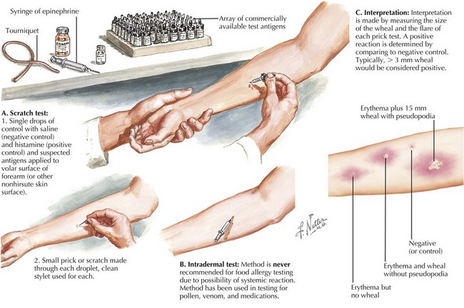
After someone has a reaction, the cause is then pursued. Often, referral to a specialist is recommended. Knowledge of positive and negative predictive values and sensitivity and specificity of various tests is important. False-positive and false-negative testing can occur; therefore, specific skin prick testing and cap radioallergosorbent testing (RAST) is often useful to confirm one’s suspicion (Figure 18-3). Of note, intradermal testing should never be used for food allergy testing. Intradermal tests are performed for allergies to pollens. The authors do not recommend broad panels of testing; however, if there is a suspicion for a specific food, then the clinician should test accordingly.

Management

For patients who have already had an anaphylactic reaction, prevention of future reactions is of highest priority. Ideally, avoidance of the inciting agent is the best prevention. However, accidental exposures may occur, so knowledge of appropriate management of allergic reactions is essential. An anaphylaxis management plan should be developed for each patient with a known anaphylactic history and should contain the following: name of patient, known allergen, type of reaction, corresponding dose and type of medication for each clinical scenario, and when to seek additional medical care. For example, a child who has a known milk allergy accidentally drinks some milk and develops facial hives. Per his anaphylaxis management plan, he is given a proper dose of diphenhydramine for his weight with resolution. He may not need to seek immediate medical attention if the reaction does not progress. Another child who has the same allergy and same exposure may develop an immediate wheeze and angioedema. Because the reaction is more severe, this child would be given epinephrine, and emergency services (911) would be called. In all cases, the family would have access to the plan to help guide their decisions during an anaphylactic reaction.

Emergency Management

Managing an anaphylactic reaction begins like any other emergent clinical scenario, with the assessment of airway, breathing, and circulation. Supplemental oxygen should be administered as needed, and the Trendelenburg position is recommended when possible. The treatment of choice in a patient with anaphylaxis is epinephrine (1 : 1000 solution) given in a dose of 0.01 mL/kg up to a maximum of 0.3 mL/dose for children and 0.5 mL/dose for adults. The dose should be given intramuscularly (IM) because absorption of epinephrine is faster if given IM compared with the subcutaneous route. Epinephrine can be readministered every 5 minutes with a maximum of three doses. Epinephrine infusions, along with other vasopressors such as dopamine, have been used in severe cases. An intravenous (IV) line must also be placed as soon as possible to aid with fluid replacement because patients with severe reactions, such as anaphylactic shock, may need rapid administration of large volumes of fluids.

Antihistamines, including H1 and H2 blockers, have been used for the treatment of anaphylaxis. Medications such as diphenhydramine (1-2 mg/kg; maximum, 50 mg) can be given orally or IV depending on the severity of the reaction. H2 blockers, such as ranitidine (1 mg/kg; max IV, 50 mg/dose; max PO, 150 mg/dose), have been used in combination with H1 blockers in the most severe cases. For those with history of asthma or who have persistent wheezing despite epinephrine, β-2 adrenergic agents, such as albuterol, should be used. The exact role of corticosteroids in the management of anaphylaxis is unclear, but they may prevent or ameliorate the late phase reaction by acting on inflammatory mediators. There is also no recommended dose or drug of choice, but agents such as prednisone or prednisolone can be given orally (1-2 mg/kg; maximum 60 mg/day) or methylprednisolone can be given IV (1-2 mg/kg; maximum 125 mg).

Hospital Management

Anaphylaxis in a hospitalized patient is treated the same way. The offending agent, if known and applicable (e.g., an antibiotic infusion), should be stopped. If the antigen has been injected, as with allergy immunotherapy, use of a tourniquet proximal to injection site is recommended. Care should be taken to release the tourniquet every 5 minutes, and it should be left no longer than 30 minutes. For patients with previous reaction to IV radiograph contrast media, premedicating with corticosteroid and diphenhydramine is recommended. If one has a known reaction to a drug and there are no other alternatives, then a consultation with an allergist is warranted. Antibiotic desensitization protocols have been established and successful in many patients with IgE-mediated reactions. Of note, during the desensitization protocol, the patient remains at risk for anaphylaxis, and after a patient is desensitized to the antibiotic, he or she must stay on protocol. If that patient misses a dose, he or she may need to repeat the protocol again. Also, if the antibiotic is needed in the future, the protocol must be repeated again.

Patients taking β-blocker therapy present a special challenge to treatment of anaphylaxis. These patients may have reactions that are resistant to standard therapy for anaphylaxis. Hypotension and bradycardia are often difficult to treat. Glucagon is the drug of choice in these patients because it has both inotropic and chronotropic effects on the heart. Atropine is useful only for patients with bradycardia.