Anaesthesia for Vascular, Endocrine and Plastic Surgery

Published on 27/02/2015 by admin

Filed under Anesthesiology

Last modified 22/04/2025

Print this page

rate 1 star rate 2 star rate 3 star rate 4 star rate 5 star
Your rating: none, Average: 0 (0 votes)

This article have been viewed 1429 times

31

Anaesthesia for Vascular, Endocrine and Plastic Surgery

MAJOR VASCULAR SURGERY

Many aspects of vascular surgery have changed during the last two decades, largely as a result of advances in radiological practice and cardiology. Examples include improvements in the treatment of myocardial infarction, the development of endovascular aortic surgery and lower limb angioplasty; such progress is likely to continue. However, anaesthesia for major vascular surgery remains a challenging area of practice. In addition to general considerations, the specific features of the commoner vascular procedures are described in this chapter: elective and emergency repair of abdominal aortic aneurysm (AAA), endovascular AAA repair, lower limb revascularization and carotid endarterectomy.

General Considerations

Peripheral vascular disease is a manifestation of generalized cardiovascular disease, and therefore coronary artery disease is present to some degree in almost all patients presenting for major vascular surgery. Most patients are elderly and have a high incidence of other coexisting medical disease, in particular:

Most of these are considered independent risk factors for perioperative cardiac complications after major surgery (see Ch 18).

The broad aims of preoperative evaluation before vascular surgery are to:

Vascular surgery is associated with a high morbidity and mortality, resulting mostly from cardiac complications (myocardial infarction, arrhythmias and cardiac failure) (see Chs 18 and 43). It is therefore vital that cardiac function is assessed preoperatively and that the risks of surgery are evaluated and discussed with the patient. Although the outcome of subsequent vascular surgery is improved in those who have previously undergone coronary revascularization by coronary artery bypass grafting (CABG), this is associated with additional risks. Percutaneous coronary interventions (insertion of bare metal or drug-eluting intracoronary stents (ICS)) are being used increasingly as an alternative to CABG in suitable patients. There is a risk of stent thrombosis after ICS so patients receive dual antiplatelet therapy (aspirin and clopidogrel) for 6 weeks after bare metal and 1 year after drug-eluting stent insertion. The risks of perioperative cardiac events or stent thrombosis are very high if surgery is performed during the period when dual antiplatelet drugs are needed because of increased haemorrhage if antiplatelet therapy is continued, or acute thrombotic events if it is interrupted. The most recent guidelines suggest that elective surgery should be postponed for at least 4–6 weeks after bare metal stent insertion (and ideally delayed for 3 months) and for 1 year after drug-eluting stent insertion, unless surgery is more urgent. It should be remembered that coronary revascularization should be performed only if indicated because of the severity of coronary disease and is not justified simply to improve outcome from subsequent vascular surgery.

Preoperative Medical Therapy in Vascular Surgical Patients

The preoperative assessment clinic is an ideal opportunity to assess concurrent medication. Drugs particularly relevant to the patient with vascular disease are β-blockers, antiplatelet drugs and statins.

β-Blockers are used extensively in patients with angina, and improve the long-term survival after myocardial infarction and in patients with heart failure. Some studies have shown beneficial effects of perioperative β-blockade in high-risk patients undergoing major vascular surgery although the benefits in lower risk patients are less clear. Current guidelines suggest that β-blockers may be commenced in patients with inducible ischaemia (documented on preoperative stress testing), or proven coronary artery disease, who are undergoing major vascular surgery. The optimum duration, dose and role of individual regimens for β-blockade are not established, but if they are used, bisoprolol 2.5–10 mg or atenolol 25–100 mg daily should be started 1–2 weeks preoperatively, titrated towards a heart rate of 60–70 beats min–1. Conversely, the perioperative discontinuation of β-blockers may be harmful, and they should be continued when already being used to control angina, arrhythmias or hypertension.

Current UK recommendations are that all patients undergoing major vascular surgery should be receiving antiplatelet therapy with aspirin unless there is a contraindication. Clopidogrel or dipyridamole are alternatives. Some clinicians consider that clopidogrel should be stopped 5 days before aortic surgery, but this should be discussed with the surgeon and prescribing physician. Aspirin should be continued throughout the perioperative period. Statin therapy should also be considered in all vascular surgical patients, and if prescribed it should be continued throughout the perioperative period.

Minimum investigations before major vascular surgery should include ECG, chest X-ray, full blood count and serum urea and electrolyte concentrations, but more invasive or specialized tests may be required (see Ch 18), including cardiopulmonary exercise testing where available. Some assessment of exercise tolerance should be made in all patients because it is a useful indicator of functional cardiac status, although many vascular patients are limited by intermittent claudication or old age and may have a sedentary lifestyle. In this case, a patient with severe coronary artery disease may have no symptoms of angina and a normal resting ECG. In some patients (e.g. those with limited functional capacity or life expectancy because of severe intractable coexistent medical conditions), the risks of elective vascular surgery may outweigh the overall potential benefits and invasive surgery may not be appropriate.

Abdominal Aortic Aneurysm

Abdominal aortic aneurysms (AAAs) occur in 2–4% of the population over the age of 65 years, predominantly in males. Approximately 90% of AAAs arise below the origin of the renal arteries and they tend to expand over time. The risk of rupture increases exponentially when the aneurysm exceeds 5.5 cm in diameter and elective surgery is then indicated. The mortality from elective AAA repair is decreasing and is now 5–8%, but overall mortality from a ruptured AAA is up to 90%, and is 50% in those who survive until emergency surgery can be performed. Consequently, screening programmes are in place to identify patients with a small asymptomatic aneurysm and to offer intervention when the aneurysm reaches a diameter of 5.5 cm. Open surgery involves replacing the aneurysmal segment with a tube or bifurcated prosthetic graft, depending on the extent of iliac artery involvement. In all cases, the aorta must be cross-clamped (see below) and a large abdominal incision is required. Surgery is prolonged and blood loss may be substantial. Patients are usually elderly, with a high incidence of coexisting disease. These factors contribute to the high morbidity and mortality of this procedure. Endovascular aortic aneurysm surgery avoids some of these problems (see below).

Elective Open AAA Repair

Preoperative evaluation and risk assessment are paramount. All vasoactive medication (except perhaps ACE inhibitors and angiotensin-II receptor blockers) must be continued up to the time of surgery and an anxiolytic premedication may be advantageous. The patient may have recently undergone arteriography and the injection of large volumes of radiopaque dye may cause renal dysfunction. Maintenance of hydration with intravenous crystalloids is advisable the night before surgery. Some patients with severe chronic obstructive pulmonary disease may benefit from regular nebulizers and chest physiotherapy before surgery to decrease the incidence of respiratory complications.

An intra-arterial and two large intravenous cannulae should be inserted before induction of anaesthesia, with monitoring of ECG and pulse oximetry. Cardiovascular changes at induction may be diminished by preoperative hydration and careful titration of the intravenous induction agent. After neuromuscular blockade, the trachea is intubated (see below) and anaesthesia continued using a balanced volatile/opioid technique. Perioperative epidural analgesia is useful and may be undertaken before or after induction of anaesthesia.

Several important considerations apply to patients undergoing aortic surgery (Table 31.1). The following are required:

TABLE 31.1

Major Anaesthetic Considerations for Patients Undergoing Aortic Surgery

High incidence of coexisting cardiovascular and respiratory disease

Cardiovascular instability during induction of anaesthesia, aortic cross-clamping and declamping

Large blood loss and fluid shifts during and after surgery

Prolonged major surgery in high-risk patients

Marked heat and evaporative fluid losses from exposed bowel

Potential postoperative impairment of respiratory, cardiac, renal and gastrointestinal function

In the more compromised patient, e.g. with ischaemic heart disease and poor left ventricular function, an additional cardiac output monitor (e.g. transoesophageal echocardiography or other non-invasive device) should be used to monitor cardiac index and guide fluid management. A pulmonary artery catheter may be indicated in some patients. All possible measures should be undertaken to maintain body temperature, including heated mattress and overblanket, warmed intravenous fluids, and warmed and humidified inspired gases. The ambient temperature should be warm and the bowel may be wrapped in clear plastic to minimize evaporative losses.

Three specific stimuli may give rise to cardiovascular instability during surgery:

image Tracheal intubation. Laryngoscopy and tracheal intubation may be accompanied by marked increases in arterial pressure and heart rate which may precipitate myocardial ischaemia in susceptible individuals. This response may be attenuated by the i.v. administration of a β-blocker (e.g. esmolol 1.5 mg kg–1) or a rapidly-acting opioid (e.g. alfentanil 10 μg kg–1) before intubation.

image Cross-clamping of the aorta. Clamping of the aorta causes a sudden increase in aortic impedance to forward flow and hence left ventricular afterload. This increases cardiac work and may result in myocardial ischaemia, arrhythmias and left ventricular failure. Arterial pressure proximal to the clamp increases acutely even though left ventricular ejection fraction and cardiac output are reduced. The effects on preload are variable. The degree of cardiovascular disturbance is greater when the clamp is applied more proximally (greater at the supracoeliac > suprarenal > infrarenal levels). A vasodilator, e.g. glyceryl trinitrate (GTN), is often infused just before clamping (and continued up to clamp release) to obviate these problems. Deepening of volatile anaesthesia or an additional dose of an opioid may also be used at aortic clamping. While the aorta is clamped, blood flow distal to the clamp decreases, and distal organ perfusion is largely dependent on the collateral circulation. The lower limbs, pelvic and abdominal viscera suffer variable degrees of ischaemia during which inflammatory mediators are released from white blood cells, platelets and capillary endothelium. These mediators include oxygen free radicals, neutrophil proteases, platelet activating factor, cyclo-oxygenase products and cytokines.

image Aortic declamping. Declamping of the aorta causes sudden decreases in aortic impedance, systemic vascular resistance and venous return with reperfusion of the bowel, pelvis and lower limbs and redistribution of blood. Inflammatory mediators are swept into the systemic circulation causing vasodilatation, metabolic acidosis, increased capillary permeability and sequestration of blood cells in the lungs. This is a critical period of anaesthesia and surgery because hypotension after aortic declamping may be severe and refractory unless circulating volume has been well maintained. If relative hypervolaemia is produced during the period of clamping by infusion of fluids to produce a CVP of greater than 12–14 mmHg (and perhaps administration of GTN until shortly before clamp release), declamping hypotension is less of a problem and metabolic acidosis may be diminished. Declamping hypotension usually resolves within a few minutes but vasopressors or positive inotropes are often required; these can be given before clamp release in anticipation. Good communication with the surgeon and slow or sequential clamp release helps the anaesthetist to manage aortic declamping. Renal blood flow decreases even when an infrarenal cross-clamp is used, and steps to maintain renal function are often required. The single most important measure is the maintenance of extracellular fluid volume (i.e. CVP > 12–14 mmHg). Prophylactic mannitol 0.5–1.0 g kg–1 i.v. or furosemide may also be administered, although the evidence for their efficacy is conflicting.

Bleeding is a problem throughout the operation, often after aortic clamping when back bleeding from lumbar vessels occurs, but may be particularly severe at aortic declamping as the adequacy of vascular anastomoses is tested. A red cell salvage device should be used routinely in aortic surgery when it is available, but other modes of autologous blood transfusion (predonation, normovolaemic haemodilution) may be valuable. In addition to red cells, specific clotting factors are often required. It is often preferable to reserve the use of clotting factors until the anastomoses are complete and most of the anticipated blood loss has occurred. Many surgeons request the administration of heparin (usually 5000 units i.v.) before insertion of the graft and occasionally it may be appropriate to reverse its effects with protamine (0.5 mg per 100 units of heparin). Point-of-care haemostatic testing (thromboelastography or thromboelastometry) are very useful to diagnose coagulopathy and guide the use of clotting products.

Epidural analgesia is usually provided through a catheter placed at the mid-thoracic level, unless there is a contraindication. There is some debate as to whether epidural local anaesthetics are best administered during surgery (to attenuate cardiovascular and stress responses) or at the end of surgery (because sympathetic blockade may cause hypotension and make cardiovascular management more difficult during the procedure). A popular technique is to use combined volatile general anaesthesia with boluses of fentanyl or an infusion of remifentanil for intraoperative analgesia; epidural analgesia is then established after aortic declamping and once cardiovascular stability is ensured, using a combination of local anaesthetic and fentanyl.

Most patients are elderly and are unable to tolerate the large heat loss occurring through the extensive surgical exposure, which necessitates displacement of the bowel outside the abdominal cavity. Hypothermia causes vasoconstriction, which may cause myocardial ischaemia, delayed recovery and difficulties with fluid management during rewarming, because large volumes of intravenous fluid may be required. Therefore, all measures should be taken to prevent hypothermia.

Emergency Open Repair

The principles of management are similar to those discussed above. However, the patient may be grossly hypovolaemic and arterial pressure is often maintained only by marked systemic vasoconstriction and the action of abdominal muscle tone acting on intra-abdominal capacitance vessels. Resuscitation with intravenous fluids before the patient reaches the operating theatre should be judicious; permissive hypotension (maintaining systolic pressure at 80–100 mmHg) limits the extent of haemorrhage and improves outcome. The patient is prepared and anaesthesia induced on the operating table. While 100% oxygen is administered by mask, an arterial and two large-gauge i.v. cannulae are inserted under local anaesthesia. The surgeon then prepares and towels the patient ready for surgery and it is only at this point that anaesthesia is induced using a rapid-sequence technique. When muscle relaxation occurs, systemic arterial pressure may decrease precipitously and immediate laparotomy and aortic clamping may be required. Thereafter, the procedure is similar to that for elective repair.

The prognosis is poor for several reasons. There has been no preoperative preparation and most patients have concurrent disease. There may have been a period of severe hypotension, resulting in impairment of renal, cerebral or myocardial function. Blood loss is often substantial and massive transfusion of red cells and clotting factors is usually required. Postoperative jaundice is common because of haemolysis of damaged red cells in the circulation and in the large retroperitoneal haematoma which usually develops after aortic rupture. In addition, postoperative renal impairment and prolonged ileus often occur. Artificial ventilation and organ support are required for several days and the cause of death is usually multiorgan failure.

Endovascular Aortic Aneurysm Repair

Endovascular aortic aneurysm repair (EVAR) is now an established alternative to open surgery. A balloon-expandable stent-graft is inserted under radiological guidance via the femoral or iliac arteries into the aneurysm to exclude it from the circulation. It is performed via groin incisions and the aortic lumen is temporarily occluded from within, rather than being cross-clamped. The cardiovascular, metabolic and respiratory consequences are reduced in comparison with conventional open surgery. Perioperative blood loss, transfusion requirements, postoperative pain, hospital stay and morbidity are lower compared with open surgery. Perioperative morbidity is 1–2% although long-term (> 8 years) survival after EVAR is similar to that after open surgery because of deaths from cardiovascular and respiratory diseases. Despite advances in stent-graft technology, the morphology of the aneurysm in some patients renders it unsuitable for EVAR (based on the site, shape, degree of angulation and the size of iliac arteries). Repeated radiological procedures (e.g. angioplasty) are required in up to 20% of patients. The procedure usually takes 1–2 h and may be performed by radiologists and/or surgeons but the patients have the same coexisting diseases and some of the anaesthetic considerations are similar. In some cases, EVAR may be preferred as a less invasive technique in patients judged unfit for open surgery. Access to the iliac vessels is often possible using infra-inguinal incisions in the groin and postoperative pain is therefore minimal compared to open surgery. In many centres, EVAR is performed in the radiology suite, in which case the anaesthetist must ensure that anaesthetic facilities for high-risk patients are adequate.

EVAR may be performed under general, regional or local anaesthesia with or without sedative adjuncts. In all cases, direct arterial pressure monitoring is mandatory because rapid fluctuations in arterial pressure may occur during stent-graft deployment. In awake patients, hyoscine 20 mg i.v. may be useful to decrease bowel motility during stent-graft placement. CVP monitoring is not usually necessary unless dictated by the patient’s medical condition (e.g. moderate/severe cardiac disease). Short periods of apnoea are needed during insertion of the device; this is easy when ventilation is controlled but requires the patient’s co-operation if a regional or local anaesthetic technique is used. The devices are positioned under angiographic control and large volumes of radiocontrast may be used, predisposing to contrast-induced nephropathy (CIN). It is important to avoid hypotension and hypovolaemia, both of which can contribute to CIN; sodium bicarbonate, N-acetylcysteine or mannitol may be administered, although the evidence of their benefit is limited. Brisk haemorrhage is unusual during EVAR and, although bleeding may be significant, it is usually insidious. However, large-diameter cannulae should be inserted and vasoactive drugs readily available because if endovascular repair is not technically feasible, conversion to open surgery may be required. EVAR may also be used to repair contained ruptured or leaking AAAs, and has an increasing role in the management of thoracic aortic aneurysms.

Surgery for Occlusive Peripheral Vascular Disease

Peripheral reconstructive surgery is performed in patients with severe atherosclerotic arterial disease causing ischaemic rest pain, tissue loss (ulceration or gangrene), severe claudication with disease at specific anatomical sites (aorto-iliac, femoropopliteal, popliteal or distal) or failure of nonsurgical procedures. Most patients are heavy smokers, suffer from chronic pulmonary disease and have widespread arterial disease. Most patients present with intermittent claudication. Consequently, exercise tolerance is limited and severe coronary artery disease may be present despite few symptoms. Surgical revascularization is performed to salvage the ischaemic limb, but arterial angioplasty is a less invasive alternative and is increasingly performed as a first-line procedure in suitable patients. Patients presenting for surgical reconstruction are often those in whom angioplasties have failed and who may have more severe vascular disease. Short-term mortality after lower limb revascularization is comparable to that following AAA repair and long-term outcome is worse as a consequence of associated cardiovascular disease. Acute limb ischaemia which threatens limb viability requires rapid intervention comprising full anticoagulation, intrathrombus thrombolysis after arteriography, analgesia and revascularization via embolectomy, angioplasty or bypass surgery as indicated. The clinical findings of sensory loss and muscle weakness necessitate intervention within 6 h and therefore preoperative evaluation and correction of risk factors may be limited.

Peripheral Arterial Reconstruction

The commonest procedures involve the insertion of an autologous vein or synthetic vascular graft between axillary and femoral, or femoral and popliteal, arteries. Axillofemoral bypass surgery is performed in those not considered fit for open aortic surgery, and these patients are often particularly frail. All these operations are prolonged and an IPPV/relaxant balanced anaesthetic technique is suitable. A meticulous anaesthetic technique is paramount, with particular attention to the maintenance of normothermia and administration of i.v. fluids. Hypothermia or hypovolaemia may cause peripheral vasoconstriction, compromising distal perfusion and postoperative graft function. Blood loss through the walls of open-weave grafts may continue for several hours after surgery and cardiovascular status should be monitored closely during this time. Epidural analgesia may be used alone or as an adjunct to general anaesthesia for lower limb procedures. Despite theoretical advantages, epidural anaesthesia has no effect on graft function per se but it does provide effective postoperative analgesia. However, i.v. heparin is usually administered during and after surgery (see below) and the risks of epidural haematoma should be considered. Oxygen therapy should be continued for at least 24 h after surgery, and monitoring in a high-dependency unit is often required.

Carotid Artery Surgery

Despite advances in the medical treatment of patients with stroke, it remains a significant cause of death and disability. Carotid endarterectomy is performed to prevent disabling embolic stroke in patients with atheromatous plaques in the common carotid bifurcation, or internal or external carotid arteries. Most patients are elderly, with generalized vascular disease. Cerebral autoregulation may be impaired and cerebral blood flow is therefore much more dependent upon systemic arterial pressure. The main risk of surgery is the production of a new neurological deficit (which may be fatal or cause permanent disability), although cardiovascular complications account for 50% of the overall morbidity and mortality.

Carotid endarterectomy is unusual in that it is a preventative operation with well-defined indications based on the results of large-scale, randomized studies performed in Europe and the USA. In patients with a previous stroke and a carotid stenosis > 70%, the benefits of surgery outweigh the risks, whereas in those with mild stenosis (< 30%), the risks outweigh the benefits and medical treatment with antiplatelet drugs is preferred. Therefore, the patients presenting for surgery are those with the most severe disease, and the potential benefits are only realized if the overall perioperative mortality and morbidity are low (< 5%). Specific perioperative risk factors are age > 75 years, female sex, systolic hypertension, peripheral vascular disease (probably as a marker for coronary artery disease), experience of the surgeon and ipsilateral cerebral symptoms. Longer-term outcome is also worse in smokers and those with diabetes or hyperlipidaemia. Carotid artery angioplasty is a less invasive alternative to surgical endarterectomy, although its place is yet to be established. It is now clear that the risks of major stroke are highest within the first few days after a transient ischaemic attack (TIA) or minor stroke. Consequently, carotid endarterectomy should be performed as soon as is feasible (within 1 week and ideally within 48 h) after a minor stroke or TIA when indicated (embolic stroke, significant carotid stenosis). This limits the time available for preoperative preparation, investigation and risk reduction.

During surgery, the internal, external and common carotid arteries are clamped and the atheromatous plaque removed. During application of the clamps, cerebral perfusion is dependent on collateral circulation via the circle of Willis. Many surgeons insert a temporary shunt to bypass the site of obstruction, minimizing the period of potential cerebral ischaemia. Several methods are available to assess cerebral blood flow during clamping, before proceeding with the endarterectomy; if flow is adequate, some surgeons prefer not to use a temporary shunt. Monitoring of neurological status in an awake patient is considered by many to be the ‘gold standard’, but other methods used in practice include:

Although most strokes related to surgery are associated with thromboembolism rather than hypo- or hypertension, and the majority of these are caused by inadvertent technical surgical error, the anaesthetist has a crucial role in the maintenance of cardiovascular stability before, during and after surgery. Rapid swings in arterial pressure are common because of the direct effects of surgical manipulation, plaque removal and carotid cross clamping in patients with impaired baroreceptor function due to carotid atheroma and cardiovascular disease.

The main aims of anaesthesia for carotid endarterectomy are maintenance of oxygen delivery to the brain, cardiovascular stability, airway protection, provision of neurological protection and rapid recovery. Most intraoperative strokes are apparent on recovery from anaesthesia and early postoperative neurological assessment is important. Any residual postoperative effects of anaesthesia may confuse the diagnosis of intraoperative embolism or ischaemic change, so a technique that permits rapid return of function is required. These aims may be achieved using general, local or regional anaesthetic techniques, with or without sedative or analgesic adjuncts. In all cases, an intra- arterial cannula is mandatory for monitoring of arterial pressure, which should be maintained particularly during carotid clamping, and attention paid to maintain normothermia.

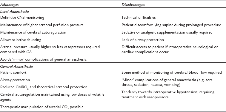
Local infiltration of the surgical field may be used alone or in combination with superficial and intermediate or deep cervical plexus blockade. Superficial cervical plexus block is performed by infiltration of local anaesthetic along the entire length of the posterior border of the sternomastoid muscle, using 10–15 mL local anaesthetic (e.g. levobupivacaine 0.25%). The intermediate block involves injection of 5 mL of local anaesthetic 1–2 cm deep to the midpoint of the sternomastoid. Advantages of locoregional techniques include definitive neurological monitoring (therefore allowing selective use of shunts), preservation of cerebral and coronary autoregulation, and the maintenance of higher cerebral perfusion pressures during the procedure (Table 31.2). These techniques rely on good cooperation between the patient, the surgeon and the anaesthetist. Many patients find it difficult to lie still and supine for the duration of the procedure, particularly those with heart failure or respiratory disease; this may be compounded by diaphragmatic compromise because phrenic nerve paralysis may accompany deep cervical plexus blockade. Sudden loss of consciousness or seizures may occur if cerebral perfusion is inadequate after clamping, and subsequent airway control may be very difficult because access is limited.

Performing surgery under general anaesthesia avoids these problems. Both propofol and volatile agents (at low doses) preserve cerebral and coronary autoregulation, reduce cerebral oxygen requirements and, in theory, may provide some degree of neuroprotection. General anaesthesia with artificial ventilation allows PaCO2 to be manipulated but hypotension may be more common compared with regional anaesthesia. The airway is not accessible during surgery and tracheal intubation with a well-secured reinforced tracheal tube is advisable. Anaesthesia should be induced cautiously using an i.v. agent and maintained with a balanced technique using an inspired oxygen concentration of 50% in air or nitrous oxide (100% inspired oxygen produces cerebral vasoconstriction) with isoflurane, sevoflurane or desflurane. All anaesthetic agents should be short-acting, and remifentanil, alfentanil or low-dose fentanyl (100–200 μg) are useful adjuncts. Hypotension may potentially occur after induction and during the placement of cerebral monitoring, but it should be treated promptly. Vasopressors (e.g. ephedrine 3–6 mg or phenylephrine 25–50 μg increments) are frequently required and should be drawn up before induction of anaesthesia. A high PaO2, normocapnia and normothermia should be maintained. Blood loss and fluid requirements are usually modest. Postoperatively, significant pain is unusual and the combination of wound infiltration with local anaesthetic with a nonsteroidal anti-inflammatory analgesic during surgery is effective.

Data from the GALA trial (general anaesthesia vs. local anaesthesia) and systematic reviews have shown no difference in overall outcome with any specific anaesthetic technique.

Patients should be monitored in a high-dependency environment for several hours postoperatively. Hypertension is common in the early postoperative period because of impaired circulatory reflexes; pain from the wound or from bladder distension may also contribute. Hypertension is associated with adverse neurological outcomes because it may compromise the graft or cause intracranial haemorrhage. Arterial pressure should be controlled to achieve systolic pressures < 165 mmHg and diastolic pressures < 95 mmHg, accounting for the range of individual preoperative values. Intravenous α- or β-blockers or an infusion of a vasodilator (e.g. GTN or hydralazine) may be required as prophylaxis or treatment.

The other main postoperative complication is the development of a haematoma. Initial treatment involves local pressure and reversal of heparin with protamine. However, local oedema and the presence of a large haematoma may cause airway compromise and hypoxaemia requiring urgent surgical exploration. Induction of general anaesthesia in these circumstances is particularly hazardous and evacuation of the haematoma under local infiltration is usually preferable. Recurrent laryngeal nerve damage is a recognized complication of carotid endarterectomy. In most cases, this simply causes a hoarse voice but in patients who have had a previous contralateral carotid endarterectomy, specific preoperative evaluation of vocal cord function should be performed before surgery.

Cardioversion

Direct current (DC) cardioversion is an effective treatment for some re-entrant tachyarrhythmias, which may produce haemodynamic instability and myocardial ischaemia and which do not respond to other measures. Atrial fibrillation of less than 6 months’ duration, atrial flutter, supraventricular tachycardia and ventricular tachycardia may be converted to sinus rhythm, although maintenance of sinus rhythm depends usually on subsequent antiarrhythmic drugs. Cardioversion has little effect on contractility, conductivity or excitability of the myocardium, and has a low incidence of side-effects or complications.

Pre-Anaesthetic Assessment

Patients may present with a chronic arrhythmia for elective cardioversion or as an emergency in extremis with a life-threatening arrhythmia. They may have other serious cardiovascular pathology such as rheumatic disease, ischaemic heart disease, recent myocardial infarction or cardiac failure. Digoxin therapy predisposes to postcardioversion arrhythmias; it is often withheld for 48 h before cardioversion. If DC cardioversion is required in a patient receiving digoxin, the initial DC dose should be low (e.g. 10–25 J) and increased if necessary. In some patients there is a significant risk of embolic phenomena, e.g. those with:

These patients should receive prophylactic anticoagulants for 2–3 weeks before cardioversion, and anticoagulation should be continued afterwards. Accurate knowledge of the medical and drug history and thorough clinical examination are essential before anaesthesia.

Cardioversion

Direct current (DC) electrical discharge passed through the heart depolarizes all excitable myocardial cells and interrupts abnormal pathways and foci. The electrodes are usually positioned on the anterolateral chest with the patient supine, but the anteroposterior arrangement, with the patient in the lateral position, is sometimes used. The paddles should not be sited over the scapula, sternum or vertebrae and the skin must be protected with electrolyte jelly, saline-soaked gauze or any type of conducting pad.

The ECG monitoring lead chosen should demonstrate a clear R wave in order to synchronize the discharge away from the T wave and thus reduce the risk of development of ventricular fibrillation. If the arrhythmia does not convert after the first 50 J discharge, further shocks are given, using an increased energy discharge of up to 200 J.

Despite the use of synchronized discharge, ventricular fibrillation may be produced in the presence of hypokalaemia, ischaemia, digoxin toxicity and QT prolongation (e.g. caused by quinidine or tricyclic antidepressants).

Anaesthesia

Treatment should be carried out only in areas specifically designed for the purpose and with a full range of drugs, resuscitation and monitoring equipment available. These must be checked by the anaesthetist, and patients prepared as for a surgical procedure.

ECG monitoring, pulse oximetry and measurement of arterial pressure are instituted. A vein is cannulated and the patient’s lungs are preoxygenated before i.v. induction of anaesthesia. The choice of drug is determined by the cardiovascular stability and recovery period required. If the patient is clinically shocked, precautions to prevent aspiration of gastric contents should be taken and a rapid-sequence induction with cricoid pressure and tracheal intubation should be used. However, many patients are admitted for elective cardioversion on a day-case basis, and a technique using i.v. propofol and spontaneous ventilation is suitable.

As soon as the patient is unconscious, the airway is secured and oxygenation maintained with a suitable breathing system. Before activation of the defibrillator, it is important to check that the patient is not in contact with any person or metal object. If repeated shocks are required, incremental doses of the anaesthetic may be given. The patient should be monitored carefully both during anaesthesia and after recovery of consciousness, in particular for evidence of recurrent arrhythmia, hypotension, pulmonary oedema, or systemic or pulmonary embolism.

SURGERY FOR TUMOURS OF THE ENDOCRINE SYSTEM

Amine precursor uptake and decarboxylation (APUD) cells originate from neuroectoderm and are distributed widely throughout the body. They synthesize and store neurotransmitter substances, including serotonin, ACTH, calcitonin, melanocyte-stimulating hormone (MSH), glucagon, gastrin and vasoactive intestinal polypeptide (VIP). Neoplastic change within these cells produces the group of tumours termed apudomas, e.g. carcinoid, pancreatic islet cell tumour, pituitary and thyroid adenoma, medullary carcinoma of thyroid and small cell carcinoma of the lung. These may be orthoendocrine or paraendocrine – the former produce amines and polypeptides associated normally with the constituent cells, while the latter secrete substances produced usually by other organs. Two orthoendocrine apudomas in particular may produce significant problems for the anaesthetist.

Carcinoid Tumour

Carcinoid tumours are rare tumours derived from enterochromaffin cells of the intestinal tract, most commonly the small bowel or appendix. However, they may arise at any site in the gut and rarely in the gallbladder, pancreas or bronchus. They are usually benign. Malignant change occurs in 4% and may produce hepatic metastases. Carcinoid tumours may secrete a number of vasoactive peptides and amines (e.g. serotonin, histamine, kinins and prostaglandins) which have a variety of effects on vascular, bronchial and gastrointestinal smooth muscle activity. These compounds are normally metabolized in the liver and carcinoid tumours are usually asymptomatic unless the mediators reach the systemic circulation from hepatic metastases, an extra-abdominal primary (e.g. bronchus), or if the tumour is large and hepatic metabolism is exceeded. In these cases, the clinical symptoms of carcinoid syndrome occur. These are variable but include flushing, increased intestinal motility, abdominal pain, bronchospasm and dyspnoea. Flushing, bronchospasm, increased intestinal motility, hypotension and oedema are related to the production of kallikrein, which is metabolized to bradykinin, a potent vasodilator. Adrenergic stimulation and alcohol ingestion increase the production of bradykinin. Serotonin (5-hydroxytryptamine, 5-HT) causes abnormal gut motility, diarrhoea and bronchospasm. It has positive inotropic and chronotropic effects and produces vasoconstriction. It may cause endocardial fibrosis, leading to pulmonary and tricuspid stenosis or regurgitation (although bronchial carcinoid tumours may lead to left-sided cardiac valvular lesions). Histamine secretion may cause bronchoconstriction and flushing. Acute attacks of carcinoid syndrome may also be precipitated by fear or hypotension.

Diagnosis is confirmed by high urinary excretion of 5-hydroxyindoleacetic acid (5-HIAA), a metabolite of 5-HT. Urinary 5-HIAA concentrations correlate with tumour activity and perioperative complications.

Primary and secondary tumours are localized by CT, MRI, ultrasound, combined PET/CT scans or radionuclide scans. Although medication may alleviate some symptoms, the definitive treatment of carcinoid tumours is surgery, including excision of the primary tumour and resection or radiofrequency ablation of hepatic metastases. The main anaesthetic considerations are perioperative prevention of mediator release and preparation for control of carcinoid crises. Systemic release of carcinoid mediators can be exacerbated or precipitated by anxiety, tracheal intubation, inadequate analgesia, tumour manipulation or the administration of catecholamines or drugs which cause histamine release. In severe cases, acute intraoperative cardiovascular instability (arrhythmias and extreme fluctuations in arterial pressure) and resistant bronchospasm may occur.

Management

Patients may be taking drugs to diminish symptoms of diarrhoea, flushing and bronchospasm, but specific agents are used to inhibit synthesis, prevent release or block the actions of the mediators released by the tumour. The most important drug is the somatostatin analogue octreotide, which improves both symptoms and biochemical indices, and which is useful in the prevention and management of perioperative hypotension and carcinoid crisis. Somatostatin (half-life 1–3 min) is secreted naturally by the pancreas and regulates gastrointestinal peptide production by inhibiting the secretion of growth hormone, thyroid-stimulating hormone (TSH), prolactin and other exocrine and endocrine hormones. Octreotide, the octapeptide analogue of somatostatin, has a longer half-life, high potency and low clearance, and may be given i.v. or s.c. The usual s.c. dose is 50–200 μg every 8–12 h. It is useful for symptom relief in other conditions, notably acromegaly, VIPoma and glucagonoma. It may cause gastrointestinal side-effects, gallstones and impaired glucose tolerance.

5-HT antagonists (ketanserin, methysergide) and antihistamines, e.g. ranitidine, chlorphenamine (chlorpheniramine), are also used. Cyproheptadine has both antihistamine and anti 5-HT actions.

Conduct of Anaesthesia

Perioperative management should be in close cooperation with both physician and surgeon, and the patient’s regular medication should be continued up to the time of surgery. The possibility of cardiac valvular lesions should be considered. Hypovolaemia and electrolyte disturbance should be corrected before operation. Anxiolytic premedication with minimal cardiovascular disturbance is desirable; an oral benzodiazepine is often used alone or together with an antihistamine, although oversedation should be avoided. Octreotide must be continued as premedication 50–100 μg s.c. 1 h preoperatively. It may also be administered during surgery as an i.v. infusion at 50–100 μg h–1. A smooth anaesthetic technique is essential, and techniques which may cause hypotension, including epidural and subarachnoid block, should be used with extreme caution. Drugs which release histamine (e.g. thiopental, morphine, pethidine, atracurium, mivacurium) should be avoided.

Continuous monitoring of ECG and direct arterial pressure should be started before careful induction of anaesthesia with etomidate or propofol, accompanied by measures to obtund the potentially exaggerated pressor response to tracheal intubation. Succinylcholine is best avoided because it may cause peptide release and nondepolarizing muscle relaxants with minimal histamine release (e.g. rocuronium or vecuronium) are preferable. Anaesthesia should be maintained with opioids (e.g. fentanyl or remifentanil), inhaled nitrous oxide and a volatile agent. Total intravenous anaesthesia with propofol has also been used. Bronchospasm may be severe and should be treated with octreotide or aminophylline rather than adrenaline, and a flow-generator type of ventilator capable of delivering the inspired gases at high pressure should be used. Major fluid shifts may occur during surgery and the effects of circulating peptides may distort the physiological response to hypovolaemia. Central venous pressure measurement is advisable when large blood loss is likely and pulmonary artery catheterization may be required in patients with cardiac complications. Intraoperative hypotension may be severe and should be treated with intravenous fluids and octreotide 100 μg i.v. Sympathomimetic drugs may cause α-mediated peptide release and are not recommended for the treatment of bronchospasm or hypotension. Hypertension is usually less severe and usually responds to increased depth of anaesthesia, β-blockade or ketanserin.

Close cardiovascular monitoring and good analgesia are required postoperatively and the patient should be observed in a high-dependency or intensive therapy unit. The use of epidural analgesia is controversial, but an epidural infusion of fentanyl alone or with bupivacaine 0.1% has been used successfully.

Phaeochromocytoma

Phaeochromocytomas are derived from chromaffin cells which secrete catecholamines (predominantly noradrenaline, but also adrenaline and occasionally dopamine) and occur in less than 0.1% of hypertensive patients. The majority present in middle-aged adults but they may be found in childhood. Most are found as a single benign tumour of the adrenal medulla, but 10% occur in ectopic sites, e.g. paravertebral sympathetic ganglia. Approximately 10% of phaeochromocytomas are malignant, and 10% are bilateral. Genetic factors are frequently involved and they may be associated with multiple endocrine neoplasia (MEN) and other syndromes (Table 31.3).

TABLE 31.3

Associations of Phaeochromocytoma with Other Syndromes

Von Hippel-Lindau disease (retinocerebral haemangioblastoma)

MEN IIa (Sipple’s syndrome)

 Phaeochromocytoma

 Medullary cell thyroid carcinoma

 Hyperparathyroidism

MEN IIb (mucosal neuroma syndrome)

 Phaeochromocytoma

 Medullary cell thyroid carcinoma

 Mucosal neuromata

 Marfanoid habitus

Phaeochromocytoma-paraganglionoma syndrome

Von Recklinghausen’s disease (multiple neurofibromatosis)

The clinical features depend on the quantity of hormones secreted and on which is predominant, although episodes may be paroxysmal and clinical findings may be normal between attacks. Noradrenaline-secreting tumours tend to cause severe refractory hypertension, headaches and glucose intolerance; circulating blood volume is reduced and vasoconstriction occurs. Adrenaline-secreting tumours trigger palpitations, anxiety and panic attacks, sweating, hypoglycaemia, tachycardia, tachyarrhythmias and occasionally high-output cardiac failure. Malaise, weight loss, pallor and psychological disturbances may occur, and end-organ damage (e.g. retinopathy, nephropathy, dilated cardiomyopathy) may arise as a consequence of hypertension. They present several problems to the anaesthetist (Table 31.4).

TABLE 31.4

Anaesthetic Considerations in Patients with Phaeochromocytoma

Preoperative

Hypertension

Hypovolaemia (vasoconstriction with reduced circulating volume)

Pharmacological stabilization

 α-blockade

 β-blockade

 Control of catecholamine synthesis

End-organ damage

Anxiolytic/sedative premedication

Intraoperative

Severe cardiovascular instability, particularly:

 at induction of anaesthesia and tracheal intubation

 during pneumoperitoneum (laparascopic procedures)

 during tumour handling

 following ligation of venous drainage

Hypoglycaemia after tumour removal

Postoperative

Hypotension

Hypoglycaemia

Somnolence, opioid sensitivity

Hypoadrenalism

LV dysfunction

Diagnosis

Diagnosis is important because the mortality of patients undergoing unrelated surgery with an unsuspected phaeochromocytoma is up to 50%. Diagnosis is confirmed by measurement of high plasma and urine concentrations of free catecholamines. Random 1-h or 24-h urinary excretion of catecholamine metabolites (metanephrines and 3-methoxy-4-hydroxymandelic acid (HMMA; also known as vanillylmandelic acid [VMA])), are an alternative. In some cases a clonidine suppression test may be required to distinguish between hypertensive patients and those with phaeochromocytoma. MRI of the abdomen is probably the investigation of choice to localize tumours greater than 1 cm in diameter. Computed tomography (performed without intravenous contrast media, which may precipitate release of hormone) is an alternative. Confirmation of the identity and position of an adrenal mass is by uptake of [131I] m-iodobenzylguanidine (MIBG) monitored by gamma camera. MIBG scanning or 18 F-fluorodopamine positron emission tomography may be useful for the localization of small or extra-adrenal tumours not detected by other means.

Preoperative Preparation

Medical treatment of the effects of the tumour must be achieved before surgery. α-Adrenergic antagonists counteract the increased peripheral vascular resistance and reduced circulating volume, and phenoxybenzamine (noncompetitive, nonselective antagonist), prazosin and doxazosin (α1-selective, competitive antagonists) have been used successfully. Noncompetitive α-antagonists are preferable because surges of catecholamine concentrations, occurring particularly during tumour handling, do not overwhelm the effects of a noncompetitive drug. Phenoxybenzamine is given in increasing titrated doses over 2–3 weeks before surgery, starting from 10 mg b.d. up to a usual dose of 40–50 mg b.d. In this way, the circulating volume expands gradually with normal oral intake of fluid. Adverse effects include initial postural hypotension, tachycardia, blurred vision and nasal congestion. A β-adrenergic antagonist may be required later to control tachycardia, but acute hypertension, cardiac failure and acute pulmonary oedema may occur if β-blockade is introduced first because of unopposed α-mediated vasoconstriction. Propranolol, metoprolol and atenolol are useful agents if β-blockade is required. Labetalol is favoured by some physicians, but its β-effect predominates and α-antagonists should be administered first. Occasionally, phenoxybenzamine or phentolamine may be given by i.v. infusion (e.g. for 48–72 h preceding surgery). In this event, intravascular volume must be monitored by measurement of CVP and i.v. colloids are often required to maintain a normal circulating volume. Alternatively, catecholamine synthesis may be suppressed actively by administration of alpha-methyl-p-tyrosine, a tyrosine hydroxylase inhibitor. This drug may be very successful in controlling catecholamine effects but may cause severe side-effects, including diarrhoea, fatigue and depression and is usually reserved for long-term medical treatment in patients considered unsuitable for surgery.

Preoperative investigations depend on the patient’s physical condition; the presence of end-organ damage should be determined. Nephrectomy may be required to remove the tumour completely and renal function should be assessed preoperatively. Echocardiography may also be useful.

Conduct of Anaesthesia

Sudden, severe hypertension (due to systemic release of catecholamines) may occur during tumour mobilization and handling, particularly if preoperative preparation has been inadequate. Severe hypotension may occur after ligation of the venous drainage of the tumour (when catecholamine concentrations decrease acutely). Marked fluctuations in arterial pressure may also occur during induction of anaesthesia and tracheal intubation.

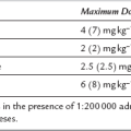
Sedative and anxiolytic premedication is useful and both α- and β-adrenergic antagonists should be continued up to the day of surgery. Monitoring of ECG, CVP and direct arterial pressure must be started before induction of anaesthesia. Intraoperative monitoring should include temperature, blood gas tensions and glucose concentration; transoesophageal echocardiography or pulmonary artery catheterization may be required if significant cardiomyopathy is present. Anaesthetic drugs should be selected on the basis of cardiovascular stability and agents which have the ability to provoke histamine (and hence catecholamine) release are best avoided (Table 31.5). The exact choice of individual anaesthetic drugs is less important than careful conduct of anaesthesia, which may be induced by slow administration of thiopental, etomidate or propofol and maintained with nitrous oxide in oxygen, supplemented by sevoflurane or isoflurane. Desflurane has the theoretical disadvantage of causing sympathetic stimulation if the inspired concentration is increased too rapidly. The use of moderate doses of an opioid (e.g. fentanyl 7–10 μg kg–1) may aid cardiovascular stability. Drugs should be immediately available to treat acute hypertension (e.g. SNP, phentolamine or nicardipine), tachycardia or arrhythmias (e.g. esmolol). Hypotension is treated with fluids initially but vasopressors (e.g. ephedrine, phenylephrine or noradrenaline) may be required. Intravenous magnesium sulphate may be useful: it suppresses catecholamine release from the tumour and adrenergic nerve endings, is a direct-acting vasodilator and has antiarrhythmic effects but has a narrow therapeutic window and plasma Mg2 + concentration should be monitored. Perioperative epidural analgesia may attenuate some of the cardiovascular responses, except during tumour handling, and is useful for postoperative analgesia. However, it should be used judiciously to avoid hypotension. Postoperative problems may include hypoglycaemia, somnolence, opioid sensitivity, hypotension and hypoadrenalism. Invasive monitoring should be continued for 12–24 h after surgery and the patient must be nursed in a high-dependency or intensive care unit.

TABLE 31.5

Drugs Which Should be Avoided in Patients with Phaeochromocytoma

Atropine

Succinylcholine

d-Tubocurarine

Atracurium

Pancuronium

Droperidol

Morphine

Halothane

Laparoscopic adrenalectomy is now the surgical treatment of choice for adrenal phaeochromocytoma. It is performed via the transperitoneal or retroperitoneal routes. These laparascopic techniques are associated with less postoperative pain, and earlier mobilization and recovery, compared with open surgery. Overall, cardiovascular disturbance may be less, but the creation of a pneumoperitoneum during transperitoneal laparoscopy may cause large surges in catecholamine concentrations in addition to those occurring during tumour mobilization. Consequently, similar anaesthetic considerations apply as for open surgery.

PLASTIC SURGERY

Plastic surgery includes the reconstitution of damaged or deformed tissues (congenital abnormalities or resulting from trauma, burns or infection), removal of cutaneous tumours or cosmetic alteration of body features. Division or removal of the abnormality often necessitates skin grafting. Major plastic surgery includes the formation and repositioning of free and pedicle grafts and the movement of skin flaps.

General Considerations

Many of these procedures have important common features. Patients may be physically deformed and attention should be directed to their psychological state. This is influenced by long periods of confinement and rehabilitation, concern over disfigurement or loss of limb function, and occasionally chronic pain. The presence of local or generalized infection and the patient’s state of nutrition are important factors in postoperative outcome and should be considered. Conversely, cosmetic surgery of the face, tattoo removal, breast augmentation and removal of unwanted adipose tissue are usually performed on healthy patients. Surgery is often prolonged, requiring special attention to blood and fluid replacement therapy, and maintenance of body temperature. Pain is usually peripheral in origin but may be severe, particularly from donor skin graft sites; local anaesthetic techniques (nerve or plexus blockade, or local infiltration) are very effective.

Anaesthesia for prolonged procedures should be administered using humidified gases in a warmed theatre environment, employing a technique which minimizes protracted recovery from anaesthesia. A remifentanil-based technique supplemented by a relatively insoluble volatile agent (e.g. isoflurane, desflurane or sevoflurane) is effective. Alternatively, a total intravenous technique may be employed, although the vasodilatation produced by volatile agents may be beneficial to surgical outcome. Nitrous oxide may produce bone marrow depression with exposure of more than 8 h duration and an oxygen/air mix should be substituted. Fluid balance should be maintained scrupulously. Significant haemorrhage is common during plastic surgery. Blood transfusion is frequently required, although microvascular flow is optimal with a haematocrit of approximately 0.3 and overtransfusion should be avoided. The outcome of microvascular surgery depends on adequate blood flow through a patent graft. Volatile anaesthetic agents and regional or sympathetic blockade cause vasodilatation, which may be helpful. However, graft blood flow may be impaired by hypotension, venous congestion or vasoconstriction caused by hypovolaemia, hypothermia, hypocapnia or pain. Therefore, maintenance of normal arterial pressure, circulating volume and cardiac output, normothermia, and provision of good analgesia are important to maximize peri- and postoperative perfusion of the surgical site. Vascular spasm may be diminished by the use of local vasodilators.

The patient must be positioned to avoid ligament strain, and lumbar support is useful during long procedures. Pressure areas should be protected with soft padding to prevent pressure injury, particularly over bony prominences, and a pressure-relieving mattress used. Measures should be taken to prevent DVT formation. When surgery has been completed, wound dressing and bandaging may be lengthy procedures. Bandages may be applied around the trunk and the patient must be lifted carefully to avoid injury.

Head and Neck

Tracheal intubation using a reinforced tube is recommended for surgery in this area. Tumours or scarring of the neck, deformity of facial bones and cleft palate can make tracheal intubation particularly difficult. The airway should be assessed carefully before anaesthesia, any difficulties anticipated and a complete range of equipment should be available. The administration of muscle relaxants in such patients may be unwise before the airway is secured by intubation; awake fibreoptic intubation under local anaesthesia or an inhalational technique should be considered. The method of maintenance is determined by the condition of the patient, the type and duration of surgery (frequently prolonged) and the experience and preference of the anaesthetist. Venous drainage is improved and bleeding reduced in head or neck surgery if the patient is positioned in a 10–15° head-up tilt. Hypotensive techniques may also be indicated, in which case an arterial cannula is advisable for measurement of arterial pressure. It is important to protect the eyes from pressure, the ears from blood and other fluids and the tracheal tube and anaesthetic tubing from dislodgement. It may be difficult to monitor chest movement, and access to the arms may be impossible. An i.v. infusion with extension tubing is essential; there should be access to a three-way tap for injection of drugs.

Limbs

Local anaesthesia (e.g. by blockade of nerve plexuses in the neck, axilla or groin) may be an advantage in terms of analgesia and vasodilatation for surgery on upper or lower limbs. The duration of some plastic surgical operations and the use of a surgical tourniquet to provide a bloodless field may preclude some techniques, but prolonged neural blockade may be achieved using a catheter technique and by selection of an agent with a prolonged duration of action (e.g. bupivacaine or ropivacaine). Intravenous sedative drugs or light general anaesthesia are useful adjuncts to help the patient tolerate a prolonged procedure. Specific nerve blocks may be useful; for example, blockade of the femoral and lateral cutaneous nerve of the thigh provides good analgesia for skin graft donor sites during and after operation. Bier’s block is of limited value because of tourniquet pain, and cuff deflation may be required by the surgeon to identify bleeding points.

Surgical techniques of reimplantation and microsurgical repair of the limbs are well established and make specific demands upon the anaesthetist. These include maintenance of general anaesthesia for up to 24 h, control of vascular spasm and provision of optimum conditions for postoperative recovery.

BURNS

Thermal burn injuries are common, and despite improvements in outcome over the last few decades, can still result in significant morbidity and mortality. Factors associated with death include increased age, the surface area and depth of the burn and the presence of inhalational injury. The anaesthetist may be involved with victims of thermal burns at an early stage during basic resuscitation and airway management, during transfer to, or management in, a critical care or specialized burns unit, or for the provision of general anaesthesia for:

Surgeons now perform debridement of burns with escharotomies at an earlier stage because infection and sepsis are reduced, and cosmetic results are better.

Burns are classified according to the depth of burn and percentage of body surface area (BSA) involved. Partial-thickness burns may be confined to the epidermis (superficial epidermal or first-degree burns), or extend to the superficial or deeper layers of the dermis (superficial or deep dermal, second-degree burns). Typical features of first-degree burns include severe pain, lack of blistering and erythema which blanches on palpation. They heal spontaneously in a few days and should not be included in estimates of burn size. Superficial epidermal burns include blistering and also blanch, but usually heal naturally within 2 weeks. Deep dermal burns also cause blistering but do not blanch and are less painful than superficial burns. Full-thickness (third-degree) burns extend through the dermis into the subcutaneous tissues and appear white, red, brown or black, and do not blanch. They usually cause sensory loss because of superficial nerve injury. Both deep dermal and full-thickness burns require excision and grafting. The Wallace ‘rule of nines’ may be used to assess the area of burns in order to guide fluid management but is inaccurate in children. Lund and Browder charts are more accurate and are widely available. In clinical practice, the area of significant burn injury is often overestimated.

Pathophysiology

Early death in victims of fire is usually caused by hypoxaemia, resulting either from a reduction in inspired oxygen concentration in a smoke-filled atmosphere or from poisoning by products of combustion, e.g. carbon monoxide, hydrogen cyanide, hydrogen sulphide and ammonia. The affinity of carbon monoxide for haemoglobin is 200 times greater than that of oxygen, and in the presence of high carboxyhaemoglobin concentrations, arterial oxygen content is reduced. The oxygen dissociation curve is also distorted and shifted to the left, resulting in reduced oxygen delivery to the tissues. Inhalation of hot gases causes direct thermal upper airway burns with supraglottic oedema which may lead to airway obstruction within a few hours. Inhalation of smoke particles and toxic products of combustion can cause an inhalational injury comprising mucosal oedema, mucociliary damage, bronchospasm and loss of surfactant with the development of pneumonitis over the next 1–4 days. In addition to the local inflammatory response at the site of the burn, major burns also cause the widespread systemic release of cytokines, notably TNFα, interleukin-1 (IL-1) and IL-6, and reactive oxygen species. These may mediate the development of the systemic inflammatory response syndrome and contribute to the acute lung injury. Persistently high IL-6 concentrations are associated with a poor prognosis.

Cardiovascular changes after burn injuries include increased microvascular permeability with extravasation of plasma proteins, reduced plasma oncotic pressure and interstitial oedema. This is most marked at the site of the burn but also occurs throughout the vasculature, leading to marked hypovolaemia within hours. Myocardial contractility and cardiac output decrease independently of the reduction in circulating volume because of circulating depressant factors and diastolic dysfunction. Systemic vascular resistance is increased. If resuscitation is adequate, cardiac output may increase markedly after the first 24 h. Plasma potassium and urea concentrations increase initially because of cell tissue necrosis and haemolysis; muscle breakdown results in rhabdomyolysis. Renal failure may occur early after major burns primarily because of inadequate fluid resuscitation, but haemolysis or rhabdomyolysis may contribute.

The sympatho-adrenal response to burns includes enormous increases in plasma concentrations of catecholamines, aldosterone, ACTH and arginine vasopressin (AVP). These result in marked retention of sodium and water, with increased excretion of potassium, calcium and magnesium, so that hypokalaemia and anaemia are common after the first 48 h post-burn. There is also a hypercatabolic state with tachycardia, hyperpnoea and hyperpyrexia which may persist for several weeks or months. Muscle breakdown occurs in association with decreased protein synthesis, increased lipolysis and glycogenolysis so that nutritional and calorie requirements are increased markedly. Sepsis frequently develops following severe burns, often leading to widespread metabolic derangement, multiorgan failure and death. The outcome of sepsis in patients with burns is worse than in sepsis after trauma.

Recovery from burns trauma may be protracted. The anaesthetist must be aware of the probable requirement for multiple administrations of general anaesthesia, frequent use of opioid analgesics in the early stages and the importance of psychological support throughout the patient’s stay in hospital.

Initial Management

The initial assessment and management of acute burn injuries are similar to those applied to other victims of trauma and are summarized in Table 31.6. The aim of immediate treatment is to secure the airway and administer high-flow humidified oxygen through a non-rebreathing system; tracheal intubation and IPPV with 100% oxygen may be required to maintain an adequate PaO2. Fluid resuscitation should be started. Intravenous fluids guided by a formal protocol are required if the burn exceeds 10–15% of total BSA and central venous access is usually required with burns greater than 20% BSA. Early warning signs of upper airway burns include facial or intra-oral burns, singed facial or nasal hair, carbonaceous sputum, hypoxaemia, dyspnoea, cough or wheeze, although these are not universally present in the early stages and airway obstruction may develop later. A history of burns in an enclosed space is highly suggestive. A high index of suspicion for airway burns should be maintained in all cases and prophylactic tracheal intubation is often justified, particularly in children or if interhospital transfer is required. However, the decision to secure the airway by tracheal intubation may be difficult and a senior anaesthetist should be involved. Hoarseness or stridor may indicate impending airway obstruction and tracheal intubation in these cases is mandatory. Pulse oximeters are unreliable in the presence of carboxyhaemoglobin because they cannot distinguish between oxyhaemoglobin and carboxyhaemoglobin and therefore overestimate true oxygen saturation; arterial blood gas analysis is required. Burns are extremely painful and carefully titrated intravenous opioids should be administered. Indications for ICU admission include potential airway problems, burns involving > 20% BSA and the presence of other injuries.

TABLE 31.6

Initial Management of Patients with Severe Burns

1.History – time, extent and mechanism of burn, age and weight of patient, brief medical history

2.Airway assessment

3.Breathing – administer 100% humidified oxygen via a non-rebreathing mask

4.Circulation – establish two large-bore i.v. cannulae and commence fluid resuscitation

5.Assess neurological status

6.Exposure with environmental control

7.Analgesia – i.v. opioids

8.Formally assess burn area and re-evaluate fluid requirements

9.Monitoring – vital signs, urine output

10.Investigations – ABG, COHb, U&E, FBC, clotting screen, cross-match blood, ECG, CXR

11.Secondary survey to exclude other injuries

12.Burns dressings

ABG, blood gas analysis; COHb, carboxyhaemoglobin; U&E, urea and electrolyte concentrations; FBC, full blood count; CXR, chest X-ray.

Tissue burns produce rapid fluid shifts and oedema formation, particularly during the first 36 h. The resulting depletion of intravascular volume is greatest in the first few hours and it is essential that a fluid replacement regimen is started as early as possible to avoid hypovolaemic shock and acute renal failure. Crystalloid-based regimens such as the Parkland formula are used commonly (Table 31.7) although mixed colloid–crystalloid regimens based on that of Muir and Barclay are sometimes used. Many advocate administration of crystalloid solutions only in the first 24 h, with colloids added thereafter. However, the pathophysiology of fluid shifts is complex and these formulae should be used only as a guide to fluid therapy. Recent studies have shown that the type of resuscitation fluid used (crystalloid or colloid) does not affect mortality. Vital signs, urine output and body temperature should be observed closely, and volume replacement titrated to achieve a urine output of 0.5–1.0 mL kg–1 h–1 (1.0–1.5 mL kg–1 h–1 in children). Non-invasive methods of cardiac output monitoring may also be used to guide fluid replacement and resuscitation. Haematocrit, base deficit and serum lactate, and urea and electrolyte concentrations should be monitored. It is important to be aware that excessive volumes of fluid for resuscitation can predispose to some complications including abdominal and extremity compartment syndromes. Bladder pressure monitoring (to detect intra-abdominal hypertension) has been recommended for all patients with major burns of > 30% BSA.

TABLE 31.7

Fluid Regimens for Burned Patients

1. Estimate/measure weight

2. Estimate percentage area of burn using ‘rule of nines’ for adults and ‘rule of tens’ for children

3. Proceed with regimen if > 15% burns in adults or > 10% in children

4. Parkland formula:

 Requirements in first 24 h (mL) = body weight  ×  % burn  ×  4

 Fluids given as Ringer’s lactate alone, 50% within first 8 h, 50% between 8 and 24 h

 Colloids administered only after first 24 h

5. Muir & Barclay formula:

Requirements in each time period (mL) = body weight (kg)  ×  % burns  ×  0.5

Fluids (human albumin solution 4.5%) according to formula in each of the following periods:

 0–4 h

 4–8 h

 8–12 h

 12–18 h

 18–24 h

 24–36 h

In addition, water as 5% dextrose is required at 1–2 mL kg–1 h–1.

N.B. These formulae should only be used as a guide to fluid requirements and individual prescriptions should be adjusted according to response.

Anaesthetic Problems

Airway

It is vital to secure the airway in a patient with head and neck burns but this may be extremely challenging. In the initial stages, airway obstruction may occur, particularly in children where the airway is smaller. Many anaesthetists prefer to use an inhalational induction in the conscious patient, although raw, painful tissues may render proper application of a face mask difficult. Awake fibreoptic intubation may be preferable in adults. A rapid-sequence induction using succinylcholine may be inadvisable (see below). In all cases, facilities and expertise for emergency cricothyroidotomy or tracheostomy should be available, although elective tracheostomy is generally undesirable because of the risk of subsequent pulmonary sepsis and local infection in damaged skin. Later, as soft tissues fibrose and distort, the range of movement in the neck and temporomandibular joints may become grossly restricted and render laryngoscopic intubation impossible.

It may be difficult to secure the tracheal tube in patients with facial burns. Marked facial and neck swelling may occur which may result in dislodgement of tracheal tubes. Tubes should generally be left ‘long’. Several ingenious methods have been devised, such as suspension of the anaesthetic breathing system from the ceiling, the use of umbilical tape to tie the tube in place and wiring the tube to the upper teeth. Tracheal intubation is often necessary for several days until airway oedema subsides. In this situation, or after prolonged surgery, the pharynx should be examined closely before tracheal extubation because laryngopharyngeal oedema may cause respiratory obstruction when the tracheal tube is removed.

Ventilation

Mechanical ventilation should be used in the severely burned patient and careful monitoring of ventilation is required. Humidification of inspired gas, physiotherapy and bronchial toilet are mandatory; bronchodilators and PEEP may be necessary. Acute lung injury developing over the first 4 days after burns causes alveolar oedema and hypoxaemia. This may be exacerbated by the administration of large volumes of fluid during resuscitation, but fluid restriction is associated with a worse outcome. Newer techniques include bronchial lavage, nebulized heparin, tissue plasminogen activator, acetyl cysteine and nitric oxide. The hypermetabolic state in large burns results in large increases in oxygen consumption and carbon dioxide production; i.v. nutrition increases the latter. The principles of artificial ventilation in burns patients are identical to those in acute lung injury from other causes: low tidal volume and minimal airway pressure (plateau pressure < 35 cmH2O, positive end-expiratory pressure titrated to maintain oxygenation) should be used and permissive hypercapnia tolerated if necessary; a sophisticated ventilator may be required for patients undergoing relatively simple surgical procedures. If significant inhalational injury has occurred, other ventilator modes such as airway pressure release ventilation or high frequency oscillation may be used.

Anaesthetic Drugs

Personal preference and the problems of repeated administration of anaesthesia govern the choice of anaesthetic agent. An inhaled nitrous oxide/oxygen mixture (Entonox) and i.v. ketamine are useful for analgesia during burns dressings. However, it is not safe to assume that the airway is preserved during ketamine anaesthesia and antisialagogue premedication is useful to diminish salivation. Diazepam may control the emergence hallucinations suffered by some patients who receive ketamine. Intravenous opioids (by infusion, boluses or patient-controlled analgesia) are effective alternatives. Supplementary analgesics are required in the short- and long-term, e.g. paracetamol, non-steroidal anti-inflammatory drugs. Adjunctive therapies include clonidine, tricyclic antidepressants, topical and systemic local anaesthetics or transcutaneous electrical nerve stimulation. For surgical procedures, a balanced technique using a volatile agent and opioid is indicated. The disposition and action of many drugs are affected following burns, e.g. there is marked resistance to the effects of nondepolarizing muscle relaxants from 1 week after major burns.

Succinylcholine should be not be administered from 24 to 48 h after the burn. In the presence of muscle damage, it may cause acute hyperkalaemia in concentrations sufficient to cause cardiac arrest. The mechanism involves upregulation of cholinergic receptors with the proliferation of immature receptor isoforms and extrajunctional receptors. The most dangerous period in this regard is probably between 4 days and 10 weeks after thermal injury.