Chapter 10
An Overview of the Brainstem
Basic Divisions of the Brainstem
Ventricular Spaces of the Brainstem
Cranial Nerve Nuclei and Their Functional Components
Contemporary View of the Functional Components of Cranial Nerves
Functional Components of Cranial Nerves and Their Associated Nuclei
The term brainstem (sometimes written brain stem) can mean either the portion of the brain that consists of the medulla oblongata, pons, and midbrain or the portion that consists of these structures plus the diencephalon. This book follows the former convention. For our purposes, therefore, the brainstem consists of the rhombencephalon (excluding the cerebellum) and the mesencephalon. These regions of the brainstem share a basic organization, which is the topic of this chapter. The medulla, pons, and midbrain are discussed in detail in Chapter 11 to Chapter 13.
BASIC DIVISIONS OF THE BRAINSTEM
Medulla Oblongata
At about the level of the foramen magnum, the spinal cord merges into the most caudal portion of the brain, the medulla oblongata or myelencephalon, commonly called the medulla. The foramen magnum marks the approximate location of the motor (pyramidal) decussation of the medulla (Fig. 10-1A). The medulla is slightly cone shaped and enlarges in diameter as it extends rostrally from the medulla–spinal cord junction toward the pons-medulla junction. On the posterior (dorsal) aspect of the medulla-pons continuum, this junction is represented by the caudal edge of the middle and inferior cerebellar peduncles, whereas anteriorly (ventrally) this border is formed by the caudal edge of the basilar pons (Fig. 10-1).
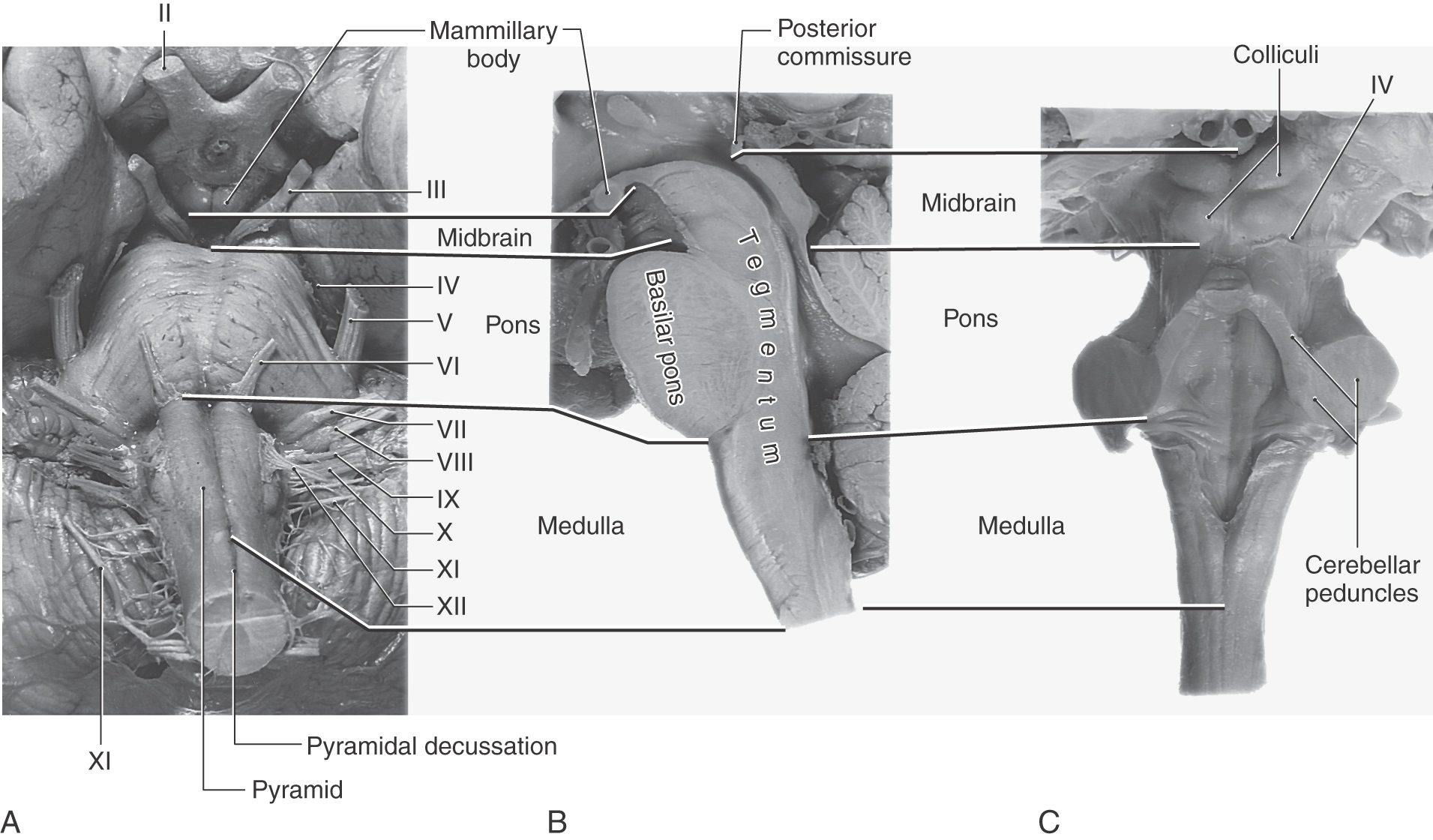
The cranial nerves associated with the medulla include the hypoglossal (XII, motor) and parts of the accessory (XI, motor), vagus (X, mixed), and glossopharyngeal (IX, mixed) nerves (Fig. 10-1A). The nuclei of the hypoglossal, vagal, and glossopharyngeal nerves as well as portions of the nuclei of the trigeminal nerve are located in the medulla.
The abducens (VI, motor), facial (VII, mixed), and vestibulocochlear (VIII, sensory) nerves are frequently called the cranial nerves of the pons-medulla junction because they exit the brainstem at this particular location (Fig. 10-1A).
Although the medulla does not have regions that are specifically regarded as tegmental or basilar (as is the case for the pons and midbrain), it does have regions that function in the same way and are rostrally continuous with these respective regions of the pons (Fig. 10-2). For example, the central regions of the medulla contain the cranial nerve nuclei affiliated with the medulla. This medullary area is rostrally continuous with the pontine tegmentum, which contains the cranial nerve nuclei associated with the pons. In similar manner, the pyramids of the medulla (containing corticospinal fibers) are located on the anterior (“basal”) aspect of the medulla and are rostrally continuous into the basilar pons (Fig. 10-2).
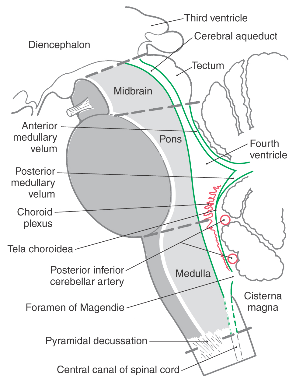
Figure 10-2. Midsagittal drawing of the brainstem. Ventricular spaces of the brainstem are outlined in green. The tegmental and basilar areas and contiguous areas of the medulla are shown in light and dark gray, respectively. Compare with Figure 10-1B.
Pons
The pons (the anterior part of the metencephalon) extends from the pons-medulla junction to an imaginary line drawn from the exit of the trochlear nerve posteriorly to the rostral edge of the basilar pons anteriorly (Fig. 10-1). What we commonly call the pons is actually composed of two portions, the pontine tegmentum (located internally; see Fig.10-1) and the basilar pons. The basilar pons is bulbous and quite characteristic of the anterior aspect of the pons. The pontine tegmentum contains portions of the trigeminal nuclei and the vestibular nuclei and, just rostral to the pons-medulla junction, the facial motor nucleus, superior salivatory nucleus, and abducens nucleus. The trigeminal nerve (V, mixed) emerges from the lateral aspect of the pons, and abducens (VI), facial (VII), and vestibulocochlear (VIII) nerves exit at the pons-medulla junction (Fig. 10-1A).
The cerebellum, although part of the metencephalon, is not part of the brainstem. It is joined to the brainstem by three large, paired bundles of fibers called the cerebellar peduncles. These are the inferior cerebellar peduncle, the middle cerebellar peduncle (or brachium pontis), and the superior cerebellar peduncle (or brachium conjunctivum), connecting the cerebellum to the medulla oblongata, basilar pons, and midbrain, respectively.
Midbrain
The midbrain (mesencephalon) extends rostrally from the pons-midbrain junction to join the diencephalon (thalamus); this interface is usually described as a line drawn from the posterior commissure posteriorly to the caudal edge of the mammillary bodies anteriorly (Fig. 10-1B). The oculomotor nerve (III, motor) exits the anterior aspect of the midbrain, whereas the trochlear nerve (IV, motor) exits its posterior aspect (Fig. 10-1A, C). The exit of the trochlear nerve is regarded as the pontomesencephalic junction on the posterior aspect of the brainstem; along with its decussating fibers, it composes the isthmus rhombencephali (the transition from pons to midbrain).
The posterior aspect of the midbrain is characterized by the superior and inferior colliculi and the anterior aspect by the crus cerebri and interpeduncular fossa.
Tegmental and Basilar Areas
The central core of the midbrain and the pons is called the tegmentum, and their anterior (ventral) parts are the basilar areas. These regions are continuous with each other and with comparable areas of the medulla (Figs. 10-1B and 10-2). The tegmentum of the pons and midbrain and the contiguous central portion of the medulla contain ascending and descending tracts, many relay nuclei, and the nuclei of cranial nerves III to XII.
The basilar part of each brainstem division is anterior to the tegmentum (of the midbrain and pons) and to the central portion of the medulla (Fig. 10-2). Consequently, these basilar structures also form a rostrocaudal continuum. Basilar structures of the brainstem include the descending fibers of the crus cerebri (midbrain), basilar pons, and pyramid (medulla) and specific populations of neurons in the midbrain and pons that originate from the alar plate of the embryonic brain.
VENTRICULAR SPACES OF THE BRAINSTEM
The ventricular spaces of the brainstem are the cerebral aqueduct in the mesencephalon and the fourth ventricle in the rhombencephalon (Fig. 10-2). The cerebral aqueduct is a narrow channel, 1 to 3 mm in diameter, that connects the third ventricle (the cavity of the diencephalon) with the fourth ventricle (the rhombencephalic cavity). The cerebral aqueduct contains no choroid plexus; its walls are formed by a continuous mantle of cells collectively called the periaqueductal gray. The roof of the midbrain is the tectum.
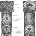
The fourth ventricle is the cavity of the rhombencephalon. Its rostral portion lies between the pons and cerebellum, and its caudal part is located in the medulla (Fig. 10-2). The fourth ventricle is continuous rostrally with the cerebral aqueduct, caudally with the central canal of the caudal medulla and cervical spinal cord, and laterally with the subarachnoid space via the midline foramen of Magendie and the two lateral foramina of Luschka. The foramen of Magendie is located in the caudal roof of the ventricle and opens into the dorsal cerebellomedullary cistern (cisterna magna) (Fig. 10-2). The lateral recesses of the fourth ventricle extend around the brainstem at the pons-medulla junction and end at the foramen of Luschka, which opens into the lateral cerebellomedullary cistern (Fig. 10-4; see also Fig. 6-9).
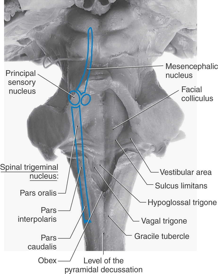
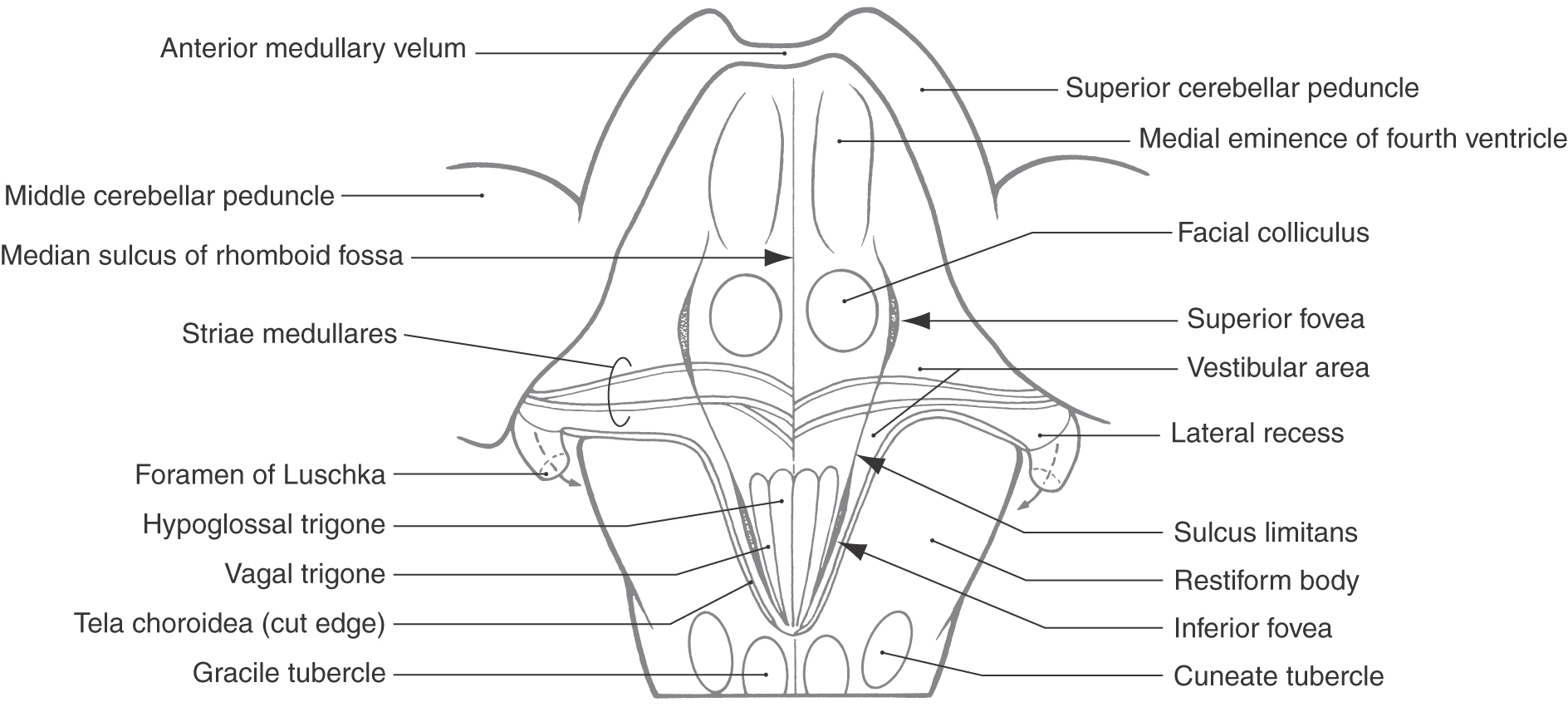
Figure 10-4. The rhomboid fossa (floor of the fourth ventricle), elevations and depressions in the floor, and structures bordering on the fossa. Compare with Figures 10-1C and 10-3. (From Haines DE: Neuroanatomy: An Atlas of Structures, Sections, and Systems, 8th ed. Baltimore, Lippincott Williams & Wilkins, 2012.)
The roof of the fourth ventricle is formed mainly by the anterior (or superior) medullary velum rostrally, by the thin membranous tela choroidea caudally, and by a small part of the cerebellum in the middle (Figs. 10-1B and 10-2). From rostral to caudal, the walls of the fourth ventricle are formed by the superior cerebellar peduncles, the middle and inferior cerebellar peduncles, and the attachment of the tela choroidea to the medulla (Figs. 10-3 and 10-4). The tela arises from the inferior surface of the cerebellum and sweeps caudally to attach to the V-shaped edges of the medullary portion of the ventricular space. The choroid plexus of the fourth ventricle is suspended from the inner surface of the tela, and parts of it protrude outward through the foramina of Luschka (see Fig. 6-4 and Fig. 6-9).
Rhomboid Fossa
The floor of the fourth ventricle, the rhomboid fossa, is divided into halves by a deep median sulcus, and each half is traversed rostrocaudally by the sulcus limitans (Figs. 10-3 to 10-6). There are two slight depressions along the course of the sulcus limitans, somewhat like deep spots within this sulcus. The rostral depression, the superior fovea, is laterally adjacent to the facial colliculus, and the caudal depression, the inferior fovea, is laterally adjacent to the vagal and hypoglossal trigones (Figs. 10-3 and 10-4). In some surgical procedures involving the fourth ventricle or medulla, the sulcus limitans and foveae represent important landmarks. The striae medullares of the fourth ventricle are a series of fiber bundles running from the midline laterally into the lateral recess (Fig. 10-4). The rostral edge of these fibers is generally regarded as the pons-medulla junction in the floor of the fourth ventricle.
Elevations in the floor of the fourth ventricle indicate the locations of underlying cranial nerve nuclei and associated fiber bundles (Figs. 10-4 to 10-6). In general, the cranial nerve nuclei that are located between the median sulcus and the sulcus limitans are motor in function, whereas those located lateral to the sulcus are sensory in function (Figs. 10-5 and 10-6). Medial to the sulcus limitans, the hypoglossal and vagal trigones represent the underlying hypoglossal and dorsal motor vagal nuclei. In the caudal pontine region, the facial colliculus, located medial to the sulcus limitans, marks the location of the underlying abducens motor nucleus and the internal genu of the facial nerve. Lateral to the sulcus limitans in the medulla and caudal pons is a flattened region called the vestibular area, which marks the location of the vestibular nuclei.

CRANIAL NERVE NUCLEI AND THEIR FUNCTIONAL COMPONENTS
Cranial nerves, like spinal nerves, contain sensory or motor fibers or a combination of these fiber types. These various fibers are classified on the basis of their embryologic origin or common structural and functional characteristics. Primary sensory fibers, somatic motor neurons, and preganglionic and postganglionic visceromotor neurons that exhibit “like anatomical and physiological characters so that they . . . act in a common mode” (C.J. Herrick) are classified as having a specific functional component. For example, fibers conveying sharp pain, a specific type of input, from widely separated body parts (the foot, hand, and face) have the same functional component. This principle, already introduced in relation to spinal nerves (see Chapter 9), is also directly applicable to cranial nerves.
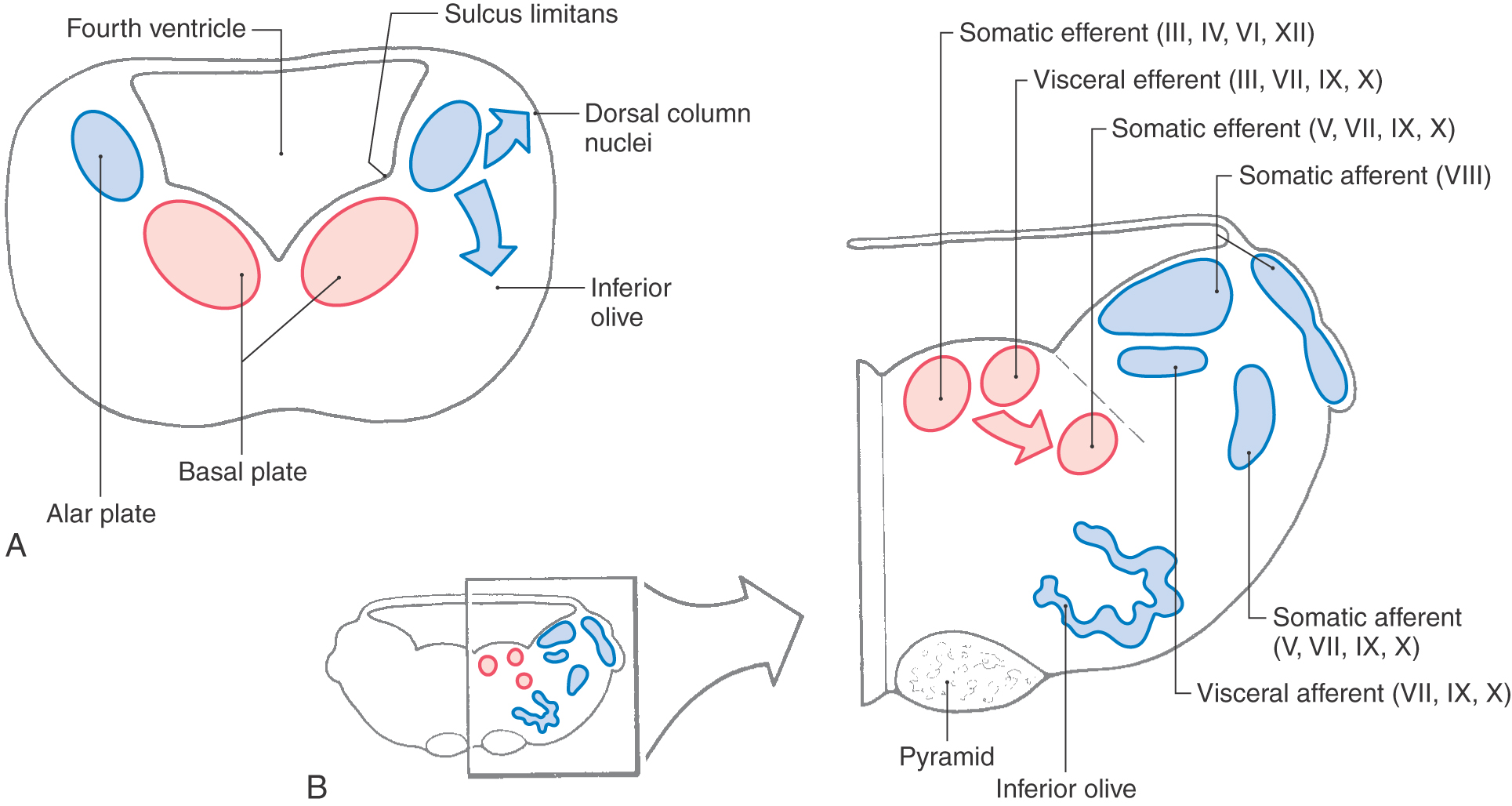
Early in development, the rostrocaudally oriented cell columns forming the alar and basal plates of the spinal cord essentially extend throughout the brainstem. As development progresses, maturing neurons in alar and basal plates begin to migrate to form their adult structures, and the caudocephalic continuity of the cell columns may be disrupted. In this respect, the primitive cell column retains its relative position as it differentiates, but it may become discontinuous as the individual nuclei derived from the same column are formed (Figs. 10-5 and 10-7). Motor nuclei of cranial nerves arise from basal plate neurons, whereas the nuclei that receive primary sensory input via cranial nerves originate from the alar plate.
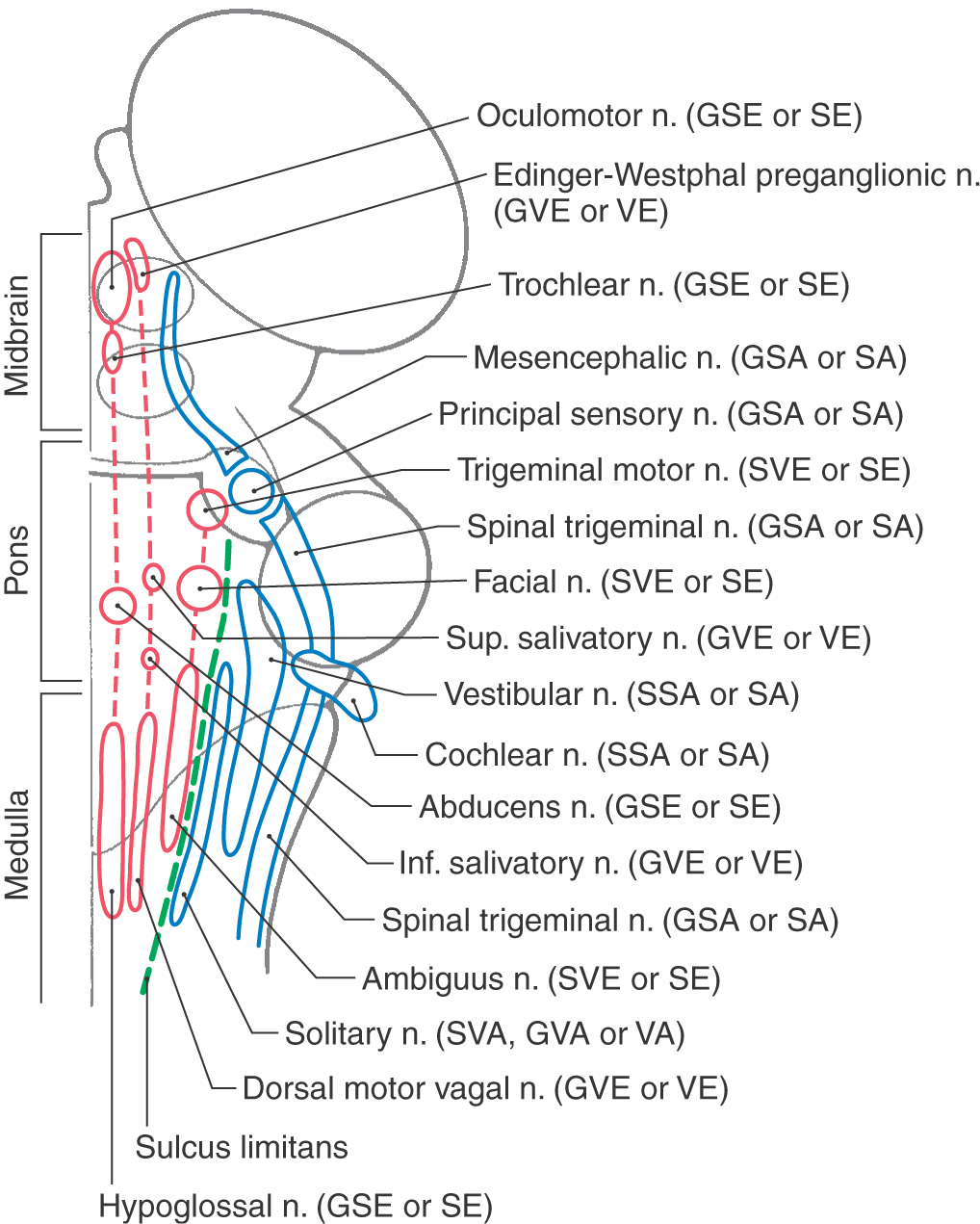
Figure 10-7. Diagram showing the cranial nerve nuclei of the brainstem and their functional components in the brainstem. The various nuclei are unrolled onto a single plane (see also Fig. 10-5B) with basal plate derivatives (red) and alar plate derivatives (blue) located medially and laterally, respectively, to the sulcus limitans. The traditional and contemporary functional components (e.g., GSE or SE and GVE or VE) are listed side-by-side.
In the caudal medulla, the rostral continuation of the central canal is small; therefore, basal and alar plates are located anterior and posterior, respectively, to this space (Fig. 10-5). As the fourth ventricle flares open at the level of the obex, the alar plate shifts laterally and the basal plate retains an anterior (and now medial) position (Figs. 10-5 and 10-6). Rostrally, as the fourth ventricle funnels into the cerebral aqueduct of the midbrain, the alar plate rotates back to a posterior position and the basal plate again assumes an anterior position (Fig. 10-5). The sulcus limitans, an embryologic landmark that persists in the medulla and pons of the adult, separates structures derived from the basal plate from those derived from the alar plate.
Contemporary View of the Functional Components of Cranial Nerves
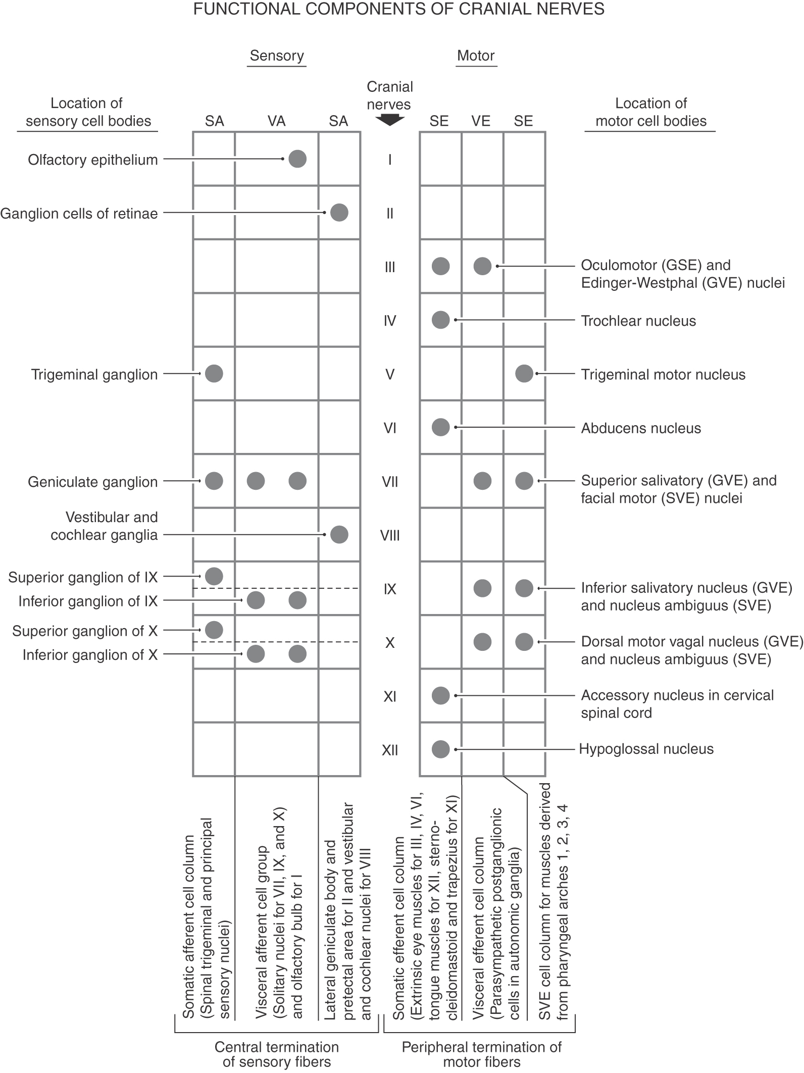
The sensory and motor functional components associated with spinal nerves (somatic afferent, SA; visceral afferent, VA; somatic efferent, SE; visceral efferent, VE) are also found on certain of the cranial nerves (Table 10-1). This is especially the case for cell columns in the spinal cord that are continuous with functionally similar cell columns of the brainstem. For example, SE cell groups of the spinal cord are in line with the motor nuclei of cranial nerves III, IV, VI, and XII. The same is the case for spinal cells that give origin to VE outflow and the parasympathetic cells of cranial nerves III, VII, IX, and X and for areas of the posterior horn receiving SA input and the spinal trigeminal nucleus (Fig. 10-7). Consequently, the four functional components found in spinal nerves are also associated with these cranial nerves of the brainstem that have a comparable function (Table 10-1).
Table 10-1 Comparison of the Traditional and Contemporary Designation of Functional Components
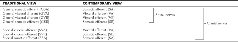
The functional component traditionally associated with the innervation of muscles that presumably arose from the mesoderm of pharyngeal arches was special visceral efferent (SVE). However, contemporary developmental studies have revealed that all striated muscles innervated by cranial nerves (i.e., III-VI, VII, IX, X, and XII) arise from the paraxial mesoderm and then migrate toward their respective adult positions. The mesoderm that will become extraocular muscles and the tongue musculature migrates to the region of the orbit and into the base of the tongue; this mesoderm becomes muscles that will be innervated by cranial nerves III, IV, VI, and XII (Fig. 10-8). The mesoderm that will become the masticatory muscles, muscles of facial expression, and pharyngeal and laryngeal musculature originates from the paraxial mesoderm and initially migrates into arches I, II, III, and IV; from these sites, the pharyngeal arch mesoderm continues to develop into the striated muscles innervated by cranial nerves V, VII, IX, and X (Fig. 10-8; Table 10-2). Because all of these striated muscles innervated by these cranial nerves originated from a common embryologic source (paraxial mesoderm), the functional component SE can be applied to all of these motor neurons and their axons. In similar fashion, and recognizing developmental principles, the functional component for the eighth cranial nerve can be consolidated from SSA to SA. This contemporary view does not negate the fact that some cranial nerves convey special sense, such as taste (VA) and auditory and vestibular functions (SA). Whereas the contemporary and simplified version is used here, either the contemporary or traditional version can be used in a teaching environment to describe the sensory and motor components of cranial nerves.
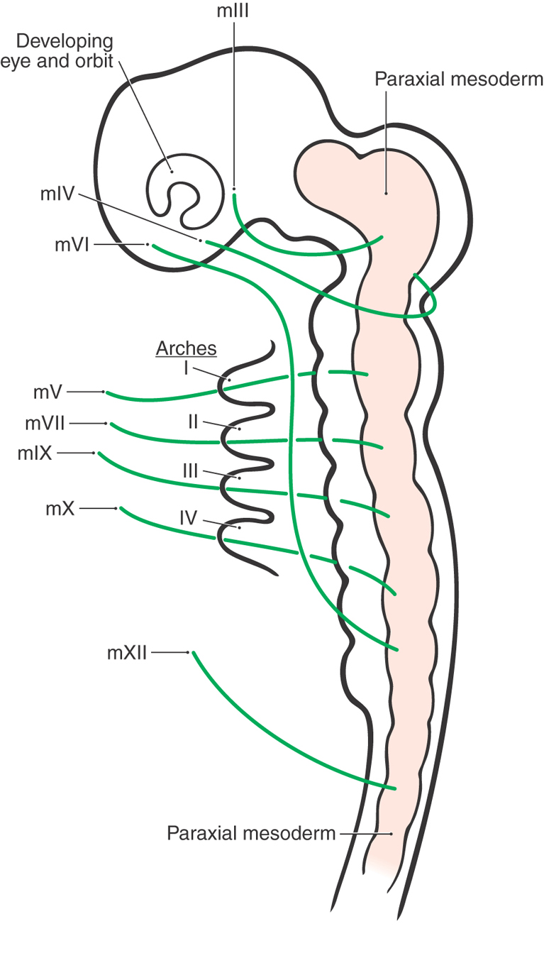
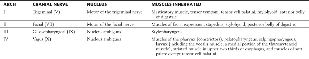
Table 10-2 The Pharyngeal Arches and Associated Cranial Nerves, Motor Nuclei, and Muscles Innervated
Functional Components of Cranial Nerves and Their Associated Nuclei
Motor
The striated muscles that are forming in the head anlage receive their innervation from motor neurons that are derived from the basal plate (Figs. 10-5 to 10-7). In addition, smooth muscle and glandular epithelium of the developing head receive innervation via preganglionic visceromotor neurons that also arise from the basal plate.
Early in development, some of these basal plate neurons remain immediately adjacent to the midline in the floor of the ventricular space and segment into the SE nuclei of the oculomotor (III), trochlear (IV), abducens (VI), and hypoglossal (XIII) nerves (Figs. 10-6 and 10-7). In general, these motor neurons innervate muscles that develop in the area of the orbit and in the base of the tongue from the paraxial mesoderm that has migrated into these respective areas (Fig. 10-8).
A second group of basal plate neurons moves ventrolaterally from the midline and segments into the SE nuclei of the trigeminal (V), facial (VII), glossopharyngeal (IX), and vagus (X) nerves (Figs. 10-6 and 10-7). The last two cranial nerves have their SE cells in the nucleus ambiguus. The striated muscles that are innervated by these four cranial nerves (V, VII, IX, and X) originated as paraxial mesoderm that migrated into pharyngeal arches I to IV; the muscles of the adult arise from these respective sites.
A third group of basal plate neurons move laterally to take up positions in the medulla, caudal pons, and midbrain (Fig. 10-6). These cell groups form the VE nuclei that contain VE preganglionic parasympathetic cells that form the Edinger–Westphal nucleus and associated cells (via the oculomotor nerve), superior salivatory nucleus (via the facial nerve), inferior salivatory nucleus (via the glossopharyngeal nerve), and dorsal motor vagal nucleus (via the vagus nerve). All of these preganglionic cells project to peripheral ganglia, which in turn send postganglionic fibers to innervate visceral structures (Fig. 10-9).
Sensory
The alar plate of the brainstem is located lateral to the sulcus limitans in the floor of the rhomboid fossa; this is the brainstem area that gives rise to the sensory receiving nuclei of the adult brainstem. As the alar plate differentiates, it gives rise to the solitary tract and nucleus, the vestibular and cochlear nuclei, and the sensory nuclei of the trigeminal nerve (Fig. 10-6).
The VA receiving area for the brainstem is the solitary tract and nucleus; regardless of what cranial nerve conveys VA input, it all arrives at the solitary nucleus (Figs. 10-7 and 10-9). Taste input is carried on cranial nerves VII, XI, and X; it has sensory cell bodies in the ganglia of these nerves, enters the solitary tract, and terminates in the rostral portion of the solitary nucleus (this is specifically called the gustatory nucleus). Visceral sensation from the salivary glands, viscera of the thorax and gut, and other sources follows the same route; it has cell bodies in the ganglia of these nerves but terminates in the caudal part of the solitary nucleus (this is the cardiorespiratory nucleus).
The vestibular nuclei receive SA input from receptor cells of the vestibular apparatus, which has its bipolar cells in the vestibular ganglion (Scarpa ganglion), and these central fibers terminate in the vestibular nuclei (Fig. 10-9). In similar manner, receptors in the organ of Corti have their SA cell bodies in the spiral ganglion and their central processes terminate in the cochlear nuclei. Vestibular afferents are concerned with balance and equilibrium and can be classified as proprioceptive SA input; cochlear afferents are concerned with hearing and may be called exteroceptive SA input.
In a plan similar to that of the solitary tract and nucleus being the VA center for the brainstem, the sensory nuclei of the trigeminal nerve are the SA center of the brainstem for all sensation arising from the face, forehead, cornea, oral cavity, anterior two thirds of the tongue, maxillary and frontal sinuses, teeth, and portions of the dura (Figs. 10-6, 10-7, and 10-9). Even though SA information is conveyed on the trigeminal (V), facial (VII), glossopharyngeal (IX), and vagus (X) nerves, this sensory input terminates in trigeminal sensory nuclei. Centrally, many of the fibers conveying pain and thermal sense coalesce to form the spinal trigeminal tract and terminate in the medially adjacent spinal trigeminal nucleus. Other fibers conveying discriminative touch will synapse in the principal sensory nucleus; those fibers conveying proprioceptive information from the masticatory and extraocular muscles and the periodontal ligament form the mesencephalic tract. In this latter situation, the primary sensory cell bodies form the immediately adjacent mesencephalic nucleus.
The spinal trigeminal nucleus extends caudally from about midpontine levels to the spinal cord–medulla junction. On the basis of its cytoarchitecture and connections, the spinal trigeminal nucleus is divided into a pars caudalis (between the level of the cervical spinal cord and obex), a pars interpolaris (between the level of the obex and the rostral end of the hypoglossal nucleus), and a pars oralis (rostral to the level of the hypoglossal nucleus) (Fig. 10-3).
The blood supply to the brainstem originates from branches of the vertebral and basilar arteries. As we shall see in the next three chapters, branches of the vertebrobasilar system serve not only the medulla, pons, and most of the midbrain but also the entire cerebellum.
HERNIATION SYNDROMES RELATED TO THE BRAINSTEM
The brainstem contains the nuclei of most of the cranial nerves, important nuclei that influence the spinal cord, heart rate and respiration, and all the ascending and descending tracts that connect the forebrain with the spinal cord. In this respect, there are numerous clinical events that may arise in which damage to the brainstem causes deficits that may vary from mild to severe or may cause death, in some cases suddenly. These lesions will be explored in more detail in later sections in this text. At this point, we briefly consider four herniation syndromes that are specifically related to the brainstem.
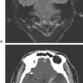
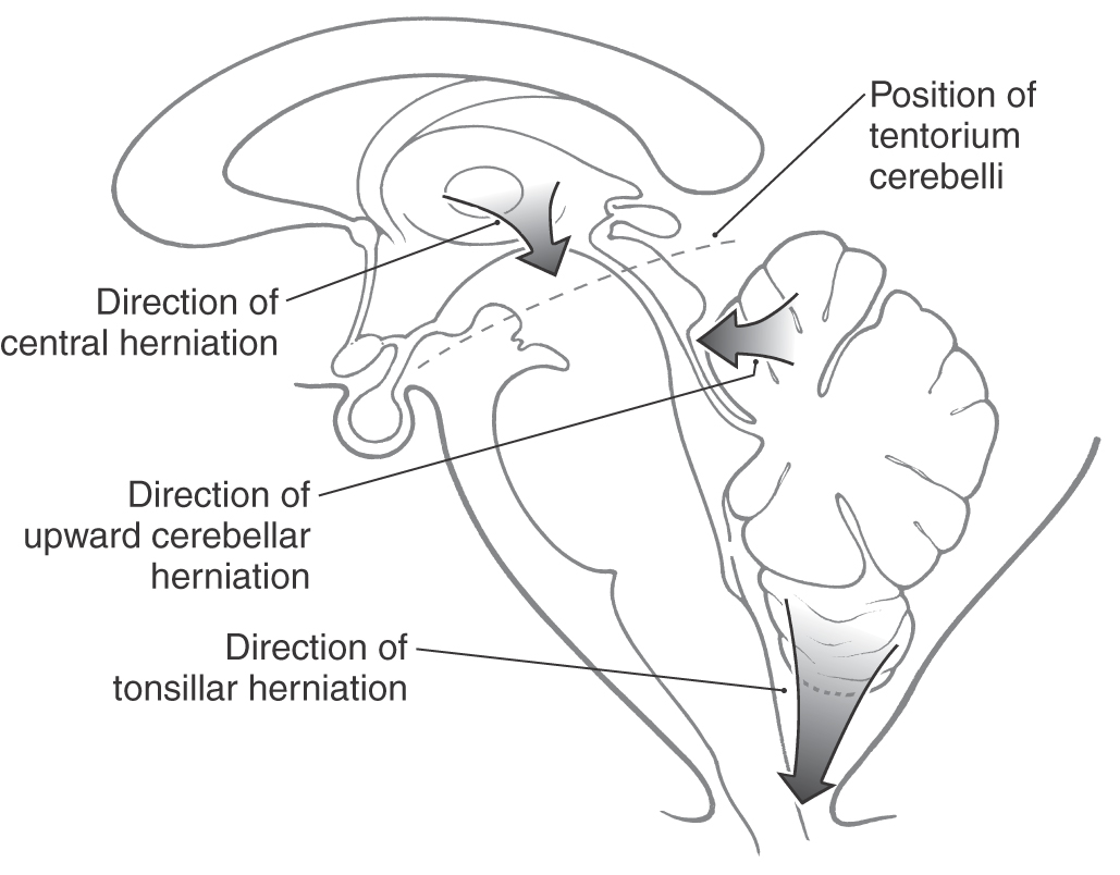
Herniation is best described as the protrusion of one anatomic structure into the territory of another (Figs. 10-10 and 10-11) with the result of causing displacement, damage, destruction, and neurologic deficits. In the case of the central nervous system, the causes of herniation are usually related to an increase in intracranial pressure (mass lesion—tumor; edema—brain swelling; large infarcts).
Central Herniation
Central herniation (also called transtentorial herniation) is the case in which a space-occupying lesion in the hemisphere (supratentorial compartment) elevates intracranial pressure and forces the diencephalon downward through the tentorial notch and into the brainstem (Fig. 10-10). Initially, there may be a change in respiration, eye movements are irregular, and the pupils may be moderately dilated. As the damage progresses downward (caudally) into the brainstem, there is significant change in respiration (Cheyne-Stokes respiration with intermittent tachypnea and apnea), a profound loss of motor and sensory functions, and a probable loss of consciousness. This is a serious neurologic event, and immediate measures should be taken to decrease intracranial pressure.
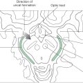
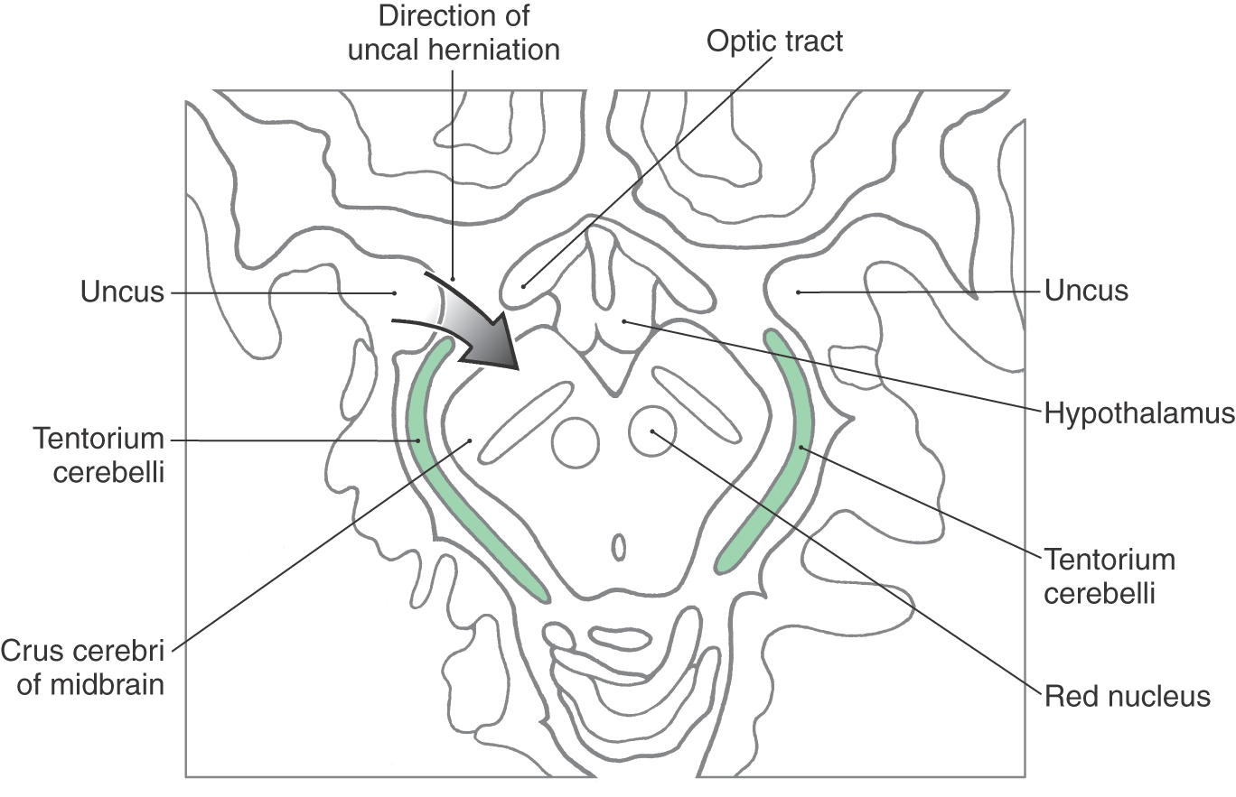
Uncal Herniation
The most common cause of uncal herniation, the movement of the rostromedial edge of the temporal lobe (the uncus) downward over the edge of the tentorium cerebelli (Fig. 10-11), is typically an expanding hemorrhagic lesion in the hemisphere. Uncal herniation initially compresses the midbrain, but if unchecked, the damage may extend into lower brainstem levels. Early signs include a dilated pupil and abnormal eye movements (oculomotor nerve involvement) with double vision ipsilateral to the herniation followed by weakness of the extremities (corticospinal fiber involvement) opposite to the dilated pupil. As the herniation progresses, respiration is affected, abnormal reflexes appear, and there is a potentially rapid decline.
Upward Cerebellar Herniation
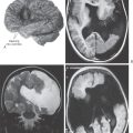
A mass in the posterior fossa may force portions of the cerebellum upward through the tentorial notch (upward cerebellar herniation) and compress the midbrain (Fig. 10-10). The result may be occlusion of branches of the superior cerebellar artery with resultant infarction of cerebellar structures or obstruction of the cerebral aqueduct and hydrocephalus. The latter is seen as signs characteristic of an increase in intracranial pressure (vomiting, headache, lethargy, decreased levels of consciousness).
Tonsillar Herniation
Pressure in the posterior fossa may force the cerebellar tonsils downward into and possibly through the foramen magnum; this is tonsillar herniation (Fig.10-10). This may result in rapid compression of the medulla with a potentially catastrophic neurologic outcome. The medulla is damaged by mechanical compression or distortion, and the vessels serving the medulla are simultaneously compressed and occluded. This vascular insult results in infarction of essential respiratory and cardiac centers in the medulla; there may be a rapid loss of respiration and a failure of medullary cardiac activity.
Sources and Additional Reading
Readings for the brainstem chapters are listed at the end of Chapter 13.