Amenorrhea, Oligomenorrhea, and Hyperandrogenic Disorders

Published on 10/03/2015 by admin

Filed under Obstetrics & Gynecology

Last modified 22/04/2025

Print this page

rate 1 star rate 2 star rate 3 star rate 4 star rate 5 star
Your rating: none, Average: 0 (0 votes)

This article have been viewed 2308 times

Chapter 32 Amenorrhea, Oligomenorrhea, and Hyperandrogenic Disorders

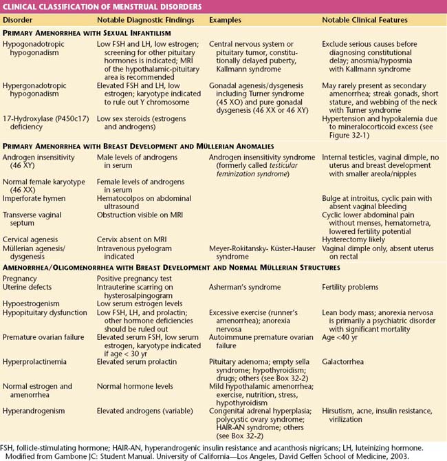
Amenorrhea, or the absence of menses, is a common symptom of several pathophysiologic states. This condition traditionally has been divided into primary amenorrhea, in which menarche (the first menses) has not occurred, and secondary amenorrhea, in which menses has been absent for 6 months or more. A more functional or clinical division of menstrual disorders based on initial history and physical examination would be as follows: primary amenorrhea with sexual infantilism, primary amenorrhea with breast development and müllerian anomalies, and amenorrhea and oligomenorrhea with breast development and normal müllerian structures. The last group includes disorders causing primary as well as secondary amenorrhea, oligomenorrhea, and the hyperandrogenic states (Table 32-1).

image Primary Amenorrhea

The diagnosis of primary amenorrhea is made when no spontaneous uterine bleeding has occurred by the age of 16 years. The workup should be initiated earlier if there is no evidence of breast development (thelarche) by age 14 years or if the patient has failed to menstruate (menarche) spontaneously within 2 years of thelarche. The presence of normal breast development confirms gonadal secretion of estrogen but not necessarily the presence of ovarian tissue. The presence of normal amounts of pubic and axillary hair confirms gonadal or adrenal secretion of androgens as well as the presence of functional androgen receptors.

PRIMARY AMENORRHEA WITH SEXUAL INFANTILISM

Patients with primary amenorrhea and no secondary sexual characteristics (sexual infantilism) display the absence of gonadal hormone secretion. The differential diagnosis is based on whether the defect is the result of a lack of gonadotropin secretion (hypogonadotropic hypogonadism) or an inability of the ovaries to respond to gonadotropin (hypergonadotropic hypogonadism due to gonadal agenesis or dysgenesis). The distinction can be made by the measurement of a basal serum follicle-stimulating hormone (FSH).

Hypogonadotropic Primary Amenorrhea and Sexual Infantilism

Patients with hypogonadotropic hypogonadism have low FSH levels, whereas patients with hypergonadotropic hypogonadism (e.g., gonadal dysgenesis) have elevated FSH levels in the menopausal range (>20 or 40 mIU/L, depending on the assay used). The measurement of serum luteinizing hormone (LH) is of limited additional diagnostic value. The absence of breast development is indicative of inadequate secretion of estrogen.

Hypogonadotropic hypogonadism may be caused by lesions of the hypothalamus or pituitary gland or by functional disorders that result in inadequate gonadotropin-releasing hormone (GnRH) synthesis and release. Because patients with sexual infantilism caused by hypogonadotropic hypogonadism may have a craniopharyngioma or other central nervous system tumor, magnetic resonance imaging (MRI) or computerized tomography (CT) of the hypothalamic-pituitary area is recommended.

Hypogonadotropic hypogonadism resulting in primary amenorrhea and sexual infantilism may also be the result of lesions of the pituitary, including prolactin-secreting adenomas, or a general process of pituitary failure. These patients should be screened for other pituitary hormonal deficiencies by testing for thyroid-stimulating hormone (TSH), growth hormone, and adrenocorticotropic hormone (ACTH).

Finally, apparent hypogonadotropic hypogonadism may actually represent constitutionally delayed puberty. This delay in the normal onset of puberty is generally attributed to undefined hereditary factors because there is commonly a history of late puberty in family members. Constitutional delay of puberty is a diagnosis of exclusion.

Hypergonadotropic Primary Amenorrhea and Sexual Infantilism

Patients with hypergonadotropic hypogonadism have some form of failed gonadal development or premature gonadal failure and will have elevated FSH levels. These patients may have gonadal agenesis (the absence or early disappearance of the normal gonad). Examples in males who may appear to be female in some cases are pure gonadal dysgenesis, or the testicular regression syndrome. These patients have an apparently normal 46 XY karyotype but lack testicular development. If fetal testicular regression occurs between 8 and 10 weeks of gestation, they may have female external genitalia with or without ambiguity in addition to a lack of gonads, a hypoplastic uterus (secondary to absent secretion of antimüllerian hormone), and rudimentary genital ducts (Swyer syndrome). Regression of the testes after 12 to 14 weeks results in variable development of male external genitalia. Anorchia, or streak gonads, occurs with testicular regression syndrome.

Other individuals with hypergonadotropic primary amenorrhea and sexual infantilism may have gonadal dysgenesis, or the presence of an abnormally developed gonad due to chromosomal defects. The differential diagnosis includes 45 XO (Turner syndrome), a structurally abnormal X chromosome, mosaicism with or without a Y chromosome, and pure gonadal dysgenesis (46 XX and 46 XY). Although most affected patients show no signs of secondary sexual characteristics, occasionally an individual with mosaicism or Turner syndrome will have sufficient ovarian follicular activity and secrete enough estrogen to cause breast development, menstruation, ovulation, and rarely even pregnancy.

In individuals with the presence of a Y chromosome, there is a risk for developing a gonadoblastoma (a benign germ cell tumor of the gonad) and eventually dysgerminoma (a malignant germ cell tumor). All patients with hypergonadotropic hypogonadism should have a karyotype performed. Because it is important to identify mosaicism, a greater number of white blood cells (>35) should be karyotyped.

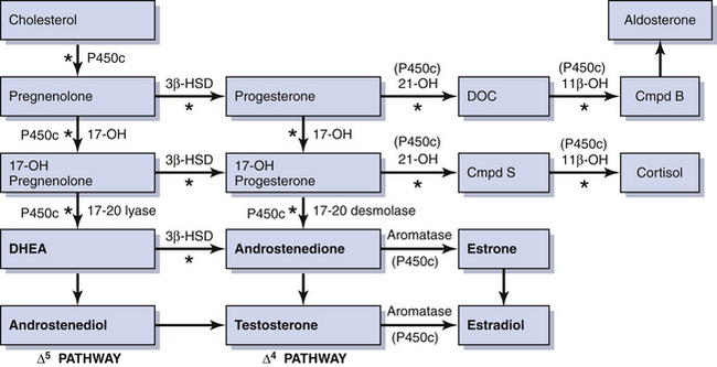
Rarely, some patients with primary amenorrhea and sexual infantilism have a defect of estrogen and androgen production. One example of this is a 17-hydroxylase (P450c17) deficiency, which prevents the synthesis of these sex steroids (Figure 32-1). These individuals have hypertension and hypokalemia caused by mineralocorticoid excess. Other patients, such as those with a 46 XY karyotype and Leydig cell agenesis, may lack the cells necessary for sex steroid production. Because the Leydig cells in the testicle are responsible for producing testosterone, these individuals are born with female external genitalia.

Patients with sexual infantilism may be treated to stimulate breast development by very gradually increasing estrogen doses. One commonly used regimen is to start with 0.3 mg conjugated estrogen every other day and slowly increase over 3- to 6-month intervals. This treatment should be guided by the presence or absence of mastalgia and the rate of breast development. The estrogen can be safely increased to 0.6 mg or more daily if necessary.

Individuals with persistent hypogonadotropic hypogonadism who seek fertility require either human menopausal gonadotropin injections or pulsatile GnRH administered by an infusion pump. Patients with gonadal dysgenesis and 17-hydroxylase deficiency who have a normal uterus and cervix can achieve pregnancy only by in vitro fertilization using donor oocytes.

PRIMARY AMENORRHEA WITH BREAST DEVELOPMENT AND MÜLLERIAN ANOMALIES

Patients with primary amenorrhea, breast development, and some defect of müllerian structures fall into two categories: those with complete androgen insensitivity syndrome (AIS), formerly called testicular feminization, and those with müllerian dysgenesis or agenesis. The distinction between these two diagnoses can be made by the measurement of a serum testosterone level and determination of the karyotype.

Müllerian Dysgenesis or Agenesis

Patients with primary amenorrhea, breast development, and a 46 XX karyotype have levels of testosterone appropriate for females. This clinical diagnosis may be caused by müllerian defects that cause obstruction of the vaginal canal (e.g., imperforate hymen or a transverse vaginal septum) or by the absence of a normal cervix or uterus and normal fallopian tubes. An imperforate hymen should be suspected in adolescents who report monthly dysmenorrhea in the absence of vaginal bleeding (see Figure 18-7, pg 237). Clinically, these patients often present with a vaginal bulge and a midline cystic mass on rectal examination. Ultrasonography confirms the presence of a normal uterus and ovaries with a hematocolpos. These patients should be treated with hymenectomy.

Alternatively, women may present with similar symptoms but without a vaginal bulge. When ultrasonography confirms a normal uterus and ovaries, a transverse, obstructing vaginal septum (see Figure 18-8, pg 237) or cervical agenesis should be suspected. MRI is the diagnostic procedure of choice in these patients. If the MRI scan confirms a transverse septum, surgical correction is indicated. Surgical construction of a functional cervix is extremely difficult. In general, it is recommended that these women undergo hysterectomy.

Finally, rectal examination and ultrasonography may be a sign of the absence of a uterus indicating müllerian agenesis or Meyer-Rokitansky-Küster-Hauser syndrome. This syndrome is characterized by a failure of the müllerian ducts to fuse distally and to form the upper genital tract. These patients may have unilateral or bilateral rudimentary uterine tissues (anlagen), fallopian tubes, and ovaries. It is uncommon to have functional endometrial tissue within the anlagen. On occasion, the ovaries are not visible on ultrasonography because they have not descended into the pelvis. In these cases, CT or MRI may identify them well above the pelvic brim. Currently, the pathophysiology leading to müllerian dysgenesis defects is not known.

Creation of a neovagina can be accomplished using one of two general approaches. The Frank method of vaginal dilation uses dilation of the vaginal pouch with vaginal forms (usually thermoplastic acrylic resin [Lucite] dilators) over the course of weeks to months. Alternatively, a McIndoe vaginoplasty, which involves the surgical creation of a neovaginal space using a split-thickness skin graft, may be performed. Both of these methods should be initiated and performed close to the time when the patient anticipates having vaginal intercourse.

Congenital anatomic abnormalities of the uterus or vagina, or both, are often associated with renal abnormalities such as a unilateral solitary kidney or a double renal collecting system, among others. Therefore, these patients should have an intravenous pyelogram or other diagnostic study to confirm a normal urinary system.

image Amenorrhea and Oligomenorrhea with Breast Development and Normal Müllerian Structures

Disorders in which the patient has breast development and a demonstrable cervix and uterine fundus on physical examination may cause primary as well as secondary amenorrhea, or may present as oligomenorrhea (menstrual cycles at greater than 35- to 45-day intervals).

All patients with menstrual bleeding disorders should be tested for pregnancy. Once pregnancy has been excluded, these individuals can be characterized as shown in Table 32-1. Initial history taking should include questions about the timing of thelarche, pubarche, and menarche. The timing and development of the menstrual disorder (present since puberty or new), significant weight change, strenuous exercise activities, dietary habits, sexual activity, concomitant illnesses or complaints, abnormal facial or body hair growth, scalp hair loss, acne, and the presence or absence of hot flashes and vaginal dryness should be noted. A comprehensive list of medications and dietary supplements taken should be obtained.

In addition to a pregnancy test, the initial investigation of the amenorrheic patient should include an FSH level and a progestin challenge test. Failure of the patient to have withdrawal bleeding after receiving a progestational agent indicates significant hypoestrogenism or hyperandrogenism, a uterine defect, or pregnancy. The absence of a withdrawal bleed after the administration of a progestational agent due to a uterine defect can be ruled out by the presence of withdrawal bleeding following sequential estrogen and progestin therapy. Progestogens used include medroxyprogesterone acetate, 5 to 10 mg/day orally for 5 to 14 days; norethindrone acetate, 2.5 to 5 mg/day orally for 5 to 14 days; oral micronized progesterone, 100 to 300 mg/day for 5 to 14 days; or progesterone in oil, 100 mg intramuscularly. Some clinicians prefer to order a serum estradiol (E2) instead of a progestin challenge to evaluate estrogen status.

AMENORRHEA AND OLIGOMENORRHEA ASSOCIATED WITH HYPOESTROGENISM

The differential diagnosis for patients with amenorrhea associated with low levels of estrogen includes hypothalamic-pituitary dysfunction (hypothalamic amenorrhea), premature ovarian failure, and hyperprolactinemia. Women in the first group have low FSH and prolactin levels, women in the second group have high FSH and normal prolactin levels, and women in the third group have high prolactin and low FSH levels.

AMENORRHEA AND OLIGOMENORRHEA WITH HYPERPROLACTINEMIA AND GALACTORRHEA

The principal action of prolactin is to stimulate lactation. Hypersecretion of prolactin leads to gonadal dysfunction by interrupting the secretion of GnRH, which inhibits the release of LH and FSH and, in turn, impairs gonadal steroidogenesis. The primary influence on prolactin secretion is tonic inhibition of dopamine input from the hypothalamus. Any event disrupting this inhibition can result in a rise in prolactin levels.

The consequences of hyperprolactinemia that are clinically significant include menstrual disturbances and galactorrhea. About 10% of women with amenorrhea have elevated prolactin levels, and a prolactin level should be measured in all cases of amenorrhea of unknown cause. Potential causes of elevated serum prolactin are noted in Box 32-1. Normal serum prolactin levels are less than 20 ng/dL, depending on the laboratory used. In patients with prolactin-secreting tumors, levels are usually more than 100 ng/dL. An elevated prolactin level should be confirmed by a second test, preferably in the fasting state because food ingestion may cause transient hyperprolactinemia. At the same time that the repeat prolactin level is drawn, a TSH level should be obtained to test for hypothyroidism because hyperprolactinemia may be seen in hypothyroid conditions.

A biologically inactive complex of prolactin and immunoglobulin, called big prolactin, can give a physiologically insignificant elevation. Therefore, the presence of a clinical abnormality should initiate the decision to test for hyperprolactinemia. If clinically significant hyperprolactinemia is not explained by hypothyroidism or drug use, CT or MRI of the sella turcica should be performed.

Galactorrhea is the most frequently observed abnormality associated with hyperprolactinemia. The secretion of milk may occur spontaneously or only after breast manipulation. Both breasts should be examined gently by palpating the gland moving from the periphery to the nipple. To confirm galactorrhea, a smear may be prepared and examined microscopically for the presence of multiple fat droplets (indicating milk). Besides galactorrhea, hyperprolactinemia frequently causes oligomenorrhea or amenorrhea.

Pharmacologic Agents Affecting the Secretion of Prolactin

A number of drugs may cause hyperprolactinemia and nonphysiologic galactorrhea (see Box 32-1).The mechanism of drug-induced hyperprolactinemia is secondary to reduced hypothalamic secretion of dopamine, depriving the pituitary of a natural inhibitor of prolactin release. When clinically indicated, patients with hyperprolactinemia caused by medications should be encouraged to discontinue the medication for at least 1 month. If hyperprolactinemia persists, or if the patient cannot interrupt the medication, a complete evaluation is indicated.

Treatment of Galactorrhea and Hyperprolactinemia

The objectives of therapy include the elimination of lactation, the establishment of normal estrogen levels, and the induction of ovulation when fertility is desired. The recommended forms of management are periodic observation, medical therapy, and surgery.

MEDICAL THERAPY

Patients with hyperprolactinemia may have galactorrhea and anovulation with resulting infertility. In more severe cases, they may be hypoestrogenic, which places them at risk for developing osteoporosis. Anovulatory patients without demonstrable tumors by MRI, and for whom the only issues are prevention of osteoporosis and menstrual cycle regulation, may be treated medically with combination hormonal contraceptives.

The ergot compounds, bromocriptine and cabergoline, act as dopamine agonists to reduce prolactin secretion and allow for the restoration of cyclic, physiologic estrogen secretion. Bromocriptine has a high initial incidence of side effects such as headache, nausea, and orthostatic hypotension. As a consequence, it should be started at a dose of 1.25 to 2.5 mg at bedtime and slowly increased in divided doses to tolerance and restoration of normal prolactin levels. Some patients tolerate bromocriptine better when it is given vaginally. Cabergoline is taken in twice-weekly doses beginning at 0.25 mg and increasing to a maximum of 1 mg twice weekly. It is better tolerated and more convenient to take than bromocriptine, but it is also more expensive.

Ninety-five percent of women without radiographic evidence of an adenoma require 5 mg/day of bromocriptine, whereas about 50% of patients with adenomas require higher doses to resume regular menses. Bromocriptine normalizes the secretion of prolactin in 82% of women with microadenomas and restores menses and fertility in more than 90%. Usually, menses resume, and galactorrhea resolves after about 6 weeks of bromocriptine therapy in women without adenomas. If an adenoma is present, it takes another 3 or 4 weeks for bromocriptine to become effective. Return of ovulation requires an average of 10 weeks without a tumor and 16 weeks with a microadenoma. Restoration of normal menstrual cycles and pregnancy may occur without complete normalization of the serum prolactin level. Discontinuation of therapy usually results in the return of hyperprolactinemia, leading to galactorrhea and amenorrhea.

Patients with macroadenomas (≥10 mm diameter) should have visual field testing and screening for other pituitary hormone deficiencies. A repeat MRI is done 6 months after the full therapeutic dose of bromocriptine is reached. As long as shrinkage of the adenoma is demonstrated, bromocriptine therapy is continued. Surgery should be performed for patients with significant visual field defects or symptoms that cannot be relieved by medical therapy.

Bromocriptine therapy is usually discontinued as soon as a pregnancy is confirmed. The risk for symptomatic enlargement of a microadenoma during pregnancy is only about 1%. When a macroadenoma is confined to the sella turcica, it is also unlikely to enlarge significantly during pregnancy. If there is extension of a macroadenoma beyond the sella turcica, there is a 15% to 30% risk for enlargement during pregnancy. If possible, these larger lesions should be debulked before conception, and bromocriptine treatment should be initiated. Pregnant patients with macroadenomas should have visual fields evaluated in each trimester. When abnormalities in visual fields develop, bromocriptine treatment should be reinstituted or increased and maintained for the rest of the pregnancy. There is no increase in fetal malformations as a result of bromocriptine treatment, and the drug can be discontinued after the completion of pregnancy to allow for breastfeeding. Cabergoline has not been adequately evaluated for use in pregnancy.

image Amenorrhea and Oligomenorrhea with Hyperandrogenism

Hyperandrogenism is the clinical manifestation of elevated levels of male hormones in women. Features may range from mild unwanted excess hair growth and acne to alopecia (hair loss), more extensive hirsutism, and masculinization and virilization. Hirsutism is the presence of male-like hair growth caused by conversion of vellus to terminal hairs in areas such as the face, chest, abdomen, or upper thighs. Figure 32-2 illustrates a scoring system for hirsutism. Signs of masculinization include loss of female body fat and decreased breast size. Virilization is the addition of temporal balding, deepening of the voice, and enlargement of the clitoris to any of the previous signs of excess male hormone. Androgens in women are normally produced in the ovaries and the adrenal glands (see Figure 32-1, pg 357). Hyperandrogenic disorders may be divided into functional and neoplastic disorders of the adrenal or ovary (Box 32-2).

image

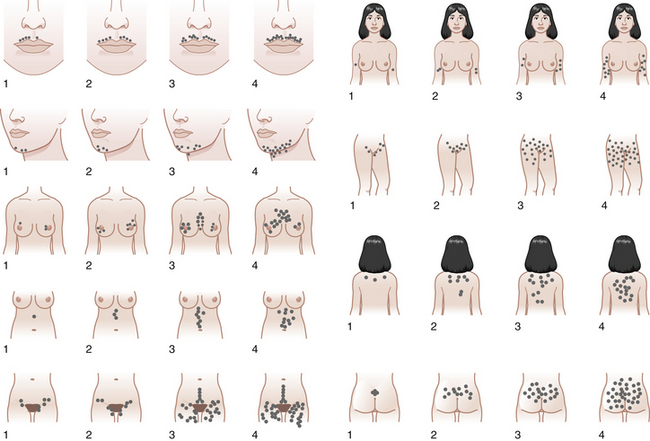
FIGURE 32-2 The Ferriman-Gallwey scoring system for hirsutism.

(Adapted from American Journal of Obstetrics and Gynecology, 140[5], Hatch R, 815-830, Copyright 1981, with permission from Elsevier.)

image Hyperandrogenic Disorders

In general, hyperandrogenic disorders can be attributed to excessive secretion of androgens by the ovaries, by the adrenals, or both.

ADRENAL DISORDERS

OVARIAN DISORDERS

Polycystic Ovary Syndrome

Six to 10% of women of reproductive age have some form of PCOS. This syndrome is a chronic condition that has been defined as anovulation or oligo-ovulation with clinical or laboratory evidence of hyperandrogenism and without evidence of any other underlying condition. Its onset is usually at the time of puberty. There is a heritable aspect to PCOS with an increased chance that first-degree female relatives are affected.

Clinically, the most common signs of PCOS are hirsutism (90%), menstrual irregularity (90%), and infertility (75%). Hirsutism is less likely in women who have used combined hormonal contraceptives for most of their postpubertal lives and for women of East Asian ethnicity. Although many patients with PCOS demonstrate abdominal obesity, the prevalence of obesity varies widely by country of origin, with the United States having the highest prevalence of obesity in women with PCOS (about 60%).

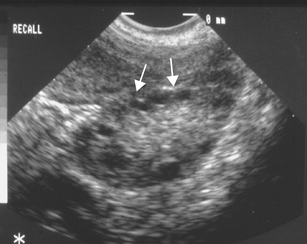
In most patients with PCOS, the ovaries contain multiple follicular cysts that are inactive and arrested in the mid-antral stage of development. The cysts are located peripherally in the cortex of the ovary (Figure 32-3). The ovarian stroma is hyperplastic and usually contains nests of luteinized theca cells that produce androgens. About 20% of hormonally normal women may also have polycystic-appearing ovaries.

The hyperandrogenism of PCOS results from an overproduction of male hormones by the ovary and often the adrenal gland. It is not clear what the ultimate underlying pathophysiology of PCOS is or even whether it is a single clinical entity. Patients with PCOS exhibit increased LH pulse frequency, usually resulting in higher circulating levels of LH. It is likely that these patients exhibit increased LH levels because of increased GnRH secretion from the hypothalamus and increased pituitary sensitivity to GnRH.

The increased LH level promotes androgen secretion from ovarian theca cells, leading to elevated levels of ovarian-derived androstenedione and testosterone. This then leads to atresia of many developing follicles and interferes with the normal development of a dominant or preovulatory ovarian follicle much of the time. Peripheral conversion of androgen to estrogen results in tonic estrogen levels that are higher than those found normally in the early follicular phase and that suppress FSH release from the pituitary gland. The normal pattern of stimulated estrogen rise is disrupted, and the midcycle LH surge does not occur with resulting anovulation and progesterone production. Some PCOS patients have excessive androgen production from the adrenal glands as well as the ovaries. The mechanism for the excess adrenal androgen production in PCOS is unclear.

In women with PCOS there is an association between abnormal androgen production and insulin resistance with hyperinsulinism. In about 60% to 70% of patients with PCOS, insulin sensitivity is decreased, leading to insulin hypersecretion. This hyperinsulinemia results from direct insulin stimulation of theca cells resulting in androgen secretion. Elevated androgen and insulin levels in PCOS also reduce the hepatic production and secretion of SHBG. When SHBG production is suppressed, the amount of free testosterone may be dramatically increased, even though the overall increase in total testosterone is moderate or small. Thus, the physical manifestations of hyperandrogenism in PCOS may seem dramatic in relation to the level of total testosterone.

In the long term, the insulin resistance associated with PCOS may lead to an increased risk for metabolic syndrome (diabetes and heart disease). The unopposed estrogens in women with PCOS may cause hyperplasia of the endometrium and occasionally endometrial carcinoma.

The diagnosis of PCOS continues to be somewhat controversial because of disagreement about diagnostic criteria. PCOS is a diagnosis of exclusion. It is also a syndrome, rather than a distinct and readily definable disease. The European Society for Human Reproduction and Embryology defines PCOS as being present when patients demonstrate irregular ovulation (usually with clinically evident oligomenorrhea), signs of hyperandrogenism, or polycystic ovaries, after other causes of these signs have been ruled out.

image Evaluation of Patients with Signs of Hyperandrogenism

PHYSICAL EXAMINATION

The degree of hirsutism (see Figure 32-2), acne, or androgenic alopecia should be assessed and the thyroid palpated for enlargement. Patients should be expressly asked about excess facial hair because they may conceal their hirsutism by waxing or electrolysis and be too embarrassed to volunteer the information. Evidence of cushingoid features should be noted. Acanthosis nigricans (see Figure 32-4) is a frequent marker of insulin resistance and hyperinsulinemia. A bimanual pelvic examination may identify ovarian enlargement. Asymmetric ovarian enlargement associated with the rapid onset of virilization can indicate a rare androgen-producing tumor.

LABORATORY EVALUATION

The laboratory evaluation of patients with virilization or significant hirsutism, or both, is aimed primarily at exclusion of serious disorders.

A basal 17-hydroxyprogesterone level is useful to exclude 21-hydroxylase–deficient CAH. With concentrations greater than 2 ng/mL, ACTH stimulation testing measuring 17-hydroxyprogesterone is the definitive method of diagnosis. When Cushing’s syndrome is suspected, either a 24-hour measurement of free urinary cortisol or an overnight dexamethasone suppression test should be performed. For the latter test, 1 mg of dexamethasone is given orally at bedtime, and serum cortisol is measured in an 8:00 AM fasting specimen; normal values are less than 5 g/dL.

Measurement of prolactin and TSH levels excludes hyperprolactinemia with or without thyroid dysfunction. In patients with unclear signs of hyperandrogenism, the measurement of serum levels of total and free testosterone and DHEA-S may be helpful. Confirmed values of DHEA-S in excess of 7000 ng/mL or total testosterone in excess of 200 ng/dL is highly suspicious for an adrenal or ovarian androgen-producing tumor. However, the best predictor of an androgen-secreting neoplasm, rare as it is, is the clinical presentation. Signs of virilization are present in 98% of patients with tumors, regardless of the peripheral level of testosterone.

A pelvic ultrasound should be obtained whenever any high-risk features are present to exclude an ovarian tumor. Androgen-secreting tumors of the adrenal gland can be detected by CT or MRI. If clinical or laboratory findings indicate the presence of an androgen-secreting tumor, and it cannot be located by imaging studies, selective venous catheterization may be carried out and androgens measured in the venous blood from each adrenal gland and ovary.

In patients with PCOS and the HAIR-AN syndrome, evaluation of their metabolic status should be performed. Although a fasting glucose level is adequate screening for diabetes mellitus in many women, in patients with PCOS, optimal screening should include a 2-hour oral glucose tolerance test, measuring both glucose and insulin. Lipid levels should be measured in patients with PCOS, at least in those over the age of 35 years, and in younger patients with evidence of metabolic dysfunction (e.g., HAIR-AN syndrome).

TREATMENT OF HYPERANDROGENISM

Treatment should be guided by the nature of the underlying disease, the severity of clinical symptoms and signs, and the desires of the patient. In the rare instance that an ovarian or adrenal neoplasm exists, surgical removal of the tumor is indicated. In premenopausal women, unilateral salpingo-oophorectomy is sufficient for an ovarian tumor and preserves future childbearing potential. In postmenopausal women, the treatment is usually a total abdominal hysterectomy and bilateral salpingo-oophorectomy. In patients with Cushing’s syndrome, treatment is surgical removal of the source of excess cortisol or ACTH (adrenal or pituitary tumor).

PCOS is by far the most common functional ovarian disorder causing hyperandrogenism, and the management of PCOS depends on the patient’s presentation and desires. The therapy for the hirsutism in PCOS patients is ovarian suppression, which is usually achieved by administration of a combination contraceptive. This estrogen-progestin treatment suppresses gonadotropins (LH and FSH), which allows regression of the overproduction of testosterone and androstenedione by the ovary. Estrogen also stimulates SHBG production, which decreases free testosterone levels.

The treatment of hirsutism is best accomplished by the addition of an androgen blocker. The most commonly used drug for hirsutism in women in the United States is spironolactone. This aldosterone antagonist competes for testosterone-binding sites, thereby exerting a direct antiandrogenic effect at the target organ. In addition, spironolactone interferes with steroid enzymes and decreases testosterone production. Because this medication opposes the action of aldosterone, serum potassium levels may rise and should be monitored. Other drugs that block the binding of androgens to its receptor include flutamide and cyproterone acetate, whereas finasteride blocks the conversion of testosterone to its more potent metabolite, dihydrotestosterone. It may take up to 6 months to begin to observe a cosmetic improvement in hirsutism, and maximum effect may not be seen for up to 2 years.

Suppression of abnormal androgen production or action generally suppresses future hair growth but does not immediately cause the existing hirsutism to disappear. To obtain good cosmetic results, some local hair removal is usually required in addition to the biochemical treatment. Local methods include shaving, depilatory creams, electrolysis, and laser hair removal. Plucking of individual hairs should be discouraged because growth of surrounding hair follicles may be stimulated by this technique.

All patients with PCOS and chronic anovulation are at risk for the development of endometrial hyperplasia and endometrial cancer. Hence, management of patients not taking combined oral contraceptives should always include scheduled progestin-induced withdrawal of the endometrium to reduce this risk. This may be accomplished with 10 mg of oral medroxyprogesterone acetate daily, 100 mg of oral micronized progesterone twice daily, or 5 mg of norethindrone acetate daily, for 12 to 14 days every other month.

The underlying insulin resistance and hyperandrogenism of many patients with PCOS may have a significant effect on their risk for diabetes mellitus and possibly cardiovascular morbidity. Women with PCOS have a threefold to sevenfold higher risk for developing type 2 diabetes mellitus. Women with PCOS and hyperandrogenism also tend to have increased levels of low-density lipoprotein cholesterol (LDL cholesterol) and reduced levels of high-density lipoprotein cholesterol (HDL cholesterol) and are at increased risk for developing hypertension. Thus, patients with PCOS and chronic anovulation should be counseled regarding weight loss, nutrition, exercise, and other lifestyle changes that will reduce their risk for developing diabetes mellitus and cardiovascular disease.

Patients with functional adrenal hyperandrogenism, such as CAH, are treated by the administration of glucocorticoids (e.g., 0.25 mg dexamethasone every other day at bedtime). However, many of these women (like those with PCOS) also require suppression of ovarian androgen secretion using combination oral contraceptives and antiandrogens.