AIRWAY MANAGEMENT IN THE TRAUMA PATIENT: HOW TO INTUBATE AND MANAGE NEUROMUSCULAR PARALYTIC AGENTS

Published on 20/03/2015 by admin

Filed under Critical Care Medicine

Last modified 22/04/2025

Print this page

rate 1 star rate 2 star rate 3 star rate 4 star rate 5 star
Your rating: none, Average: 0 (0 votes)

This article have been viewed 3494 times

CHAPTER 75 AIRWAY MANAGEMENT IN THE TRAUMA PATIENT: HOW TO INTUBATE AND MANAGE NEUROMUSCULAR PARALYTIC AGENTS

Injury is the leading cause of death in persons between the ages of 1 and 45 years in the United States and the third leading cause overall. Airway compromise is a common cause of death or severe morbidity in trauma victims. Management of the airway is a fundamental skill in trauma medicine. Obstruction of the airway has been reported in two-thirds of patients who die in the prehospital setting when death was not inevitable. Airway care is a cornerstone of resuscitation and is the first priority for patients both in the prehospital setting and in the emergency room (ER).

Over the last few decades, improvement in the management of trauma victims has helped to decrease mortality and morbidity. An organized systematic approach to treatment is particularly important and includes focus on the airway in the “primary survey,” as outlined by the American College of Surgeons in their Advanced Trauma Life Support Course and Manual (ATLS). The ATLS emphasizes the importance of management during “the golden hour” after major trauma by stressing immediate attention to life-threatening conditions as soon as they are discovered. The development of new airway equipment as well as new techniques and algorithms for managing the difficult airway has significantly contributed to improved outcomes. The goal for emergency airway intervention is to make certain that the patient’s ventilation is adequate to meet oxygen demands, thereby reducing the risk of ischemic injury to the brain, heart, and other organs as well as protecting the patient from the risks of aspiration and airway obstruction.

This review focuses on airway management of the adult traumatized patient. Specific aspects of pediatric airway management and the controversies of prehospital management of airways are not included.

AIRWAY CONSIDERATIONS IN THE TRAUMA PATIENT

Several circumstances make management of the trauma patient’s airway unique. These include the frequent need for emergent intubation, the presence of complicating injuries, fixation in neck collars, and the risk of tracheopulmonary aspiration. There is no standard definition for a difficult airway, but it is often defined in the literature as an airway that requires more than two or three attempts for successful intubation. In the emergency department, difficult intubation conditions have been reported in at least 3% of cases. During the last few decades, we have seen a marked reduction in severe airway complications related to anesthesia for surgery in the operating room (OR). This, however, is not the case for airway management outside the OR. Management of the trauma airway is considered a task for the experienced physician.

Airway and breathing are the first two components of the ABCs (airway, breathing, and circulation) of initial evaluation of trauma patients. All seriously injured patients should receive supplemental oxygen, and many require intubation. Trauma victims are frequently either unconscious or combative as a result of head trauma or intoxication. The airway is vulnerable to mechanical obstruction from loss of muscle tone and airway reflexes. In addition, the airway is frequently contaminated with debris, blood, and secretions. Direct airway trauma and facial trauma may make the situation even more complex. The fully conscious, talking patient who maintains his/her own airway may not need airway intervention initially, but it must be kept in mind that the patient’s status may change quickly. Continuous monitoring and frequent reevaluation of the airway is mandatory. Inhalation of oropharyngeal and gastric contents is always a risk in these individuals, but the actual frequency of aspiration is unknown. All trauma patients are presumed to have a full stomach and should be treated accordingly, using a technique to secure the airway that minimizes the risk of pulmonary aspiration.

The cervical spine is considered “unstable” in the trauma victim until proven otherwise. The evaluation of the spine and ruling out or diagnosing injury may be a prolonged procedure, especially in the patient with a decreased consciousness level. In the United States, 1.5%–3% of trauma victims suffer from spinal cord injury, and 55% of these injuries are located in the cervical spine. Complete spinal cord injury with loss of motor and sensory function distal to the lesion occurs in 43%–46% of cases. All trauma patients receive a rigid cervical collar to prevent secondary spine injury. However, this fixation usually makes an intubation more difficult and unpredictable.

There is an increased risk for awareness among trauma patients during airway manipulation and surgery. The incidence of awareness in the general adult patient population undergoing anesthesia is 0.1%–0.2%. Approximately 50% of these patients will have some psychological impact from their experience, and the most severe reaction is full-blown post-traumatic stress syndrome. The incidence of awareness is reported to be higher among trauma patients. These patients often have such hemodynamic instability that they tolerate only very light levels of anesthesia. It is, therefore, good practice to always consider giving amnesia-inducing drugs when neuromuscular blocking agents are used. To be paralyzed and unable to communicate is an extremely traumatic experience.

EVALUATION OF AIRWAY AND RESPIRATORY FUNCTION

Assessment of the airway as well as of the ventilatory and respiratory functions has the highest priority when a new trauma patient is encountered. Start by observing the patient’s ventilatory pattern; and then auscultate the lungs. If time permits, obtain a chest film, and evaluate for the presence of hemothorax and/or pneumothorax. If there is an emergent need for airway intervention, the time for physical examination will be limited. The vital functions of a trauma patient can deteriorate rapidly, and constant monitoring with frequent reevaluation of the airway is crucial. The goal of the evaluation is to get as clear a picture as possible of the airway anatomy and the patient’s ventilatory and respiratory functions, so that an appropriate plan for securing the airway can be established. The objective of the plan should always be a patient who is well oxygenated and ventilated and an airway that is protected after the intervention. The sophistication of the evaluation and the final plan are largely affected by the urgency of the needed intervention. It is not surprising that the incidence of difficult intubation is four to seven times higher in the emergency department than in the OR, largely due to challenging conditions in the trauma patient such as direct injury to the face and neck areas, fractures, hematomas, burns with edema, and secondary distortion of the airway.

In recent years, the development of classification systems to predict difficult intubations has reduced the incidence of airway complications in patients undergoing elective surgery. The Mallampati classification of the airway (see page 59) is commonly used and is based on assessment of tongue size in relation to other pharyngeal structures. The I to IV scoring scale predicts difficulty of intubation. The atlanto-occipital joint extension test measures the ability to extend the neck and, consequently, the ability to align pharyngeal and laryngeal axes to accommodate intubation. Measurements of the thyro-mental distance, sterno-mental distance, mandibulo-hyoid distance and inter-incisor distance are also helpful in evaluating the airway.

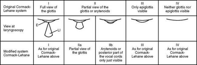
To provide high specificity and sensitivity for successful intubation and to assess the level of difficulty of an endotracheal intubation, several tests must be performed. These tests are usually correlated to the visualization of laryngeal structures and vocal cords. The gold standard for classifying the degree of exposure to the larynx entrance is the description by Cormac and Lehane (Figure 1). Grades III and IV are associated with difficult intubation. The dilemma is that all these tests are difficult to utilize in the trauma patient for many reasons, including an immobilized neck. Thus, alternative scoring systems such as the LEMON method have been proposed.

image

Figure 1 Cormack-Lehane original grading system compared with a modifi ed Cormack-Lehane system (MCLS). E, Epiglottis; LI, laryngeal inlet.

(From Yentis SM, Lee DJH: Evaluation of an improved scoring system for the grading of direct laryngoscopy. Anesthesia 82:1197-1204, 1998.)

The LEMON method was developed by the U.S. National Emergency Airway Management Course and has a maximum score of 10 points, calculated by assigning 1 point for each criterion (Table 1). It has been demonstrated that an airway assessment score based on the LEMON criteria is helpful in predicting difficult intubation in the ER. The LEMON test is designed to be a quick and easy-to-use assessment tool. A poor laryngoscopic view is more common, for example, among patients with large incisors, a reduced inter-incisor distance, and a reduced thyroid-to-floor-of-mouth distance.

Table 1 LEMON Criteria

Physical Sign Less Difficult Airway Indicators of Difficult Airway
Look at exterior No face or neck pathology Face or neck pathology, obesity, and so on
Evaluate the 3-3-2 rule Mouth opening >3F Mouth opening <3F
Hyoid–chin distance >3F Hyoid–chin distance <3F
Thyroid cartilage–mouth floor distance >2F Thyroid cartilage–mouth floor distance <2F
Mallampati Classes I and II Classes III and IV
Obstruction None Obstruction within or surrounding upper airway
Neck mobility Normal extension and flexion Limited range of motion

F, Finger-breadths.

Another important component in an emergency evaluation is assessment of conditions that may compromise mask ventilation. Mask ventilation is usually used as an intermittent bridge until final airway control is established. Difficult mask ventilation is correlated to obesity, beards, facial trauma, upper airway obstruction, and absence of teeth and is reported in up to 5% of the normal adult population. It is a useful rule to make sure that mask ventilation is possible before paralytic drugs are administered to a patient. In the emergency situation, however, there are exceptions to this rule, and the pros and cons of using muscle blockade must be assessed in each case. Furthermore, the possibility of a “can’t ventilate, can’t intubate” situation is something that should be anticipated; therefore, it is essential that equipment and competence for creating a surgical airway are immediately available. In this circumstance, the team approach and communication among team members becomes crucial in establishing the final management plan, including rescue alternatives.

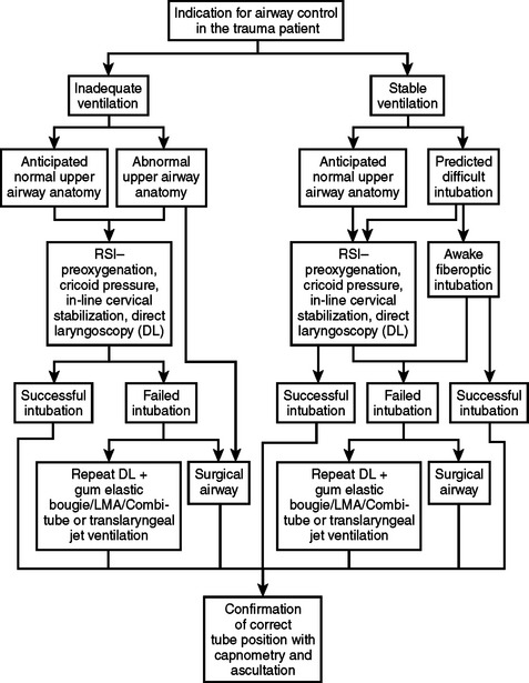
Planning an approach is the final step of the assessment (Figure 2). The team should then proceed with the airway management plan. When the patient can maintain adequate oxygenation and ventilation, and time permits, it may be beneficial to transport the patient to the OR which normally has better equipment and resources than the ER. In other situations, the right decision may be to immediately establish a surgical airway with the patient breathing spontaneously.

When reviewing the literature about airway management, it is remarkable how often the quality of professional competence/experience is mentioned. This is something that is very difficult to measure but is obviously critical to a successful outcome. All efforts should be made to have that experience accessible on short notice in a trauma organization that strives for excellence in airway management.

INDICATIONS FOR INTUBATION AND CONTROLLED VENTILATION

There are many indications for intubation and controlled ventilation in trauma patients. The decision to intubate is not always easy, but for the severely injured patient, the threshold should be low. The decision to intubate the trauma patient is usually made after a rapid assessment of injuries and identification of an indication to control the airway. Indications for intubation are:

Airway obstruction in the trauma patient can result from many conditions. Direct trauma to the larynx and trachea or maxillofacial injuries can result in severe obstruction, sometimes with delayed onset. These injuries are associated with high risk of aspiration. Cervical spine injury can be associated with hematoma formation and obstruction and also with direct paralysis of respiratory muscles. Patients with cognitive impairment, resulting from brain injury or intoxication, are prone to obstruction and secondary hypoxemia. Smoke inhalation and burns frequently result in edema formation and obstruction, which can have a delayed onset and require preventive intubation.

Hypoventilating patients need tracheal intubation. Patients with cervical spine fractures frequently hypoventilate, as do patients with cognitive impairment.

Persistent hypoxemia, despite supplemental oxygen, is also an indication for airway intervention. This condition can result from airway obstruction, hypoventilation, and lung injury, including aspiration and lung contusion. Low consciousness levels frequently result in hypoxemia that affects neurologic outcome. However, prevention of hypoxemia is associated with reduced mortality and morbidity in trauma victims.

The patient with severe cognitive impairment (Glasgow Coma Score <8) frequently has airway obstruction, is hypoventilating, and has hypoxemia. Usually, these patients have brain injury, and early intubation reduces mortality and morbidity. Other reasons for low consciousness levels include intoxication and smoke inhalation.

Trauma victims with cardiac arrest have a higher survival rate if early intubation is performed.

Patients with severe hemorrhagic shock should be intubated emergently to improve oxygenation. Immediate surgical intervention is almost inevitable.

Smoke inhalation is associated with burn injuries and inhalation of toxic products, such as carbon monoxide and cyanide. When cognitive impairment from inhalation is suspected, or airway obstruction from thermal injury is present, immediate intubation is necessary. If the patient is not intubated, close observation is required. Edema formation and airway obstruction can develop quickly, and many authors recommend routine intubation. Measurement of blood concentrations of carbon monoxide and cyanide is fundamental. Toxic carbon monoxide concentrations are indication for intubation and ventilation with 100% oxygen.

INDUCTION AGENTS AND MUSCLE RELAXANTS

A number of pharmacological agents are used to facilitate intubation of the trauma patient. Most patients will benefit from anesthesia prior to paralysis and intubation, with the exception of profoundly hypotensive and unconscious patients. Intubation of the braininjured patient without the use of anesthesia can produce a severe increase in intracranial pressure and cause herniation.

Many anesthesia induction agents have qualities that limit their usefulness in the trauma setting. The ideal induction agent should have a quick onset, provide deep hypnosis, have a short duration, and render hemodynamic stability. Intracranial and intraocular pressures should be minimally affected, and a reduction of cerebral metabolic rate is ideal. If the potential for increased blood pressure and heart rate is a concern, pretreatment with an opioid usually diminishes this response.

Many of these properties are also desirable for muscle relaxants. For example, when intubation and mask ventilation are not accomplished, the duration of neuromuscular blocking agents should ideally be so short that spontaneous ventilation is reestablished before severe desaturation occurs.

Currently, we do not have agents that satisfy all of these criteria. However, the most commonly used induction agents and muscle relaxants are discussed below.

Sodium thiopental has been one of the most commonly used induction agents for many years. Maximum concentration at the receptor site occurs within a minute, and the patient is usually unconscious within 30–45 seconds. Rapid redistribution results in a short duration after a single dose. Barbiturates act at the GABA receptor site and depress the reticular activating system. The respiratory center is depressed, and the response to carbon dioxide is reduced. In the hypovolemic patient, sodium thiopental may cause a dangerous decrease in blood pressure due to depression of the medullary vasomotor center and reduced activity of the sympathetic nervous system, causing vasodilatation and decreased venous return. If thiopental is used, it must be dosed cautiously, using a lower dose in the circulatory-compromised patient. Thiopental causes cerebral vasoconstriction as well as decreased intracranial blood volume and intracranial pressure. Therefore, it is often an excellent choice in a patient with an isolated brain injury and stable circulation, when elevated intracranial pressure is a concern. Thiopental may induce acute intermittent porphyria in susceptible patients.

Propofol is a relatively new agent which can be used for sedation and induction and maintenance of anesthesia. It is an alkylphenol with high lipid solubility. It causes marked vasodilatation, making it less suitable for induction of trauma patients. If propofol is used, the induction dose must be drastically reduced. It is attractive for continuous sedation in the circulatory-stable patient because of its short half-life and its properties that reduce cerebral metabolism and blood flow. Propofol depresses pharyngeal and laryngeal reflexes and muscle tone more than other induction agents.

Etomidate is a carboxylated imidazole that produces unconsciousness within 30 seconds after intravenous (IV) administration. The duration is short, and etomidate has very favorable circulatory properties. Its direct cardiovascular effects are minimal, but it can cause hypotension by affecting sympathetic output. Etomidate also reduces cerebral blood flow, cerebral oxygen consumption, and intracranial pressure. These traits make it very attractive for the unstable trauma patient with head injuries; and it is, therefore, considered by many to be the induction agent of choice for trauma patients. Although the circulatory properties are beneficial, careful dose reduction should be considered in the hypovolemic patient, as even etomidate may drop the cardiac output. There is also some concern that it causes adrenocortical suppression for a few hours after administration, and it may theoretically cause instability in the catecholamine-depleted patient and potentially modify the humoral stress response.

Ketamine is another choice for induction of anesthesia. It is derived from phenylcyclidine and induces a condition called dissociative anesthesia. Onset occurs within a minute after IV administration. Ketamine stimulates the cardiovascular system via endogenous release of catecholamines, which often results in tachycardia and hypertension. However, it has direct negative inotropic properties and can produce further hypotension in patients with long-standing shock and depleted endogenous catecholamine stores. This drug should probably be avoided in victims with head injuries because of its properties to increase cerebral metabolism, blood flow, and intracranial pressure. It is often the preferred induction agent in hypotensive, hypovolemic patients; and with ketamine, ventilation is preserved and airway reflexes are more or less intact. Ketamine is also a potent analgesic. All these qualities make ketamine a useful drug under field conditions. However, it is well known for its potential to produce unpleasant hallucinations and psychomimetic reactions during emergence. This risk can be reduced by pretreatment with benzodiazepines.

Muscle relaxants are used to create conditions that facilitate the intubation procedure. The larynx, for example, must be visualized as optimally as possible, and muscle tone suppression is integral to creating this condition. To help prevent hypoxemia and hypercapnia, a short onset time for muscle paralysis is important. Today, succinylcholine and rocuronium are the two drugs available that fulfill this criterion. Succinylcholine has some undesirable side effects, and substantial research has been underway to find an alternative drug. So far, however, no superior replacement has been introduced on the market, although promising drugs are under investigation. The current alternative to succinylcholine is rocuronium, but it does not match the short onset and short duration of succinylcholine.

Techniques that shorten the onset of alternative drugs have been studied. One such technique is known as “priming” and results when approximately one-tenth of the intubating dose is given 60 seconds prior to the remainder of the full dose. This partially preoccupies the receptor sites, thereby shortening the time required to achieve adequate intubation conditions. Another method, called “timing,” requires that the administration of the muscle relaxant be carefully timed before the induction agent so that the patient does not experience the unpleasant sensation of being conscious while paralyzed. Each of these techniques is unreliable, and all alternatives to succinylcholine render a prolonged duration of muscle relaxation that can be detrimental in the “can’t intubate, can’t ventilate” situation.

Succinylcholine is a depolarizing muscle relaxant. Its onset is 30–45 seconds, and its offset is about 6–12 minutes. Shortly after administration of succinylcholine, muscle fasciculations occur. Observation of these fasciculations can help to predict when the patient is sufficiently paralyzed to commence the intubation process.

A brief increase in intracranial and intraocular pressure will follow administration of succinylcholine. The increase in intracranial pressure is small and not considered important enough to avoid succinylcholine in the brain-injured patient. Furthermore, it has never been proven that this increase in intracranial and intraocular pressure is of clinical importance. After all, laryngoscopy alone increases intracranial and intraocular pressure.

An increase in intragastric pressure after succinylcholine injection has also been documented. However, this does not likely increase the risk for aspiration of stomach contents because succinylcholine increases the tone of the lower esophageal sphincter.

A transient elevation of plasma potassium concentration of up to 1 mmol/l is seen in conjunction with administration of succinylcholine. This can be more pronounced in victims of burns, spinal cord trauma, and severe soft tissue injuries and can lead to cardiac dysrhythmias or asystole. The resulting massive release of potassium is an effect of extrajunctional receptor proliferation. This is not a relevant problem in the acute situation but must be considered later during hospital treatment of these patients.

Finally, succinylcholine also triggers malignant hyperthermia and can produce severe anaphylactic reactions.

Rocuronium is a nondepolarizing muscle relaxant. It does not trigger malignant hyperthermia or other side effects associated with succinylcholine. However, allergic reactions are still a possibility. To obtain good intubation conditions, rocuronium usually requires up to 60 seconds, and its duration after an intubation dose is approximately 45 minutes. These time factors are the primary reasons that succinylcholine is still considered the muscle relaxant of choice.

INTUBATION TECHNIQUES

During all advanced airway management procedures, the patient should be monitored adequately. The standard is electrocardiogram (ECG), pulse oximetry, end tidal CO2 monitoring, and an automated blood pressure cuff. The choice of intubation technique depends on the severity of respiratory compromise, the expected difficulty of intubation, and the skills of the practitioner.

In most cases when the airway must be controlled immediately, the best technique is the modified rapid sequence approach, using preoxygenation and cricothyroid pressure to block esophageal passage of gastric contents. This technique has an acceptable success rate of 95%–97%, but is inherently risky.

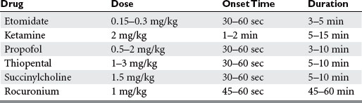
For this approach, drugs are injected at predetermined doses. Taking into consideration that the decisions regarding medications must often be made within seconds of the patient’s arrival, it is fundamental to have a thorough knowledge about drug actions in order to tailor the choice and dose and to avoid causing harm and circulatory instability (Table 2).

During the intubation process, the potential risk of spinal cord injury is always a concern if cervical fractures are present. In the past, because of this risk, blind nasal intubation of the conscious patient was frequently advocated. Today, we know more about spinal movements and different intubation techniques; and the nasal route is used mainly if the jaw is locked and the patient’s condition is relatively stable. As long as manual in-line neck stabilization is applied and axial traction is avoided to prevent distraction, rapid sequence intubation, followed by direct laryngoscopy and oral intubation, appears to be safe, even if it produces more motion in the spine compared with the use of the nasal route. Ideally, when a spine injury is present, and spine motion is the primary concern, intubation should be executed using a fiberoptic bronchoscope. This is rarely possible in an emergency situation. Preintubation maneuvers, such as jaw trust, insertion of Combitubes, and positioning of laryngeal masks, cause as much motion as some of the intubation techniques. There are no data that suggest better outcome with any particular technique, and the most immediate threat to patients with spinal cord injury is hypoxemia from hypoventilation or aspiration of gastric contents. The most severe injury to the spinal cord probably occurs at the time of the trauma; and if rapid sequence intubation is performed with care and in-line stabilization, the risk for secondary neurological injury is minimal. The most common complication of rapid sequence intubation is probably hypotension.

The rapid sequence intubation procedure can be divided into a predetermined sequence of four phases: preoxygenation, drug administration, endotracheal tube positioning, and confirmation of endotracheal tube position. Usually, the patient will present with the neck immobilized in neutral position by a cervical collar. This position should be retained throughout the intubation sequence. The patient is preoxygenated with a high flow of 100% oxygen using a non-rebreathing system before the induction agent is injected. The mask should have a tight seal. The preoxygenation process will significantly increase the time lapse until desaturation begins after apnea.

The induction agent is injected as a bolus and is immediately followed by the muscle relaxant. When the patient starts to lose consciousness, one assistant will hold the patient’s neck in in-line stabilization, and a second assistant will apply cricoid pressure using Sellick’s maneuver to compress the esophagus between the cricoid cartilage and C6, prohibiting the regurgitation of stomach contents during airway manipulation. It is permissible to open the cervical collar to facilitate intubation as long as strict attention is maintained to prevent cervical movements.

When the patient is paralyzed, intubation should proceed in a gentle and nontraumatic fashion. After the cuff is insufflated, the correct position of the tube should be verified by direct visual inspection during the intubation process, end tidal CO2 monitoring, and auscultation in both flanks and over the stomach. Indirect signs, such as chest movements and condensation in the tube, are helpful when verifying tube positioning. If the breath sounds are not bilateral and equal, an explanation must be determined. The most common explanations for this situation include mainstem intubation, pneumothorax, hemothorax, and obstruction of the airway. Since the right mainstem bronchus leaves the trachea at a steeper angle than the left, foreign bodies and endotracheal tubes that have been placed too deeply frequently end up on the right side. Absence of breath sounds and a gurgling sound over the epigastrium indicate esophageal intubation. After correct placement, the tube should be taped securely. Note the distance from the upper sixth molar. When the tube is correctly positioned, the average distance from the tip of the endotracheal tube is 21 cm in an adult female and 23 cm in an adult male.

The in-line stabilization of the patient’s head, which is required, is not optimal for visualization of the larynx during direct laryngoscopy; and it is not uncommon that suboptimal conditions are encountered during the intubation procedure and the larynx is not fully exposed. The gum elastic bougie is an excellent tool that allows tracheal intubation in most cases when the laryngeal inlet is not optimally exposed. After the bougie is inserted into the trachea, vibrations can usually be felt when its tip is sliding against tracheal cartilages. Then, the endotracheal tube is advanced over the bougie and into the trachea. Frequently, a twisting motion must be applied if the tip of the tube is trapped against the anterior larynx wall.

If intubation of the patient fails, a surgical airway usually needs to be created. Multiple intubation attempts increase the risk for edema, bleeding, and diminished visualization of the glottic opening. The incidence of hypoxemia, regurgitation of gastric contents, and cardiovascular complications are significantly increased after more than two attempts. An alternative technique should be selected before entering into the vicious cycle of multiple attempts and complications. Under some circumstances, rescue devices, such as the laryngeal mask or an esophagotracheal airway (Combitube), can be used to bridge the gap until definite control of the airway is established. Correct placement of these devices usually requires release of the cricoid pressure; and although the risk of aspiration is increased, ventilation is generally much more effective than with a face mask.

Several methods are used to create a surgical airway. Cricothyroidotomy is performed by penetrating the cricothyroid membrane to create an airway and can be executed as an open or percutaneous procedure. Tracheostomy may be selected as the primary technique under some conditions, especially if the larynx is fractured. Translaryngeal jet-ventilation can be life saving and is particularly useful if the knowledge to establish other surgical airways is not immediately present. This method is executed by inserting a catheter percutaneously into the trachea and insufflating it with oxygen. There are several potential complications, and the risk for barotraumas must be kept in mind. Once the airway is secured, it is important to keep the patient pain free and adequately sedated, especially if the patient has received long-term muscle relaxants.

SUGGESTED READINGS

Arne J, et al. Preoperative assessment for difficult intubation in general and ENT surgery: predictive value of a clinical multivariate risk index. Br J Anaesth. 1998;80(2):140-146.

Barash PG, Cullen BF, Stoelting RK. Clinical Anesthesia, 4th ed. Philadelphia, Lippincott: Williams and Wilkins, 2001.

Bell RM, Krantz BE, Weigelt JA. ATLS: a foundation for trauma training. Ann Emerg Med. 1999;34(2):233-237.

Caplan RA, et al. Adverse respiratory events in anesthesia: a closed claims analysis. Anesthesiology. 1990;72(5):828-833.

Copass MK, et al. Prehospital cardiopulmonary resuscitation of the critically injured patient. Am J Surg. 1984;148(1):20-26.

Cormack RS, Lehane J. Difficult tracheal intubation in obstetrics. Anaesthesia. 1984;39(11):1105-1111.

Cormack RS, et al. Laryngoscopy grades and percentage glottic opening. Anaesthesia. 2000;55(2):184.

Crosby ET, et al. The unanticipated difficult airway with recommendations for management. Can J Anaesth. 1998;45(8):757-776.

Eastern Association for the Surgery of Trauma (EAST). Guidelines for Emergency Tracheal Intubation Immediately Following Traumatic Injury. Allen-town, PA: Eastern Association for the Surgery of Trauma, 2002.

Foley LJ, Ochroch EA. Bridges to establish an emergency airway and alternate intubating techniques. Crit Care Clin. 2000;16(3):429-444. vi

Ghoneim MM. Awareness during anesthesia. Anesthesiology. 2000;92(2):597-602.

Hussain LM, Redmond AD. Are pre-hospital deaths from accidental injury preventable? BMJ. 1994;308(6936):1077-1080.

Ivy ME, Cohn SM. Addressing the myths of cervical spine injury management. Am J Emerg Med. 1994;15(6):591-595.

Langeron O, et al. Prediction of difficult mask ventilation. Anesthesiology. 2000;92(5):1229-1236.

Mallampati SR, et al. A clinical sign to predict difficult tracheal intubation: a prospective study. Can Anaesth Soc J. 1985;32(4):429-434.

Mort TC. Emergency tracheal intubation: complications associated with repeated laryngoscopic attempts. Anesth Analg. 2004;99(2):607-613.

Morton T, Brady S, Clancy M. Difficult airway equipment in English emergency departments. Anaesthesia. 2000;55(5):485-488.

O’Callaghan-Enrigh S, Finucane BT. Anesthetizing the airway. Anesthesiol Clin North Am. 1995;13(2):325-336.

Osterman JE, et al. Awareness under anesthesia and the development of post-traumatic stress disorder. Gen Hosp Psychiatry. 2001;23(4):198-204.

Peterson GN, et al. Management of the difficult airway: a closed claims analysis. Anesthesiology. 2005;103(1):33-39.

Practice guidelines for management of the difficult airway. A report by the American Society of Anesthesiologists Task Force on Management of the Difficult Airway. Anesthesiology. 1993;78(3):597-602.

Practice guidelines for management of the difficult airway: an updated report by the American Society of Anesthesiologists Task Force on Management of the Difficult Airway. Anesthesiology. 2003;98(5):1269-1277.

Reed MJ, Dunn MJ, McKeown DW. Can an airway assessment score predict difficulty at intubation in the emergency department? Emerg Med J. 2005;22(2):99-102.

Rose DK, Cohen MM. The airway: problems and predictions in 18,500 patients. Can J Anaesth. 1994;41(5 Pt 1):372-383.

Sakles JC, et al. Airway management in the emergency department: a one-year study of 610 tracheal intubations. Ann Emerg Med. 1998;31(3):325-332.

Sebel PS, et al. The incidence of awareness during anesthesia: a multicenter United States study. Anesth Analg. 2004;99(3):833-839.

Sekhon LH, Fehlings MG. Epidemiology, demographics, and pathophysiology of acute spinal cord injury. Spine. 2001;26(24 Suppl):S2-S12.

Tayal VS, et al. Rapid-sequence intubation at an emergency medicine residency: success rate and adverse events during a two-year period. Acad Emerg Med. 1999;6(1):31-37.

Trunkey DD. Trauma: accidental and intentional injuries account for more years of life lost in the U.S. than cancer and heart disease. Among the prescribed remedies are improved preventive efforts, speedier surgery and further research. Sci Am. 1983;249(2):28-35.

Walls RM. Management of the difficult airway in the trauma patient. Emerg Med Clin North Am. 1998;16(1):45-61.