[level-membership-for-obstetrics-gynecology-category]
Adenomyosis
Synonyms/Description
Etiology
Ultrasound Findings
Generalized Adenomyosis
Adenomyoma
Differential Diagnosis
Clinical Aspects and Recommendations
Figures
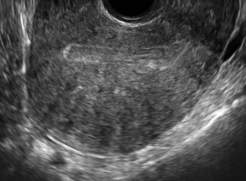
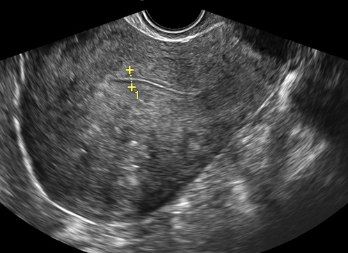
Figure A1-1 Two different patients. Typical appearance of the myometrium, which is asymmetric because of adenomyosis. Note that the endometrial echo is closer to the anterior than the posterior wall of the uterus.
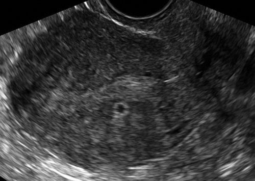
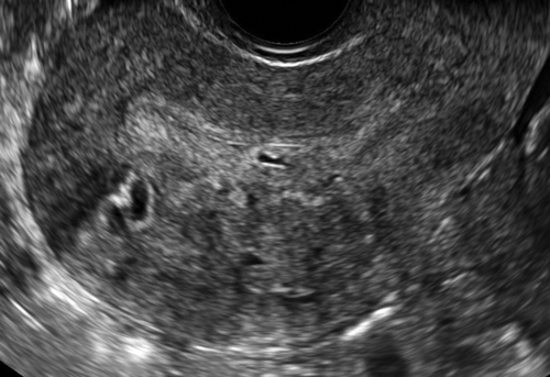
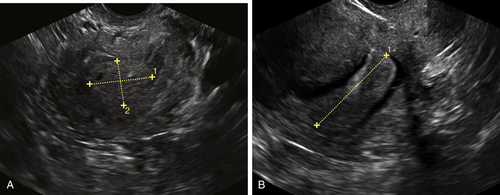
Figure A1-3 Adenomyoma projecting into the endometrial cavity from a broad base within the myometrium. A shows the mass as ill-defined within the cavity, worrisome for a malignancy, especially in a postmenopausal patient. B from the same patient shows the sonohysterogram with saline outlining the adenomyoma diagnosed by pathology.
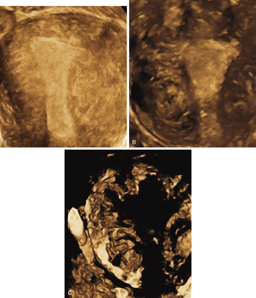
Figure A1-4 Three-dimensional ultrasound of two different patients with extensive adenomyosis. A shows the reconstructed coronal view of the uterus with a fuzzy, ill-defined junction and linear echogenicities emanating out from the edges of the endometrium. B shows a different patient with adenomyosis and a right-sided fibroid demonstrating similar echolucencies. C (same patient as B) shows the inverse mode of the 3-D image that accentuates the lack of a clear border at the junction of the endometrium and myometrium.
Suggested Reading
Bocca S.M., Oehninger S., Stadtmauer L., Agard J., Duran E.H., Sarhan A., Horton S., Abuhamad A.Z. A study of the cost, accuracy, and benefits of 3-dimensional sonography compared with hysterosalpingography in women with uterine abnormalities. J Ultrasound Med. 2012;31:81–85.
Champaneria R., Abedin P., Daniels J., Balogun M., Khan K.S. Ultrasound scan and magnetic resonance imaging for the diagnosis of adenomyosis: systematic review comparing test accuracy. Acta Obstet Gynecol Scand. 2010;89:1374–1384.
Exacoustos C., Brienza L., Di Giovanni A., Szabolcs B., Romanini M.E., Zupi E., Arduini D. Adenomyosis three-dimensional sonographic findings of the junctional zone and correlation with histology. Ultrasound Obstet Gynecol. 2011;37:471–479.
Maheshwari A., Gurunath S., Fatima F., Bhattacharya S. Adenomyosis and subfertility: a systematic review of prevalence, diagnosis, treatment and fertility outcomes. Hum Reprod Update. 2012;18:374–392.
Valentini A.L., Speca S., Gui B., Soglia G., Miccò M., Bonomo L. Adenomyosis: from the sign to the diagnosis. Imaging, diagnostic pitfalls and differential diagnosis: a pictorial review. Radiol Med. 2011;116:1267–1287.
Wéry O., Thille A., Gaspard U., van den Brule F. Adenomyosis: update on a frequent but difficult diagnosis. J Gynecol Obstet Biol Reprod.. 2005;34:633–648.
[/level-membership-for-obstetrics-gynecology-category][not-level-membership-for-obstetrics-gynecology-category]
Adenomyosis
Synonyms/Description
Etiology
Ultrasound Findings
Generalized Adenomyosis
Adenomyoma
Differential Diagnosis
Clinical Aspects and Recommendations
[/not-level-membership-for-obstetrics-gynecology-category]
