24
Acute urticaria
Non-pitting, edematous plaques erupted and were generalized. Intense itching prevented sleep. Adrenaline (epinephrine) was not effective. Hydroxyzine 50 mg every 4 h provided relief.
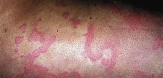
Superficial, widespread, and confluent plaques were moderately itchy. Fexofenadine (Allegra) 180 mg in the morning and hydroxyzine 25 mg q.h.s. were effective.
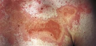
Superficial plaques with clear dusky centers. The borders are annular. Itching was moderate. Fexofenadine (Allegra) 180 mg q.d. was effective.

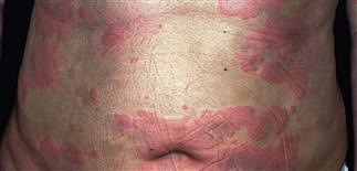
Confluent thick urticarial plaques with a polycyclic pattern. Intense hives may have a blue cyanotic appearance. Itching was intense. Hydroxyzine 25 mg every 4 h was required for control.
DESCRIPTION
By definition, acute urticaria resolves within 6 weeks. Hives lasting longer than 6 weeks are called chronic urticaria. Acute urticaria presents as non-pitting edematous plaques or wheals. Pruritus is intense and lasts up to 24 h. Urticarial vasculitis is painful and purpuric variant, and lesions last longer than 24 h. A biopsy confirms the diagnosis. Drugs and viral infections can be a cause of urticaria, as can foods, injected medications or vaccines, bites or stings.
HISTORY
• People of all ages are affected. Hives are more common in atopic individuals. • The onset is abrupt, often with intense itching. • Drugs, foods, animal hair, and airborne allergens are common causes. The etiology is not discovered in many cases. • Laboratory studies are ordered only to confirm abnormal findings on history and physical examination. Physical urticaria can be caused by vibration, cold, pressure, and sunlight.
PHYSICAL FINDINGS
• Pink-to-red, or flesh-colored non-pitting, edematous plaques may appear on any body surface. The center of the lesions may be flesh-colored, pink, or cyanotic. The borders are surrounded by a white or red halo. The size is highly variable. Lesions are round, oval, annular, or polycyclic. • The plaques change size and shape by peripheral extension and regression and may also migrate. • New lesions appear as others resolve. • Bullae or purpuric lesions appear with intense swelling. • The distribution is usually generalized in the case of food, drug or viral etiology but may be limited to the area of contact with some exposures such as natural rubber latex, animal dander, pollen, or during preparation or handling of foods. • Linear lesions suggest dermographism (physical urticaria).
TREATMENT
• Stop suspected triggers (e.g. medications, food, inhalants). • H1 blocker antihistamines are used initially. Hydroxyzine 10–25 mg every 4–6 h is the most effective but causes sedation. Liquid hydroxyzine 10 mg/5 cc works faster. Doses as high as 100 mg every 4 h may be used. Many patients adapt to the sedation after a few days of treatment. Non-sedating H1 blockers are useful for the daytime hours. • Loratadine (Claritin) 10 mg q.d. is non-prescription. • Desloratadine (Clarinex) 5 mg q.d., cetirizine (Zyrtec) 5 mg or 10 mg q.d., and fexofenadine (Allegra) 60 mg b.i.d. or 180 mg q.d. are other treatment options. • Prednisone (e.g. 60 mg for 2 days, 40 mg for 5 days, and then 20 mg for 7 days) may be effective and is used when patients are not responding to antihistamines. • Adrenaline (epinephrine) is administered for extensive, severe cases. • Cool baths with colloidal oatmeal (Aveeno Soothing Bath) relieve itching. Hot showers provide immediate relief but should be avoided, as they only worsen the pruritus afterward. • Topical steroids are minimally effective in most cases.


