[level-membership-for-opthalmology-category]18.3
Acute Syphilitic Posterior Placoid Chorioretinitis
OCT Features:
Much like the disease itself, the OCT findings can vary. Focal loss of the external limiting membrane and IS–OS/ellipsoid zone are common, the retinal pigment epithelium layer may be abnormal, and choroidal infiltration with thickening may also be visible (Fig. 18.3.1). Serous retinal detachments involving the macula are uncommon, occurring in about 10% of cases.
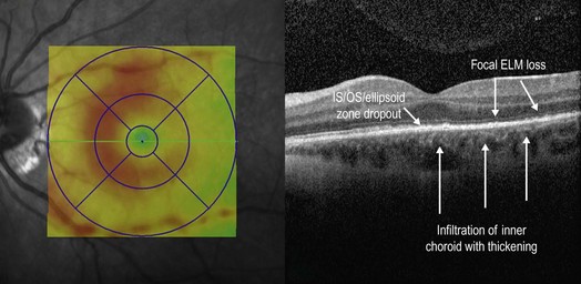
Figure 18.3.1 OCT in acute syphilitic posterior placoid chorioretinitis shows involvement of the IS–OS layer. The IS–OS/ellipsoid zone is abnormally indistinct (between black arrowheads) and then drops out completely (to right of black arrowheads). The retinal pigment epithelium layer in the region of IS–OS/ellipsoid zone dropout is irregular. There is also focal loss of the external limiting membrane and infiltration of the inner choroid with thickening. The accompanying thickness map shows abnormal thickening of the parafoveal region, particularly nasally. (Courtesy of Robin A. Vora MD.)
[/level-membership-for-opthalmology-category][not-level-membership-for-opthalmology-category]18.3
Acute Syphilitic Posterior Placoid Chorioretinitis
[/not-level-membership-for-opthalmology-category]
