Acute Retinal Necrosis Syndrome
Clinical Features:
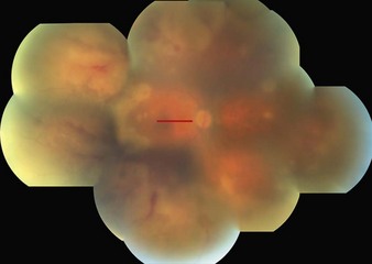
ARN commonly presents with multifocal peripheral areas of full-thickness retinal necrosis in well-circumscribed patches that rapidly coalesce in a circumferential pattern (Fig. 18.5.1). There is an associated brisk intraocular inflammatory reaction and an occlusive vasculitis that primarily affects the retinal arteries. Vitritis is universal and a mild anterior chamber reaction with keratic precipitates is typical. Elevated intraocular pressure is not unusual. Without treatment, spread is rapid and may also involve the fellow eye.
OCT Features:
In cases where the macula is not directly involved clinically, but is threatened, OCT shows subclinical disease involvement (Fig. 18.5.2), which can be useful for prognostic purposes. Disease activity beyond the clinically evident area of involvement (or leading edge) is typical of this condition. In the setting of frank necrosis involving the macula, OCT reveals attenuation of all retinal layers.
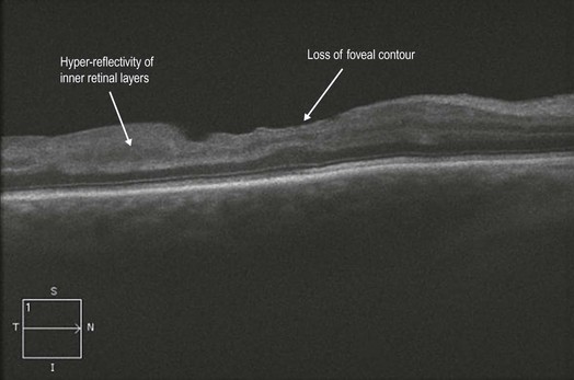
Figure 18.5.2 OCT (corresponding to Figure 18.5.1) shows significant, subclinical, disease activity that is evident within the macula. Abnormal hyper-reflectivity within the inner retinal layers is a sign of active disease. There is also loss of the normal foveal contour.


