Acute respiratory failure in a 68-year-old man

Published on 10/04/2015 by admin

Filed under Surgery

Last modified 22/04/2025

Print this page

rate 1 star rate 2 star rate 3 star rate 4 star rate 5 star
Your rating: none, Average: 0 (0 votes)

This article have been viewed 992 times

Problem 53 Acute respiratory failure in a 68-year-old man

The patient’s breathing is so laboured that he is unable to answer any of your questions.

The man’s wife tells you that he has ‘pretty bad’ emphysema. He has no known history of cardiovascular disease.

His medications are:

He has smoked heavily for most of his life, but has reduced his current cigarette consumption to about five a day. He does not drink and has no known allergies.

The ambulance officer states that haemoglobin oxygen saturation (measured by pulse oximetry, SpO2) has been around 80% throughout the journey, and that he has been wheezing loudly. He has been given 5 mg nebulized salbutamol in the ambulance.

As you examine the patient, you observe that on 6 L/min of oxygen by face mask, his SpO2 is 78%. He seems irritable, obviously dyspnoeic, and grasping the sleeve of your designer shirt, he utters ‘help …’.

You persist with the examination. His respiratory rate is 30 breaths per minute with a prolonged expiratory phase. The pulse rate is 110 bpm and regular. His temperature is 36.5°C and the blood pressure 110/75 mmHg. He is well hydrated and peripheral perfusion is adequate. His peripheral pulses are all present and of good volume. In the semi-recumbent position his jugular venous pulsation (JVP) is 3–4 cm above the base of the neck. Both heart sounds appear normal and there are no murmurs. His trachea is in the midline. He is barrel-chested and there are reduced breath sounds throughout. There is a diffuse expiratory wheeze and the percussion note is normal, there are no crepitations. His abdomen is soft and non-tender. His calves are normal and there is no pedal or sacral oedema.

An electrocardiogram is performed which shows a sinus tachycardia. His arterial blood gas (ABG) results are as follows:

Investigation 53.1 Arterial blood gas analysis

PaO2 48 mmHg
PaCO2 70 mmHg
pH 7.24
Bicarbonate 29 mmol/L

You adjust his oxygen therapy carefully. He is too dyspnoeic for a peak expiratory flow rate (PEFR) measure. You order an urgent mobile chest X-ray and go on to administer emergency medications to treat this man’s condition.

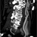
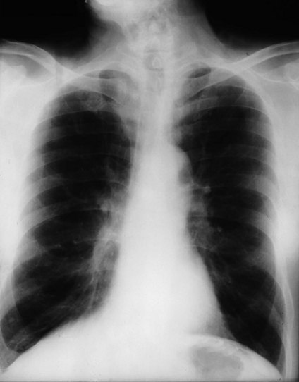
A chest X-ray is performed and is shown in Figure 53.1.

The patient starts to improve, but he is still struggling. You call the intensive care team, who admit him to the intensive care unit (ICU). He receives close monitoring of his gas exchange. He is placed on BPAP (bi-level positive airway pressure ventilation by non-invasive face mask) to support his ventilation. A radial arterial line is inserted to monitor blood gases.

The patient responds almost instantaneously with BPAP, reporting a marked reduction in dyspnoea. Oxygen therapy is carefully monitored and adjusted in the ICU. Bronchodilators are continued and he is commenced on a course of oral prednisolone beginning with 50 mg daily. His blood gases stabilize with improvement in his respiratory condition, achieving almost his baseline level of function over several days with PaO2 65 mmHg on 1 L/minute via nasal cannulae, PaCO2 53 mmHg, pH 7.38, bicarbonate 28 mmol/L.

He is discharged after 7 days in hospital to complete a 10-day course of prednisolone (dose to be tapered according to clinical progress), a programme for respiratory rehabilitation and to discontinue smoking, and follow-up appointments in the respiratory medicine clinic with pulmonary function testing.

Answers

A.1 In the absence of an obvious external cause (e.g. chest trauma, upper airways obstruction), the vast majority of cases of acute shortness of breath in adults will be due to one of four conditions:

Spontaneous pneumothorax, while much less common than the conditions above, is also an important diagnosis to consider.

Anaemia and metabolic acidosis may also cause tachypnoea and, along with pulmonary embolism, should be sought particularly in patients with normal-appearing plain chest radiograph.

A.2 You will need to have:

A.3 The most important immediate investigations are:

All are quick to perform, and can be done while the patient is being examined and treated.

ABGs provide results within minutes, can be done in many emergency departments and intensive care units, and provide information on blood gases, acid–base status, most vital electrolytes (including sodium, potassium, chloride, bicarbonate, ionized calcium), blood glucose, and haemoglobin. All of these parameters are important in the assessment of any acutely unwell patient and can immediately influence the course of treatment.

ABGs performed on room air are not necessary, and should not be performed in patients requiring supplemental oxygen to maintain adequate SpO2.

Complete blood picture, full electrolyte panel and cardiac enzymes should also be requested, but results may take some time to become available.

A.4 The ABGs show severe hypoxaemia. His acid–base status cannot be fully determined with the information available; however, the ABG result is consistent with acute on chronic respiratory acidosis.

The presence of a low arterial blood pH indicates the PaCO2 is acutely raised. As a rough guide, for every acute increase in PaCO2 of 10 mmHg, pH falls by approximately 0.07, and bicarbonate increases by about 1 mmol/L. In ‘chronic’ hypoventilation, compensatory mechanisms can bring the pH almost back to normal (takes about 2–3 days), with bicarbonate increasing by approximately 3 mmol/L for every 10 mmHg increase in PaCO2.

Working backwards accordingly, it can be estimated that this man’s baseline ‘chronic’ PaCO2 would be around 50 mmHg, and bicarbonate around 27 mmol/L. In addition to acute respiratory acidosis, he also has chronic respiratory acidosis with metabolic compensation.

Insufficient results have been provided for calculation of his anion gap; however, this is always important to calculate in order to detect a concurrent wide anion-gap metabolic acidosis (e.g. from ingested toxins, lactataemia from ischaemic bowel, alcoholic/starvation/diabetic ketoacidosis, etc.).

A.5 A major error in this patient would be to reduce the supplemental oxygen therapy. Patients do not lose their ‘hypoxic drive’ (if this phenomenon actually exists) unless they have lost their hypoxia! His PaO2 is 48, i.e. he is definitely still hypoxaemic, and this is contributing to his agitation and confusion.

A.6

A.7 The chest X-ray shows flattening of the diaphragms consistent with ‘gas trapping’, which reflects the inability of gas to escape from the lungs due to airways obstruction. The lung fields are clear, with no evidence of alveolar consolidation or oedema.

This man is having an exacerbation of his COPD.

He is starting to succumb to respiratory fatigue, and hence hypoventilate. The work of breathing is becoming too much for him. This is reflected by his climbing PaCO2, as the carbon dioxide in his alveoli is not being ‘washed out’ due to inadequate ventilation.

Those with low tidal volumes may not have prominent crepitations or wheeze, due to the low volumes of gas reaching the alveoli and low air flows. Thus, the absence of crepitations does not exclude cardiogenic pulmonary oedema, and a lack of wheeze does not exclude airways disease.

Chest tightness is a symptom commonly described by patients with bronchoconstriction as well as patients with cardiac angina.

For these reasons, acute pulmonary oedema (which may have been precipitated by cardiac ischaemia or infarction) was an important differential diagnosis in this man’s presentation, and thus the emergent importance of a CXR and ECG.

A.8

Revision Points

In acute respiratory failure:

Fatigue from sustained and severely elevated work of breathing is a common cause for rising PaCO2 in patients with acute severe exacerbation of COPD; however, a loss of ‘hypoxic drive’ (hypoxia replacing hypercapnia as a stimulus to breath) is often blamed, resulting in an inappropriate reduction in supplemental oxygen even in the presence of hypoxaemia. Nonetheless, oxygen supplementation in patients with chronic hypercapnoeic respiratory failure can result in an increase in hypercapnia. The Haldane effect (haemoglobin binding to oxygen as hypoxaemia is treated results in displacement of CO2 bound to haemoglobin, thus releasing CO2 into plasma) and V/Q (ventilation–perfusion) mismatch due to oxygen induced reduction in pulmonary vasoconstriction are important causes of hypercapnia which have been found in these patients. The significance of ‘hypoxic drive’ in COPD exacerbations is yet to be conclusively demonstrated. The most important cause from a clinical standpoint remains the inability to sustain such a high work of breathing.

Further Information

www http://www.goldcopd.org. Global strategy for the diagnosis, management and prevention of COPD, Global Initiative for Chronic Obstructive Lung Disease (GOLD) 2010

www http://www.thoracic.org/clinical/copd-guidelines/resources/copddoc.pdf. American Thoracic Society/European Respiratory Society Task Force. Standards for the diagnosis and management of patients with COPD 2004