Acute neonatal emergencies

Published on 23/06/2015 by admin

Filed under Emergency Medicine

Last modified 22/04/2025

Print this page

rate 1 star rate 2 star rate 3 star rate 4 star rate 5 star
Your rating: none, Average: 0 (0 votes)

This article have been viewed 1828 times

26.1 Acute neonatal emergencies

Essentials

Neonatal emergencies

Studies have indicated that shorter postpartum hospital stays have resulted in an increased attendance at the emergency department (ED) of newborn infants and those in the first month of life. The common presenting symptoms and signs were jaundice, poor feeding, breathing difficulties and irritability.

The underlying common pathologies were found to be physiological jaundice, feeding problems and suspected sepsis.

Maternal experience, social support, early postnatal discharge and perinatal instruction influenced presentation to the ED.

The infant with breathing difficulty

Respiratory emergencies are some of the commonest conditions presenting in the neonatal period. The increased work of breathing is manifested as:

The causes of respiratory distress are varied and are summarised in Table 26.1.1. They can be broadly divided into primary respiratory and non-respiratory causes. Primary respiratory pathology is a direct result of upper, lower or mixed airway pathology.

Table 26.1.1 General causes of respiratory distress

General causes of respiratory distress Specific conditions Respiratory disorders Hyaline membrane disease   Congenital pneumonia   Meconium aspiration syndrome   Transient tachypnoea of the newborn   Pneumothorax   Hydro/haemothorax Upper airway abnormalities Laryngomalacia   Micrognathia   Vocal cord anomalies Cardiac anomalies Heart failure   Myocarditis   Pericardial effusion   Cyanotic congenital heart conditions Structural abnormalities Diaphragmatic hernia   Congenital cystic lesions   Diaphragmatic paralysis Chest deformities Arthrogryposis   Thoracic dystrophy Haematological causes Anaemia CNS lesions Infection Metabolic conditions Metabolic acidosis

This section will discuss:

Upper airway obstruction

Clinical features

Stridor is the classic presenting sign of upper airway obstruction and is a rare phenomenon in the neonatal period. Stridor is an indication of partial obstruction of the large diameter airways, from either an intrinsic developmental defect or from secondary external compression and distortion. Stridor is the inspiratory noise that indicates this partial obstruction and early referral to an ear, nose and throat specialist should be considered. In addition to the stridor, infants often have an associated degree of respiratory distress, but may have a normal or hyper-expanded chest radiograph.

Respiratory distress attributed to lung parenchyma pathology

Clinical features

History

In respiratory distress attributed to parenchymal involvement, a clinical history is essential to elucidate potential predisposing factors. Of the causes of parenchymal disease presenting to the ED, pneumonia and extra-pulmonary cardiac failure are the two most common causes. These can often be differentiated by clinical history.

Bacterial pneumonia in the first few hours of life may be impossible to distinguish from respiratory distress syndrome or transient tachypnoea of the newborn. Therefore, respiratory distress in newborns generally should be treated as bacterial pneumonia until proven otherwise. When associated with chorioamnionitis, it is caused most commonly by Group B streptococci (GBS) or by Escherichia coli. However, Haemophilus influenzae, Streptococcus pneumoniae (pneumococcus), Group D streptococci, Listeria and anaerobes have also been described as pathogens in this setting. Infants infected with these organisms are often preterm and have very early onset of respiratory distress. Of note, infants may also develop bacterial pneumonia transnatally in the absence of maternal chorioamnionitis. Here, the causative organism is likely to be GBS, and the onset of symptoms tends to occur 12–24 hours after birth.

Neonatal pneumonia can be either congenital or acquired. Congenital pneumonia commences before birth and the most common infecting organisms include Group B Streptococcus and E. coli. Despite the majority of infants being unwell at birth, some infants do acquire these infections after birth and present with similar signs and symptoms of respiratory distress, poor feeding, fever and apnoea. The clinical history should focus on the maternal Group B Streptococcus carriage in pregnancy, length of rupture of membranes, maternal antibiotic therapy during labour and maternal fever. Acquired neonatal pneumonias are commonly viral, with respiratory syncytial virus, adenovirus and parainfluenza virus all commonly identified. A history of affected family members gives some indication of this potential.

Any infant with underlying lung pathology is at risk of air leak, and any sudden decompensation in an infant with respiratory distress should lead one to consider this diagnosis. In addition, air leak can be a spontaneous phenomenon with no identified cause.

Non-infectious causes of acquired respiratory distress include any conditions in which there is an abnormally high or low blood flow to the lungs, an increased demand for oxygen, or a decreased number of red blood cells.

The commonest non-pulmonary cause of respiratory distress seen in the ED is that of pulmonary oedema, secondary to congenital heart disease.

Congenital heart disease is one of the commonest malformations, with an incidence of 0.6%. Although 30–60% of congenital heart disease is identified antenatally, this still leaves a large percentage presenting in the postnatal period. Predischarge saturation monitoring of all newborn infants has the possibility of increasing the early identification of congenital heart disease, prior to the onset of respiratory distress, cyanosis or collapse. Cyanotic lesions generally present early but neonates with ductal-dependent systemic circulations are often well in the early neonatal period and collapse around day 4 of life. Closure of the ductus, with associated systemic collapse, is one of the commonest presentations to the ED with severe respiratory distress, secondary to associated pulmonary oedema.

Factors in the history pointing towards congenital heart disease being the cause of collapse include a family history, syndromic malformations and associated abnormalities.

In addition to predisposing factors, a history of poor feeding often predates the collapse.

Other causes of respiratory distress presenting in the ED are:

Examination

Infants with disease affecting the lung parenchyma, either primary respiratory or cardiac, generally present with the classical examination findings of respiratory distress, notably: recession of the intercostal and subcostal spaces; nasal flaring; tachypnoea (>60 bpm); and expiratory grunting.

Nasal flaring is a result of the alae nasi being the first muscles to be activated during inspiration and they aim to decrease airway resistance. The recession of the inter- and subcostal spaces is a result of the compliance of the chest wall being reduced in neonates. During inspiration the pleural pressure is reduced, but in neonates with parenchymal lung disease this needs to be reduced more than normal and thus the consequences are that the compliant chest wall may cave in as a result of these more negative pressures. The recession in conjunction with the abdominal protuberance associated with diaphragmatic descent give the characteristic seesaw pattern of neonatal respiratory distress.

Infants with respiratory infections may present in a very similar manner to those with cardiac anomalies. Specific findings for infants with pneumonia may be the presence of fever or temperature instability, feed intolerance and rhinorrhoea.

If there is a suggestion of pulmonary air leak, the clinical signs are specific. There will be reduced air entry on the side of the leak, with a reduction in chest movement on that side. Percussion, although seldom used in newborn infants, should be hyper-resonant and if the air leak is under tension there may be associated displacement of the trachea and apex beat to the contralateral side.

If a primary respiratory cause cannot be identified, non-respiratory causes should be sought. Signs indicative of congenital heart disease may include weak femoral pulses, an active praecordial impulse, hepatomegaly and a cardiac murmur.

The classic cardiac lesions presenting with respiratory distress in the neonatal period include:

With other causes of respiratory distress there may be features consistent with specific diagnoses. Metabolic conditions are often associated with hepatosplenomegaly, coma, hypoglycaemia and jaundice. Central nervous system (CNS) lesions may have seizures associated.

Investigations

Any infant in the first month of life with respiratory distress should be observed and monitored closely. This includes pulse rate, oxygen saturation, respiratory rate, temperature, blood pressure and capillary refill time.

The normal heart rate for a neonate in the first month of life is 120–160 bpm. Some newborn infants, however, have a resting heart rate below 90 bpm. Respiratory distress is generally associated with respiratory rate greater than 60 breaths per minute. Fever as a sign of sepsis is variable.

Plain radiographs of the chest are useful, but do not generally differentiate the various causes of respiratory distress. Sepsis and cardiac failure both demonstrate increased interstitial markings. In cardiac failure, fluid more specifically radiates into the interstitium from the hilum and in severe cases may be associated with an effusion. Again, in cardiac disease the size of the cardiac shadow may be increased. A cardiac silhouette greater than 60% of the transthoracic diameter is indicative of cardiac disease and needs to be investigated further, by means of an electrocardiogram and echocardiogram, and referral to a cardiologist.

The chest radiograph of an infant with pneumonia may show lobar or diffuse interstitial changes. Chest radiographs of infants who have bacterial pneumonia may exhibit a diffuse reticular nodular appearance but, in contrast to respiratory distress syndrome, they tend to show normal or increased lung volumes with possible focal or coarse densities. There may also be pleural effusions, particularly with GBS pneumonia. In the newborn who has bacterial pneumonia, blood cultures obtained before the initiation of antibiotics commonly grow the offending organism. Cultures of urine and cerebrospinal fluid should be obtained at the time of the blood culture if a newborn infant is systemically unwell. If the diagnosis is viral, mucus plugging, with over-aeration and hyper-expansion may be characteristic. Although tension pneumothorax should be a clinical diagnosis, the plain radiograph is good at demonstrating small pneumothoraces. An anterior pneumothorax may be subtle and easily missed by the unwary as appearing more lucent on the side of the pneumothorax in the absence of a lateral air meniscus.

Laboratory investigations, including full blood count, C-reactive protein (CRP) and blood cultures, are useful adjuncts to the diagnosis. The white cell count may show a leucocytosis with associated left shift or, more commonly, may show a consumptive picture with neutropenia and associated thrombocytopenia in the septic infant. The CRP is another non-specific marker of infection, but appears more useful in monitoring response to treatment of infection rather than in its diagnosis.

If a viral respiratory tract infection is suspected then a nasopharyngeal sample viewed with electron microscopy for respiratory viruses may reveal the common causes of bronchiolitis.

Arterial blood gas analysis or indirect transcutaneous monitoring may reveal arterial hypoxaemia and hypercarbia. The degree of hypoxaemia and acidosis will be a guide to the need for respiratory positive pressure support. In addition to diagnosing the severity of the respiratory acidosis, an arterial blood gas may also reveal a metabolic acidosis, making inborn errors of metabolism a potential differential diagnosis of the respiratory distress.

Management

A neonate with respiratory distress needs to be observed closely. Evaluation of airway, breathing and circulation are imperative, as outlined in the resuscitation guidelines devised by the International Liaison Committee on Resuscitation (ILCOR). Maintaining a neutral airway position and ensuring the airway is free of obstruction allows optimal oxygen to be delivered. Saturations should be maintained with either nasal cannulae oxygen, head box oxygen, continuous positive airways pressure or endotracheal intubation and ventilation. If working in a hospital with a Neonatal Intensive Care Unit or Paediatric Intensive Care Unit, support from personnel working in these areas can be invaluable in maintaining a patent airway.

If sepsis is suspected then intravenous antibiotics should be commenced, to cover both Gram-positive and Gram-negative bacteria and an evaluation made of the need for both fluid and inotropic support. Empiric treatment should be initiated as soon as possible with ampicillin 100 mg kg–1 day–1 divided every 12 hours (infants <1.2 kg) or every 8 hours (infants >1.2 kg) and cefotaxime 100 mg kg–1 day–1 divided every 12 hours or 150 mg kg–1 day–1 divided every 8 hours (infants >1.2 kg and >7 days old). Gentamicin is an alternative treatment, particularly when there is no evidence of meningitis. Treatment should be continued for at least 10 days if sepsis is present, although 14–21 days may be required, particularly for Gram-negative infections.

Viral infections are treated conservatively by respiratory and circulatory support as required. An unusual or unresponsive neonatal presentation of pneumonia warrants further evaluation. The maternal history may offer important clues. Neonatal pneumonia involving cytomegalovirus (CMV) or other viruses may be transmitted transplacentally. CMV pneumonia may not require treatment in the otherwise healthy infant. However, neonatal respiratory distress in the setting of perinatal exposure to herpes simplex virus, particularly if there is primary maternal genital infection, warrants treatment with aciclovir 30 mg kg–1 day–1 divided every 8 hours for 14–21 days until all cultures are negative. Ureaplasma urealyticum is another important organism and treatment of Ureaplasma infections in the newborn should include erythromycin 50 mg kg–1 day–1 divided every 6 hours.

If the cause of the respiratory distress is believed to be cardiac then once again supporting the airway, breathing and circulation is imperative. Added caution with fluid resuscitation should be considered so as not to exacerbate the cardiac failure. Use of prostaglandin E1 allows reopening of the ductus and increased systemic circulation. This is a temporising measure prior to the definitive surgery the infant may require.

Management of a pneumothorax requires either acute drainage with needle thoracocentesis, in the presence of a tension pneumothorax, or intercostal catheter insertion followed by appropriate underwater drainage. A repeat chest X-ray (CXR) to ensure adequate lung expansion is required prior to the removal of the chest drain.

The blue infant

Neonatal cyanosis is a result of deoxygenated blood in the systemic circulation. It is defined as an arterial saturation less than 90%.

History, examination and simple investigations available in the ED should be able to distinguish the cause of the cyanosis and this will predict the management of this condition.

Clinical features

History

Once cyanosis has been diagnosed in an infant presenting to the ED, the most important step is to differentiate between pulmonary and cardiac causes of cyanosis.

Pulmonary causes of cyanosis include pneumonia, both bacterial and viral, pneumothorax, pleural effusions and airway anomalies. If the cause of the cyanosis is felt to be pulmonary, the infant will generally have a history of worsening respiratory distress that interferes with the infant’s ability to feed successfully. The history may be indicative of an infective cause, with rhinorrhoea, fever, cough, poor feeding and worsening recession of the inter- and subcostal spaces. In addition, there may be a history of other affected family members and respiratory infections are generally more prominent in the winter months especially associated with epidemics of bronchiolitis.

Infants with cardiac causes of cyanosis may have no preceding history and generally breathe normally. A detailed antenatal history, including family history, genetic abnormalities and the results of antenatal ultrasound scans, will be important. In addition, the timing of the cyanosis may also give a clue to the diagnosis, with duct-dependent cardiac lesions generally worsening when the ductus arteriosus shuts around day 3 or 4 of life. Although congenital heart disease is generally divided into cyanotic and acyanotic, those lesions generally classified as acyanotic but duct dependent can present with severe respiratory distress and a degree of cyanosis.

In addition to congenital heart disease and pulmonary causes, an infant may present cyanosed because they have neurological depression or seizures. A history of the infant’s general activity, tone and feeding patterns will be helpful. Infants with neurological depression may be hypotonic, have abnormal autonomic responses and have poor feeding with associated failure to thrive and may indeed have seizure activity.

Once again, if seizures are suspected then a detailed family history may indicate a genetic syndrome as the cause of the seizures. A thorough history of the pregnancy and delivery is also important to rule out other causes of seizures, and early checking for hypoglycaemia is essential. Considering neonatal sepsis is always imperative and a detailed history of maternal substance abuse may indicate drug withdrawal as the cause of the seizures.

Examination

Once a detailed history has been taken to elicit possible causes of cyanosis then the infant should be examined carefully.

Infants with respiratory causes for cyanosis will generally have signs of distress, namely tachypnoea, recession of the intercostal and subcostal spaces, tracheal tug, nasal flaring and expiratory grunting. As to the precise respiratory cause of this distress, septic infants often have an associated tachycardia and may have temperature instability. They may have poor capillary return and may also have associated apnoeas, a pause in breathing of greater than 20 seconds or a pause in breathing less than 20 seconds but associated with bradycardia. Infants who are septic and cyanosed may have localised respiratory infections, such as viral bronchiolitis, or may be septicaemic.

A cyanosed infant with little or no respiratory distress is more likely to have congenital heart disease. The commonest lesions presenting with cyanosis in the neonatal period are: transposition of the great arteries; total anomalous pulmonary venous return; pulmonary atresia with an intact ventricular septum; severe pulmonary stenosis; and severe tetralogy of Fallot.

Examination of a cyanosed infant with suspected cardiac disease might reveal dysmorphic features suggesting a syndromic association with congenital heart disease. Auscultation of the lung fields and praecordium may reveal evidence of pulmonary oedema and murmurs, and an abdominal examination may reveal hepatomegaly.

To differentiate an infant with non-cardiopulmonary causes for the cyanosis, the examination would need to concentrate on the neurological system of the infant. Tone, movements and reflexes will all give important information as to the neurological status. Examination for dysmorphic features and examination of the fundi and skin may add important information as to the cause of the seizures. In the neonatal period, important causes of seizures would include hypoxia, hypoglycaemia and biochemical anomalies, narcotic withdrawal and structural brain abnormalities.

Investigations

To differentiate the various causes of cyanosis in the neonatal period a full set of observations should be performed. Oxygen saturations should be performed initially in room air to serve as a baseline. Subsequent oxygen measures should be performed in 100% oxygen, achieved by means of a headbox sealed over the baby’s head and neck. The so-called hyperoxia test may help to differentiate cyanotic heart disease from other causes of neonatal cyanosis. Infants with neurological or pulmonary causes of the cyanosis will demonstrate substantial increases in the arterial blood saturation, while infants with cyanotic congenital heart disease will show minimal elevation. As well as oxygen saturation changes, the difference should be confirmed with arterial blood gas analysis of arterial oxygen partial pressures. A partial pressure <100 mmHg in 100% inspired oxygen is more indicative of cardiac disease.

Once arterial oxygen saturations and blood gas analysis have been performed the infant should have a chest radiograph. Abnormalities of the lung fields may suggest a primary pulmonary cause for the cyanosis or a cardiac cause if the changes are suggestive of pulmonary oedema with increased vascular markings.

Assessing the cardiothoracic diameter and shape of the cardiac shadow may give some clues to a cardiac cause of the cyanosis. A typical boot-shaped heart is classically described in tetralogy of Fallot and an egg on a string appearance is described in transposition of the great arteries. Generally differentiating the cardiac causes of cyanosis is difficult and requires an echocardiogram. Additional investigations that may be useful in the diagnosis of neonatal cyanosis include a full blood count to reveal polycythaemia and a raised or depressed white blood cell count, blood cultures and a nasopharyngeal aspirate.

In addition to blood gas analysis to indicate fixed or variable hypoxia, the carbon dioxide will generally be raised in respiratory pathologies contributing to the cyanosis but may be normal in cardiac disease.

An electrocardiogram (ECG) may help differentiate cardiac disease, but the gold standard investigation to help differentiate cardiac causes of cyanosis from non-cardiac causes will be an echocardiogram combined with colour Doppler flow mapping and pulsed-wave Doppler studies. The echocardiogram should be performed as a matter of urgency in such situations to allow appropriate management of congenital heart disease. Echocardiography can be easily performed by a paediatric cardiologist in the ED or the infant may need to be transferred to a specialist centre to achieve such an investigation.

If a neurological cause is suspected for the cyanosis, urgent investigation including blood sugar level monitoring is imperative. Additional biochemical investigations may include calcium, magnesium and sodium as well as specific metabolic investigations, including a urine metabolic screen and newborn screening test if inborn errors of metabolism are suspected to be causing neurological depression. If structural lesions or intra-cerebral haemorrhage is suspected, ultrasound is an easy bedside test to perform, but magnetic resonance imaging is the gold standard for CNS investigation. Additional investigations may include electroencephalogram (EEG) and urine/meconium drug screening.

Management

Any neonate that presents with cyanosis should be appropriately resuscitated as outlined in the guidelines devised by ILCOR.

Once the presence of cyanosis is determined from the physical examination of the infant, its degree of severity should be documented immediately by oximetry and confirmed by arterial blood gas and pH determinations. Arterial blood gases and pH should be determined with the patient breathing room air (if clinically stable) and following the breathing of 100% oxygen for 5–10 minutes. If the oxygen tension rises to exceed 150 mmHg, cyanotic congenital heart disease is unlikely, although little or no change in oxygen tension strongly suggests that such a cardiac defect is the cause of the cyanosis. The gold standard for immediately ruling out non-cardiac causes of cyanosis and establishing the diagnosis of cyanotic congenital heart disease remains echocardiography. The cyanosis that results from pulmonary disease usually resolves partially, if not completely, following the administration of oxygen. Such treatment should not produce similar results in patients who have non-respiratory cyanosis. Here the cause must be determined to achieve a successful outcome.

All neonates should initially receive oxygen until a definitive diagnosis has been made. If the diagnosis is pulmonary then the addition of oxygen should resolve the cyanosis. If the respiratory distress is severe, additional support by means of positive end expiratory pressure or positive pressure ventilation may be required. If an infant requires intubation then a person skilled in this procedure should be asked to attend the ED. Following institution of airway support the infant requires intensive-care admission. Once the airway and breathing have been supported then appropriate assessment and management of the circulation is essential. In any infant with a pulmonary cause for the cyanosis, the potential for sepsis should be evaluated and treated with intravenous antibiotics.

The laboratory evaluation of most neonates who are cyanosed includes a haematocrit and haemoglobin determination, white blood cell count, differential count, blood glucose determination, and a chest radiograph. If a cardiac aetiology is likely, echocardiography is essential. In selected cases, cardiac catheterisation and angiocardiography may be necessary to define the cardiac anatomy more precisely. Electrocardiography should be performed if clinical findings suggest a tachyarrhythmia. If a CNS aetiology is suspected, appropriate scans and drug levels should be considered. The presence of methaemoglobin may be detected by placing a few drops of the patient’s blood on filter paper and comparing it with normal blood. Methaemoglobin will produce a chocolate-brown colour.

Seizures that result in cyanosis should be managed according to the results of initial investigations. Hypoglycaemia should be managed with a 5 mL kg–1 bolus of 10% dextrose, followed by an infusion of dextrose. Hypocalcaemia should be managed with intravenous calcium and other electrolyte imbalances should be corrected before trying to control the seizure. Once treatable causes of neonatal seizures have been identified and treated then appropriate anticonvulsant therapy should be administered. Phenobarbital or phenytoin are first-line anticonvulsants used in the neonatal period.

In any infant with seizures in the first month of life, sepsis should be thoroughly investigated with both blood culture and cerebrospinal fluid (CSF) cultures for both bacteria and viruses. Treatment with antibiotics and antiviral medications should be considered if there are any risk factors, symptoms or signs of sepsis.

Of all of the above aetiologies, the most common cause of cyanosis in the neonate is a cyanotic congenital heart defect. In many such infants, pulmonary blood flow depends primarily or entirely upon the patency of the ductus arteriosus. If congenital heart disease is suspected from the hyperoxia test, chest radiograph, ECG or echocardiogram then referral should be made to a paediatric cardiologist. A discussion should take place as to whether prostaglandin E1 should be commenced to either open or maintain the patency of the ductus arteriosus and the appropriate dosage should be discussed with the accepting paediatric cardiologist. This will allow mixing of deoxygenated and oxygenated blood, increasing the oxygen saturations of the infant. A dose of 0.05–0.1 mcg kg–1 min–1 IV is generally recommended, remembering that the most serious side effects of this drug are hypoventilation and apnoea.

Once a diagnosis of congenital heart disease has been made then ongoing management by a paediatric cardiologist is essential. Transfer to a paediatric cardiology centre should be organised and the infant should be fully monitored prior to transfer, including pre- and post-ductal saturations, respiratory rate, temperature, pulse, ECG monitoring and blood pressure. Transport should be provided by a team expert in neonatal resuscitation.

The infant with possible seizures

Although the incidence of seizures is higher in the first 4 weeks of life than in any other age group, the actual incidence cannot be delineated because of the large number of subtle presentations. The seizures that do present to the ED generally do so in the early neonatal period (days 1–7) and it is essential to recognise these for two reasons:

Seizures in the neonatal period are a result of an excessive depolarisation of neurones from many different causes. Disturbances of electrolytes (sodium, calcium, and magnesium) and an imbalance of excitatory and inhibitory amino acids have been identified as predisposing to neonatal seizure activity. It is important to identify the cause of the seizures, as many of them are easily treatable, but if missed there may be major long-term consequences.

Clinical features

History

The clinical history will provide essential clues to the cause of the seizures. A thorough history of the pregnancy, labour and delivery should be established as well as a detailed family history of seizures.

A perinatal history of maternal fever, prolonged rupture of membranes, low vaginal swab positive for Group B Streptococcus and associated fetal distress may be clues to an infective cause of the seizures. In addition, a history of poor Apgar scores and poor cord blood gas results, and the need for resuscitation at birth may indicate a perinatal asphyxial event.

If a perinatal cause for the seizures is not identified in the history then a postpartum cause needs to be considered. A history of maternal substance abuse may indicate seizures secondary to neonatal abstinence syndrome and a family history of seizures is suggestive of a genetic cause for the seizures.

Poor understanding of the nutritional requirements of the newborn and excessive weight loss may indicate an electrolyte imbalance as the cause of the seizures. Hypoglycaemia and hypocalcaemia may both present with a preceding history of jitteriness.

In addition to the history of the actual cause of the seizures it is also important to get a realistic picture as to the nature of the seizures. Although most neonatal seizures are partial, they can be generalised, and there are many other presenting signs of neonatal seizure activity including: jitteriness; cyanosis; apnoea; tachycardia; lethargy; and collapse.

Examination

Any infant that has presented with seizures or potential seizures needs a thorough examination. The anterior fontanelle should be soft and non-bulging in the neonatal period. A tense fontanelle is suggestive of raised intracranial pressure. This may result from an intracranial bleed or excessive swelling secondary to neonatal meningitis. If a bleed is suspected, this may be idiopathic, secondary to a bleeding tendency or a result of non-accidental injury. If a bleeding tendency is suspected other bruising or other bleeding diatheses may be identified and the stool and urine should both be examined for the presence of blood. The head should be measured and the head circumference should be plotted on a centile chart to compare with the centile that the head circumference lay on initially. This will indicate if there is progressive hydrocephalus. Once the fontanelle has been examined the rest of the nervous system should be assessed. Tone and reflexes should be elicited to indicate any CNS involvement. The eyes should be examined for retinal haemorrhages and signs that the intracerebral injury could be deliberate. Severe hydrocephalus associated with seizures may be associated with a downward deviation of the eyes.

Considering the multitude of causes of neonatal seizures there will be many potential physical signs that can be elicited. Signs of infection, either congenital or acquired, may include a petechial rash, respiratory distress and hepatosplenomegaly, or in neonatal meningitis they may be associated with severe septic shock.

It is important to recognise that a lethargic infant may be dehydrated and suffering from the effects of an electrolyte imbalance. In addition, an infant with metabolic derangements may also present shocked, acidotic and with seizures. The metabolic derangements may have a characteristic odour that, if identified, may lead to a rapid diagnosis of the seizures.

Although there are many causes of seizures in the neonatal period, the diagnosis is only made when adequate investigations are performed. A paediatric neurologist may be involved in more intractable forms of seizure.

Investigations

Any neonate presenting to the ED with possible seizures needs prompt investigation. A first line would be to perform a full set of clinical observations, including oxygen saturations, respiratory rate, pulse rate, blood pressure temperature and ECG. Once the airway and breathing have been assessed, then a blood sugar should be performed, and serum taken for calcium, magnesium and sodium levels.

In any infant, sepsis should be high on the list of differential diagnoses and thorough investigation should be performed. This should include full blood count, blood cultures and, importantly, a lumbar puncture. The cerebrospinal fluid should be examined for evidence of both bacterial and viral infections. If viral infection is suspected then polymerase chain reaction should be performed for both enteroviruses and herpes simplex virus. Glucose and protein performed on the CSF also help guide the differential diagnosis. If a congenital infection is possible, either the mother is non-immune or has risk factors for the possible development of rubella, cytomegalovirus or toxoplasmosis in pregnancy, then a TORCH screen should be performed.

In addition to biochemical and infective profiles, a metabolic screen should be performed on fresh urine collected from the infant, and certain inborn errors of metabolism can be eliminated by chasing the newborn screening test performed on all newborn infants between days 3 and 5 of life.

Once basic blood, urine and CSF tests have been collected, a cerebral ultrasound scan should be organised. This allows both sagittal and coronal views of the brain through the anterior and posterior fontanelles. This simple bedside test will demonstrate intracerebral haemorrhage and may demonstrate certain changes typical of congenital infections. If there is ongoing suspicion of intracerebral pathology an MRI or CT scan may be required. At this stage the infant would require admission and ongoing investigation in hospital.

Once a neonate is admitted to hospital with seizures, further investigations may involve an EEG, with video telemetry to differentiate a true seizure from other common diagnoses.

Acute treatment

The most important determinant of prognosis is the underlying aetiology. Thus, infants who have cerebral dysgenesis have uniformly poor outcomes, and approximately 50% of those who have moderate or severe hypoxic–ischaemic encephalopathy develop sequelae. In contrast, infants who have transient metabolic derangements and are treated promptly or who have only subarachnoid haemorrhage usually have a good outcome. Intracranial infection and inborn errors of metabolism are associated with a variable prognosis.

The vomiting infant

True vomiting in babies is best divided into two broad categories: non-bilious and bilious. Bilious vomiting occurs when bile is purged along with the gastric contents. Although some small intestinal reflux into the stomach is common with all vomiting, in non-bilious vomiting, antegrade intestinal flow is preserved, and the majority of the bile drains into the more distal portions of the intestine. If an obstruction is present, non-bilious vomiting implies that the obstruction is proximal to the ampulla of Vater. Conditions leading to bilious vomiting involve either a disorder of motility or physical blockage to this antegrade flow of proximal intestinal contents distal to the ligament of Treitz.

Non-bilious vomiting

Gastro-oesophageal reflux (GOR), although not true vomiting, is frequently included in discussions of vomiting but really only occurs as a result of failed normal oesophageal function. Normally, the lower oesophageal sphincter (LES) relaxes with swallowing and propagation of oesophageal peristalsis, allowing a food bolus to enter the stomach. Its basal contraction prevents food from re-entering the oesophagus from the stomach. Transient relaxation of the LES predisposes to GOR and is the major mechanism in infants who have GOR. The LES is aided by surrounding structures, especially the crural diaphragm, and disruption of these structures, as with a hiatal hernia, contributes to the GOR in some patients. GOR also is distinguished from true vomiting by its symptoms – the emesis of GOR is effortless and generally not associated with retching or autonomic symptoms.

Both inborn errors of metabolism and endocrine disorders can cause vomiting in neonates. The physician should consider glycogen storage disease II (Pompe’s disease), galactosaemia, urea cycle defects, phenylketonuria, Zellweger’s disease, adrenal leukodystrophy and carnitine deficiency syndromes in the sick vomiting neonate. The inborn errors of metabolism generally present in early infancy, and the vomiting is associated with symptoms of lethargy, hypo- or hypertonia, seizures, or coma. The constellation of symptoms is similar to that seen in sepsis, necessitating a high index of suspicion in the evaluation of these patients. The presence or absence of metabolic acidosis, hypoglycaemia, hyperammonaemia, or ketosis and a family history that includes possible consanguinity can help to determine the diagnosis.

Vomiting occurs in any neurological condition that involves increased intracranial pressure (ICP), such as hydrocephalus, congenital malformation, intracranial haemorrhage or mass lesion and infection. Additionally, babies who have seizures, autonomic disorders (Riley–Day syndrome), and conditions affecting the floor of the fourth ventricle without increased ICP frequently have their condition worsened by vomiting.

The anatomic and, thus, the generally surgical causes of non-bilious vomiting are those that affect the intestinal tract proximal to the point of bilious drainage (ampulla of Vater), which is proximal to the ligament of Treitz. These include oesophageal/gastric atresia, duplication/diverticulum/choledochal cyst, pyloric stenosis and web. Any infant who exhibits persistent non-bilious vomiting, with or without feeding, in the immediate newborn period must be suspected of having an intestinal atresia or a luminally obstructing lesion (pyloric stenosis, luminal band, web) proximal to the point of bile drainage An easy and rapid test to evaluate possible oesophageal atresia is the ability to pass a nasogastric tube easily into the stomach. After the tube has been passed, it is important to obtain a radiograph to ensure that the tube is in the stomach and not coiled in an atretic oesophagus. Any resistance to passage of the tube is an indication for evaluation by contrast radiograph for an obstruction. If an obstruction is present, naso-oesophageal tube drainage is important to prevent aspiration of pooled oesophageal secretions. Contrast studies are the standard for the diagnosis of these conditions.

Bilious vomiting

Although not absolute, anatomic conditions causing luminal obstruction distal to the ligament of Treitz usually cause bilious vomiting. Bilious vomiting is an ominous sign that mandates immediate evaluation. Conditions to be considered in the vomiting baby include intestinal atresia and stenosis, malrotation with or without volvulus and intestinal duplication. Also, malrotation with volvulus is a surgical emergency that is diagnosed relatively easily by gastrointestinal contrast study. It is more common in older children.

Management

Any infant who is seriously ill with vomiting requires immediate resuscitation and admission to hospital. In many cases, aggressive fluid management in addition to resuscitation following ILCOR recommendations will need to be implemented without delay.

In the case of the infant with persistent non-bilious vomiting, early consultation with a paediatrician or paediatric surgeon should occur, as in many cases further evaluation may be necessary. Where an underlying metabolic or endocrine disorder was considered in the differential diagnosis, stabilisation should occur as outlined above, with particular attention to any underlying metabolic and electrolyte derangement. Early consultation with a specialist with experience in the care of these rare metabolic conditions should occur as soon as the diagnosis is entertained. Stable infants with vomiting that is not serious and who are otherwise well, such as those with GOR, can be discharged from the ED, provided that suitable follow up and family support have been organised.

For those infants with bilious vomiting, an underlying surgical disorder must be considered until proven otherwise. Early consultation with a paediatric surgeon is mandatory in every case. If the infant is seriously ill, then resuscitation should commence immediately following ILCOR recommendations, with a particular focus on maintaining circulation and fluid status in addition to correcting underlying metabolic and electrolyte derangements. After the diagnosis has been established radiographically, the gastrointestinal tract should be decompressed with a nasogastric tube, the infant kept nil by mouth and supported with intravenous fluids until definitive surgical intervention can be undertaken.

The collapsed infant

Occasionally, a young infant will be brought to the ED because they just don’t look right to the parents. Even inexperienced parents whose first baby is just a few weeks old may notice when their child is unusually sleepy, fussy, or not eating as well as usual. To the physician in the ED, such an infant may appear quite ill, with pallor, cyanosis, or ashen in colour. The infant may be irritable or lethargic, with or without fever. In addition there may be tachypnoea or tachycardia and/or hypotension, and other signs of poor perfusion may also be apparent.

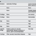
The most common entities to be considered include bacterial infection and viral syndromes. There are a number of other disorders that are uncommon, but demand diagnostic consideration because they are potentially life threatening, yet treatable (Table 26.1.2).

Table 26.1.2 Differential diagnosis of the collapsed infant

An infant who is critically ill in the first month of life should initially be presumed to have sepsis and empiric antibiotics commenced. As Escherichia coli, GBS, Listeria, and other anaerobes are the most likely causative organisms, a combination of ampicillin 200 mg kg–1 day–1 and gentamicin 7 mg kg–1 day–1 in divided doses is a reasonable starting point. In the case of suspected meningitis the addition of cefotaxime 200 mg kg–1 day–1 in divided doses may also be considered. This is a life-threatening situation; the airway, breathing, and circulation should be restored, vascular access obtained and supportive care commenced. The approach to the collapsed infant is presented in Figure 26.1.1.

image

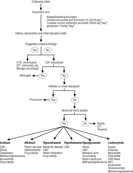
Fig. 26.1.1 Approach to the collapsing infant.

Source: Adapted from Selbst SM 1985 The septic-appearing infant. Paediatr Emerg Care 3: 160–167.

Sepsis

Sepsis should always be considered when confronted with an ill-appearing infant. The signs and symptoms of sepsis may be quite subtle. The history may vary, and some infants may seem to be ill for several days, while others deteriorate rapidly. Any one or a combination of symptoms, such as lethargy, irritability, respiratory distress, diarrhoea, vomiting, anorexia, or fever may be a manifestation of sepsis. Fever is a very unreliable finding in the septic infant as most septic infants will be hypothermic. The septic infant is often pale, ashen, or even cyanotic, with the skin often cool and mottled owing to poor perfusion. The infant may seem lethargic, obtunded, or quite irritable. If there is marked tachycardia (heart rate approaching 200 bpm) together with tachypnoea (respiratory rate >60 breaths min–1), this may herald a rapid collapse. Disseminated intravascular coagulopathy may develop, manifest as scattered petechiae or purpura. If meningitis is present there may be a bulging or tense fontanelle with a high-pitched cry. If the infection has localised elsewhere, there may be otitis media, abdominal rigidity, joint swelling or tenderness in one extremity, or possibly chest findings, such as crackles. The disease may progress rapidly, with the infant developing hypotension and/or frank shock.

A high index of suspicion is needed, as although the laboratory may be helpful in suggesting a diagnosis of sepsis, definitive cultures require time for processing. A complete blood count may reveal a leukocytosis or left shift, although this is often unreliable in infants. A coagulation profile may show evidence of disseminated intravascular coagulopathy, and blood chemistries may reveal hypoglycaemia or metabolic acidosis. Aspiration and Gram stain of urine, joint fluid, and spinal fluid, or pus from the middle ear may reveal the offending organism. Similarly, a chest X-ray may show a lobar infiltrate if pneumonia is present. A Gram stain of a petechial scraping should be considered as this will often reveal the responsible organism.

Cardiac disease

When confronted with an unwell infant, cardiac disease should be considered. An infant with a large septal defect, valvular insufficiency or stenosis, hypoplastic left heart syndrome, or coarctation of the aorta may present with congestive heart failure. The infant may arrive collapsed with clinical findings that are quite similar to those of sepsis. A chronic history of poor growth and poor feeding may suggest heart disease. The presence of a cardiac murmur is very suggestive that a structural lesion needs to be considered. The presence of a gallop rhythm, hepatomegaly, and peripheral oedema should lead to the early consideration of primary cardiac pathology. Crackles, wheezing and intercostal retractions are non-specific findings that commonly present in either heart failure or pneumonia. A CXR is the most useful investigation and will often show cardiac enlargement, pulmonary vascular engorgement or interstitial pulmonary oedema. This must be distinguished from the lobar infiltrates seen in pneumonia. An ECG may be helpful in revealing certain congenital heart lesions, in particular in hypoplastic left heart syndrome where the ECG invariably shows right axis deviation, with right atrial and ventricular enlargement. If primary cardiac pathology is considered, an early consultation with a paediatric cardiologist should occur, as an urgent echocardiogram will be essential in making the definitive diagnosis. Another important cause of the collapsed infant to consider is a tight coarctation of the aorta. In this situation the commencement of prostaglandin E1 may be life saving, stabilising the infant so that urgent transfer to a tertiary centre can take place for definitive diagnosis and operative care.

Rarely, an infant with anomalous or obstructed coronary arteries will develop a myocardial infarction and appear initially as a collapsing septic infant. Such infants present with dyspnoea, cyanosis, vomiting, pallor, and general signs of cardiac heart failure. These infants may also have cardiomegaly on CXR, and ECG changes of T-wave inversion and deep Q waves in leads I and AVL. There is a high level of urgency in transferring these infants to a tertiary centre where definitive investigation and specialist intensive care can be commenced.

An arrhythmia may cause an infant to appear quite ill. An infant with supraventricular tachycardia often presents with findings quite similar to those of a septic infant. This arrhythmia is most commonly idiopathic or may be associated with underlying heart disease drugs, fever, or infection. Infants with supraventricular tachycardia often go unrecognised at home for days, initially only exhibiting poor feeding, fussiness, and rapid breathing. If untreated, the infants will develop congestive heart failure, often presenting in a collapsed state, with a heart rate in excess of 300 bpm. The ECG will show regular atrial and ventricular beats with 1:1 conduction and the CXR may show cardiomegaly and pulmonary congestion. Management should begin with simple manoeuvres such as dunking the infant’s face in a cold water/ice bath; if this is ineffective, adenosine intravenously in increments of 50 mcg kg–1 every 2 minutes until tachycardia resolves (maximum 4 mg). In those rare situations where there is no response to therapy, and the infant remains in heart failure, cardioversion should be considered.

Additional cardiac pathologies to be considered include myocarditis and pericarditis. Such infections are now most commonly due to Staphylococcus aureus and coxsackie B virus. These are often fulminant infections, and the baby with such a condition will appear critically ill. A complete physical examination may help to distinguish these conditions from other diagnoses in that signs of heart failure may be seen. Pericarditis may produce distant heart sounds together with a friction rub. Laboratory tests may be helpful, since a chest X-ray will show cardiomegaly and a suggestion of effusion if pericarditis is present. The ECG will show generalised T-wave inversion and low voltage QRS complexes, especially if pericardial fluid is present. Also, ST-T-wave abnormalities may be seen. The echocardiogram will confirm the presence or absence of a pericardial effusion.

Gastrointestinal disorders

Gastroenteritis, even without electrolyte disturbances, can lead to severe dehydration in the infant with little reserve. Bacterial infections like Salmonella and Campylobacter and even viral agents need to be considered. A stool smear that shows polymorphonuclear leucocytes is suggestive of bacterial infection.

Pyloric stenosis is most commonly seen in male infants 4–6 weeks of age and may cause severe vomiting with significant dehydration and lethargy without fever. A careful history reveals vomiting to be the predominant feature of the illness, and there may be a positive family history for pyloric stenosis. The physical examination may reveal an abdominal mass, or ‘olive’, in up to 50% of cases; this strongly suggests the diagnosis of pyloric stenosis. Electrolytes typically show hypochloraemia and hypokalaemia, and alkalosis is prominent. An abdominal ultrasound gives a definitive diagnosis in the majority of cases.

While intussusception is rare in infants less than 5 months old, some young infants may present with vomiting, fever, or signs of abdominal pain (legs drawn up, irritability). The infant may appear to have spasms of pain during which he/she is quite fretful. This can be followed by apathy and listlessness. Diarrhoea is a late sign, as is the typical ‘redcurrant jelly’ stool. An abdominal mass may be palpated, but the diagnosis can be made using ultrasound. A plain film of the abdomen will show evidence of small bowel obstruction.

Several other unusual but important gastrointestinal disorders have to be considered in infants. Necrotising enterocolitis occurs in premature infants in the first few weeks of life and can also occur in term infants, usually within the first 10 days of life. A history of an anoxic episode at birth, or other neonatal stresses, may be risk factors for necrotising enterocolitis. These infants are quite ill, with lethargy, irritability, and anorexia, as well as distended abdomen, bilious vomiting and bloody stools. Abdominal radiographs may be very helpful and usually show pneumatosis cystoides intestinalis because of gas in the intestinal wall. Neonatal appendicitis is a rare event, but several cases have been reported to closely mimic sepsis. The mortality for this disorder is close to 80%, and perforation obviously worsens the prognosis. Thus, rapid diagnosis is essential. The most common presenting signs include irritability, vomiting, and abdominal distension on examination. There may also be hypothermia, ashen colour, and shock as the condition progresses. There may also be oedema of the abdominal wall, localised to the right flank, and possibly erythema of the skin in that area as well. The white blood cell count may be quite elevated with a left shift, and there may be a metabolic acidosis present, as well as disseminated intravascular coagulation. Abdominal radiographs may show a paucity of gas in the right lower quadrant, evidence of free peritoneal fluid, or an abnormally thickened right abdominal wall owing to oedema. Other unusual gastrointestinal emergencies to consider include volvulus, perforation due to trauma from enemas or thermometers, and Hirschsprung’s enterocolitis.

Neurological disease

The infant with botulism (Clostridium botulinum) may present with similar symptoms to an infant with collapse or sepsis. An infant with botulism will often be quite lethargic upon presentation to the ED, with a weak cry and possibly signs of dehydration. These infants are usually afebrile. If constipation has preceded the acute illness, botulism should be seriously considered. In Australia, the disease is most commonly associated with the ingestion of honey. The parents may note a more gradual progression of this illness. Infants with botulism are notably hypotonic, hyporeflexic, and may have increased secretions due to bulbar muscle weakness. Also, the presence of a facial droop, ophthalmoplegia, and decreased gag reflex are consistent with botulism, while they remain unusual findings for a septic infant. A stool specimen to identify toxins of C. botulinum may be diagnostic but requires considerable time for identification. Management is with good supportive care; an antitoxin exists but it is not readily available.

Intracranial haemorrhage secondary to non-accidental injury must be considered in the evaluation of the very ill infant. The history may or may not be helpful in establishing a diagnosis. The infant may appear gravely ill with apnoea, bradycardia, hypothermia, and bradypnoea. Careful physical examination may suggest abuse rather than any other diagnosis. The head circumference is often above the 90th percentile, the fontanelle may be full or bulging and retinal haemorrhages are found. A computer-assisted tomography scan will usually demonstrate a small posterior, interhemispheric subdural haematoma. Referral to the appropriate authority for further investigation and ongoing management is now legally mandatory and a key component of the infant’s acute care.

Resuscitation of the newborn infant

The following is based on the ILCOR recommendations for newborn infants. Resuscitation of the newborn infant presents its own set of challenges. The transition from placental gas exchange in a liquid-filled intrauterine environment to spontaneous breathing of air requires dramatic physiological changes in the infant within the first minutes to hours after birth. Up to 10% of all newborn infants may require some degree of active resuscitation at birth. In 50% of cases the need for resuscitation of the newborn infant can be predicted. However, in the ED such circumstances may arise suddenly and may occur in facilities that do not routinely provide neonatal intensive care. With adequate anticipation, it is possible to optimise the delivery setting with appropriately prepared equipment and trained personnel who are capable of functioning as a team during neonatal resuscitation.

Neonatal resuscitation can be divided into four actions: (1) basic steps, which include rapid assessment and stabilisation; (2) ventilation; (3) chest compressions; and (4) administration of medications or fluids. Tracheal intubation may be required during any of these steps. All newborn infants require rapid assessment, including examination for the presence of meconium in the amniotic fluid or on the skin; evaluation of breathing, muscle tone, and colour, and classification of gestational age as term or preterm. Newborn infants with a normal rapid assessment require only routine care (warmth, maintaining a patent airway and drying). All others receive the initial steps, including warmth, maintaining a patent airway, drying, stimulation to initiate or improve respirations, and oxygen as necessary. Subsequent evaluation and intervention are based on a triad of characteristics: (1) respirations; (2) heart rate; and (3) colour. Most newborn infants require only the basic steps, but for those who require further intervention, the most crucial action is establishment of adequate ventilation. Only a very small percentage will need chest compressions and medications (<1%).

Evaluation of the newborn

Determination of the need for resuscitative efforts should begin immediately after birth and proceed throughout the resuscitation process. An initial complex of signs (meconium in the amniotic fluid or on the skin, cry or respirations, muscle tone, colour, term or preterm gestation) should be evaluated rapidly and simultaneously by visual inspection. Actions are dictated by integrated evaluation rather than by evaluation of a single vital sign, followed by action on the result, and then evaluation of the next sign (sequential action). Evaluation and intervention for the newly born are often simultaneous processes, especially when more than one trained provider is present. To enhance educational retention, this process is often taught as a sequence of distinct steps. The appropriate response to abnormal findings also depends on the time elapsed since birth and how the infant has responded to previous resuscitative interventions.

Most newborn infants will respond to the stimulation of the extrauterine environment with strong inspiratory efforts, a vigorous cry, and movement of all extremities. If these responses are intact, colour improves steadily from cyanotic or dusky to pink, and heart rate increases. The infant who responds vigorously to the extrauterine environment and who is term can remain with the mother to receive routine care (warmth, maintenance of a patent airway and drying). Indications for further assessment under a radiant warmer and possible intervention include: meconium in the amniotic fluid or on the skin, absent or weak responses, persistent cyanosis and preterm birth.

Further assessment of the newly born infant is based on the triad of respiratory effort, heart rate, and colour. After initial respiratory efforts, the newly born infant should be able to establish regular respirations sufficient to improve colour and maintain a heart rate >100 bpm. Gasping and apnoea are signs that indicate the need for assisted ventilation. Heart rate should be consistently >100 bpm in an uncompromised newly born infant. An increasing or decreasing heart rate also can provide evidence of improvement or deterioration. An uncompromised newly born infant will be able to maintain a pink colour of the mucous membranes without supplemental oxygen. Pallor may be a sign of decreased cardiac output, severe anaemia, hypovolaemia, hypothermia, or acidosis.

Clearing the airway of meconium

Approximately 12% of deliveries are complicated by the presence of meconium in the amniotic fluid. When meconium is present a significant number (20–30%) of infants will have meconium in the trachea and the need for tracheal suctioning after delivery is indicated in depressed infants or those apnoeic at birth. If the fluid contains meconium and the infant has absent or depressed respirations, decreased muscle tone, or heart rate <100 bpm, perform direct laryngoscopy immediately after birth for suctioning of residual meconium from the hypopharynx (under direct vision) and intubation/suction of the trachea. Tracheal suctioning of the vigorous infant with meconium-stained fluid does not improve outcome. Accomplish tracheal suctioning by applying suction via a meconium aspirator to a tracheal tube as it is withdrawn from the airway. Repeat intubation and suctioning until little additional meconium is recovered or until the heart rate indicates that resuscitation must proceed without delay. If the infant’s heart rate or respiration is severely depressed, it may be necessary to institute positive-pressure ventilation despite the presence of some meconium in the airway. Babies born with meconium stained liquor who are active at birth do not require routine endotracheal suction.

Drying and maintaining a patent airway produce enough stimulation to initiate effective respirations in most newborn infants. Tactile stimulation may initiate spontaneous respirations in newly born infants who are experiencing primary apnoea. If these efforts do not result in prompt onset of effective ventilation, discontinue them because the infant is in secondary apnoea and positive-pressure ventilation will be required. If an infant remains bradycardic with a HR <100 bpm or apnoeic, after drying and airway manoeuvres, positive pressure ventilation should be commenced. This can be commenced with air but 100% oxygen should be available if CPR is required or the newborn is ever asystolic. If positive pressure ventilation is commenced with air, an oxygen saturation probe should be attached to the newborn infant’s right hand. The aim is to use blended oxygen and air to achieve saturations of 75% by 5 minutes and 90% by 10 minutes in a newborn term infant. If air is not available for use in a positive pressure system, resuscitation of term infants should continue in 100% oxygen. For cyanosed infants with regular respirations and a HR >100 bpm, free-flow oxygen can be delivered through a face mask and flow-inflating bag, an oxygen mask, or a hand cupped around oxygen tubing. The oxygen source should deliver at least 5 L min–1, and the oxygen should be held close to the face (nose) to maximise the inhaled concentration. Many self-inflating bags will not passively deliver sufficient oxygen flow (i.e. when not being squeezed). The goal of supplemental oxygen use should be normoxia. Sufficient oxygen should be administered to achieve pink colour in the mucous membranes. If cyanosis returns when supplemental oxygen is withdrawn, post-resuscitation care should include monitoring of administered oxygen concentration and arterial oxygen saturation.

Ventilation

The key to successful neonatal resuscitation is establishment of adequate ventilation. Reversal of hypoxia, acidosis, and bradycardia depends on adequate inflation of fluid-filled lungs with air or oxygen. Most newborn infants who require positive-pressure ventilation can be adequately ventilated with a bag and mask. Indications for positive-pressure ventilation include apnoea or gasping respirations, heart rate <100 bpm, and persistent central cyanosis despite 100% oxygen. Resuscitation bags used for neonates should be no larger than 500 mL and preferably self-inflating. Newer flow driven pressure limited devices, reliant on a flow of gas to create a pressure, are also recommended and can be used in the newborn and early infant period.

Although the pressure required for establishment of air breathing is variable and unpredictable, higher inflation pressures (30–40 cmH2O or higher) and longer inflation times may be required for the first several breaths than for subsequent breaths. Visible chest expansion is a more reliable sign of appropriate inflation pressures than any specific manometer reading. The assisted ventilation rate should be 40–60 breaths per minute (30 breaths per minute when chest compressions are also being delivered). Signs of adequate ventilation include bilateral expansion of the lungs, as assessed by chest wall movement and breath sounds, and improvement in heart rate and colour. If ventilation is inadequate, check the seal between mask and face, clear any airway obstruction (adjust head position, clear secretions, open the infant’s mouth), and finally increase inflation pressure and check the equipment being used is not malfunctioning. Prolonged bag–mask ventilation may produce gastric inflation and this should be relieved by insertion of an orogastric tube. If such manoeuvres do not result in adequate ventilation, endotracheal intubation should follow.

After 30 seconds of adequate ventilation, spontaneous breathing and heart rate should be checked. If spontaneous respirations are present and the heart rate is ≥100 bpm, positive-pressure ventilation may be gradually reduced and discontinued. Gentle tactile stimulation may help maintain and improve spontaneous respirations while free-flow oxygen is administered. If spontaneous respirations are inadequate, or if heart rate remains below 100 bpm, assisted ventilation must continue with bag and mask or tracheal tube. If the heart rate is <60 bpm, continue assisted ventilation, begin chest compressions, and consider endotracheal intubation. If chest compressions are commenced for bradycardia or asystole, 100% oxygen should also be used for the positive-pressure ventilation.

Endotracheal intubation

Endotracheal intubation may be indicated at several points during neonatal resuscitation: when tracheal suctioning for meconium is required; if bag–mask ventilation is ineffective or prolonged; when chest compressions are performed; when tracheal administration of medications is desired; or during special resuscitation circumstances, such as congenital diaphragmatic hernia or extremely low birth weight. The timing of endotracheal intubation may also depend on the skill and experience of the resuscitator. Perform endotracheal intubation orally, using a laryngoscope with a straight blade (size 0 for premature infants, size 1 for term infants). Insert the tip of the laryngoscope into the vallecula or under the epiglottis and elevate gently to reveal the vocal cords. Cricoid pressure may be helpful. Insert the tube to an appropriate depth through the vocal cords as indicated by the vocal cord guide line and check its position by the centimetre marking on the tube at the upper lip. Record and maintain this depth of insertion. Variation in head position will alter the depth of insertion and may predispose to unintentional extubation or endobronchial intubation.

After endotracheal intubation, confirm the position of the tube by the following: observing symmetrical chest-wall motion; listening for equal breath sounds, especially in the axillae, and for absence of breath sounds over the stomach; confirming the absence of gastric inflation; watching for a fog of moisture in the tube during exhalation and noting improvement in heart rate, colour, and activity of the infant. If available, the use of a carbon dioxide detecting device is extremely useful in neonatal intubation.

Drugs

Drugs are rarely indicated in resuscitation of the newborn infant. Bradycardia in the newly born infant is usually the result of inadequate lung inflation or profound hypoxia, and adequate ventilation is the most important step in correcting bradycardia. Administer medications if, despite adequate ventilation with 100% oxygen and chest compressions, the heart rate remains <60 bpm. Medications and fluids are easily administered via an umbilical venous catheter or a peripherally placed intravenous catheter. The intraosseous route is less commonly needed in newborns but is useful in the emergency department in the resuscitation of infants.

Administration of adrenaline (epinephrine) is indicated when the heart rate remains <60 bpm after a minimum of 30 seconds of adequate ventilation and chest compressions. Adrenaline is particularly indicated in the presence of asystole. The recommended intravenous or endotracheal dose is 0.1–0.3 mL kg–1 of a 1:10 000 solution (0.01–0.03 mg kg–1), repeated every 3–5 minutes as indicated. Higher doses have been associated with exaggerated hypertension but lower cardiac output in animals. The sequence of hypotension followed by hypertension possibly increases the risk of intracranial haemorrhage, especially in preterm infants.

Volume expanders may be necessary to resuscitate a newly born infant who is hypovolaemic. Suspect hypovolaemia in any infant who fails to respond to resuscitation. Consider volume expansion when there has been suspected blood loss or the infant appears to be in shock (pale, poor perfusion, weak pulse) and has not responded adequately to other resuscitative measures. The fluid of choice for volume expansion is an isotonic crystalloid solution such as normal saline or Ringer’s lactate. Administration of O-negative red blood cells may be indicated for replacement of large-volume blood loss. The initial dose of volume expander is 10 mL kg–1 given by slow intravenous push over 5–10 minutes. The dose may be repeated after further clinical assessment and observation of response. Higher bolus volumes have been recommended for resuscitation of older infants. However, volume overload or complications such as intracranial haemorrhage may result from inappropriate intravascular volume expansion in asphyxiated newly born infants as well as in preterm infants.

Use of sodium bicarbonate is discouraged during brief cardiopulmonary resuscitation. If it is used during prolonged arrests unresponsive to other therapy, it should be given only after establishment of adequate ventilation and circulation. Later use of bicarbonate for treatment of persistent metabolic acidosis or hyperkalaemia should be directed by arterial blood gas levels or serum chemistries, among other evaluations. A dose of 1–2 mEq kg–1 of a 0.5 mEq mL–1 solution may be given by slow intravenous push (over at least 2 minutes) after adequate ventilation and perfusion have been established.

Naloxone hydrochloride is a narcotic antagonist without respiratory-depressant activity. It is specifically indicated for reversal of respiratory depression in a newly born infant whose mother received narcotics within 4 hours of delivery. Always establish and maintain adequate ventilation before administration of naloxone and the infant should always be transferred to a neonatal or paediatric intensive care unit for observation if naloxone has been administered. Do not administer naloxone to newly born infants whose mothers are suspected of having recently abused narcotic drugs because it may precipitate abrupt withdrawal signs in such infants. The recommended dose of naloxone is 0.1 mg kg–1 of a 0.4 mg mL–1 or 1.0 mg mL–1 solution given intravenously, endotracheally, or – if perfusion is adequate – intramuscularly or subcutaneously Because the duration of action of narcotics may exceed that of naloxone, continued monitoring of respiratory function is essential, and repeated naloxone doses may be necessary to prevent recurrent apnoea.

Further reading

Alexander R., Crabbe L., Sato Y., et al. Serial abuse in children who are shaken. Am J Dis Child. 1990;144:58-60.

Brazelton T.B. Crying in infancy. Paediatrics. 1962;29:579-588.

Carey W.B. The effectiveness of parent counseling in managing colic. Paediatrics. 1994;94(3):333-334.

Forsyth B.W.C. Colic and the effect of changing formulas: A double blind, multiple-crossover study. J Paediatr. 1989;115:521-526.

Holzki J., Laschat M., Stratmann C. Stridor in the neonate and infant. Implications for the paediatric anaesthetist. Prospective description of 155 patients with congenital and acquired stridor in early infancy. Paediatr Anaestha. 1998;8(3):221-227.

ILCOR. International Liaison Committee on Resuscitation (ILCOR). Advisory statement: Resuscitation of the newly born infant. Paediatrics. 1999;103:56.

Illingworth R.S. Three month’s colic. Arch Dis Child. 1954;145:165-174.

Lucassen P.L.B.J., Assendelft W.J.J., Gubbels J.W., et al. Effectiveness of treatments for infantile colic: Systemic review. Bri Med J. 1998;316:1563-1569.

McKenzie S. Troublesome crying in infants: Effect of advice to reduce stimulation. Arch Dis Child. 1991;66:1416-1420.

Millar K.R., Gloor J.E., Wellington N., Joubert G. Early neonatal presentations to the paediatric ED. Paediatr Emerg Care. 2000;16(3):145-150.

Poole S.R. The infant with acute, unexplained, excessive crying. Paediatrics. 1991;88(3):450-455.

Selbst S.M. The septic-appearing infant. Paediatr Emerg Care. 1985;3:160-167.

Singer J.I., Rosenberg N.M. A fatal case of colic. Paediatr Emerg Care. 1992;8(3):171-172.