Acute leg pain in a 73-year-old man

Published on 10/04/2015 by admin

Filed under Surgery

Last modified 22/04/2025

Print this page

rate 1 star rate 2 star rate 3 star rate 4 star rate 5 star
Your rating: none, Average: 0 (0 votes)

This article have been viewed 1075 times

Problem 24 Acute leg pain in a 73-year-old man

When you see the patient, you find him distressed with pain. He tells you the pain in his left leg came on abruptly about 40 minutes ago. The leg now feels numb and he cannot move it. He gives no prior history of claudication.

His blood pressure is 130/80 mmHg and his pulse rate 100 (irregularly irregular). Examination of the chest is unremarkable. There are no abnormalities in his abdomen. His femoral pulses are of good volume, the pulses in his right leg can all be felt. There are no pulses to be felt below the groin on the left side. The left leg is not swollen, but pale and cool to the touch.

You are handed his in-patient notes. He had presented with 7 hours of chest pain. Work-up showed an acute anterior myocardial infarction. As his pain had subsided and he had developed Q waves in the anterior leads, he was not given any thrombolytic therapy.

He was immediately given oral aspirin and betablockers before being transferred for close monitoring in the coronary care unit. He remained there for 2 days before moving to the general ward. Now, 4 days since his admission, he has been slowly mobilizing. He was initially prescribed subcutaneous low molecular weight heparin, but you note on the drug chart that this was not continued on the ward since returning from coronary care.

His myocardial infarction was complicated by atrial fibrillation, with a rapid ventricular response rate on day 1. He had no ventricular arrhythmias and was treated with digoxin resulting in good rate control. He does not have any other significant past medical history.

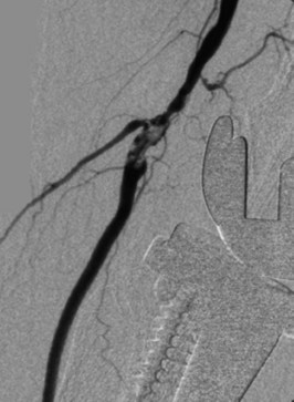
An investigation is performed. One of the results is shown in Figure 24.1.

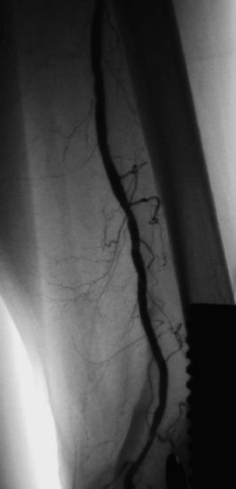
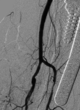
The patient underwent thrombolysis of the embolus radiologically using the ‘pulse-spray’ technique that often results in rapid clot lysis. Standard thrombolytic techniques would take 4–20 hours to achieve clot dissolution, and it was thought this patient’s leg was unlikely to be viable if revascularization took this length of time. The ‘pulse-spray’ technique is much more rapid. After 1 hour of urokinase pulsing, the angiogram was repeated (Figures 24.2 and 24.3).

The patient was anticoagulated with heparin and plans were made to continue anticoagulation on oral warfarin for at least 6 months, or possibly indefinitely depending on his atrial rhythm.

Answers

A.1 There are few possible causes of an acute severely painful leg. The most likely diagnosis is one of arterial embolus to the left leg. Other less likely possibilities include thrombosis of a pre-existing arterial atherosclerotic plaque, an extensive deep vein thrombosis of the left leg or an aortic dissection extending into the left leg arteries. A muscle haematoma if on thrombolytic or anticoagulant therapy is also a possibility.

A.2 He has acute ischaemic limb. His leg is pale, pulseless, painful and ‘perishing’ with cold. In addition he may have paraesthesia and paralysis of the affected limb – the classic six Ps.

The presence of a femoral pulse on the left indicates that the occlusion is likely to be in the superficial femoral or popliteal artery.

The sudden onset, lack of previous history and recent myocardial infarction all suggest that this is an embolic phenomenon. He has most probably thrown off an embolus from the heart which has lodged in the femoral artery. Such an embolus would have arisen from a mural thrombus at the site of infarction (in the left ventricle) or dislodged from clot in an atrium that is fibrillating. A less likely cause in this instance would be acute thrombosis of an atherosclerotic lesion in the femoral or popliteal artery.

After your initial assessment you will want to examine the notes looking for information about his post-myocardial infarction course, the timing and duration of arrhythmias and whether or not he has been anticoagulated.

A.3 You should contact the vascular surgeons immediately. Emergency arteriography or CT angiography will be required to define the site and extent of obstruction.

Unless angiography can be performed without delay, you should consider anticoagulating the patient with unfractionated intravenous heparin. Baseline coagulation studies (APTT) must be performed before starting the heparin. Heparin is dosed by weight and must be adjusted to maintain the APTT in the therapeutic range. A standard loading dose for an adult would be 5000 units, followed by an infusion of 1000 units/hour. Blood should also be collected for a complete blood picture, electrolytes and type and cross-matching. This patient is not a good candidate for general anaesthesia, but embolectomy or radiological intervention can be performed under local anaesthetic.

A.4 This is a digital subtraction angiogram, which demonstrates a filling defect in the left popliteal artery above the knee. At the level of the obstruction, there is a large collateral seen filling the distal popliteal vessel. This is consistent with a popliteal embolus. The proximal vessels are normal.

A.5 This patient has a threatened ischaemic leg requiring revascularization within 4 hours (see Revision points at end of chapter).

Management options include:

A.6 The angiograms performed after 1 hour of thrombolysis demonstrated a good radiological result. The clot had lysed and there is satisfactory revascularization of the distal vessels. The filling defect was no longer visible.

Revision Points

Acute Lower Limb Ischaemia