Cholelithiasis, sonographic Murphy sign, and GB wall thickening
• CT findings


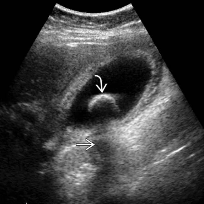
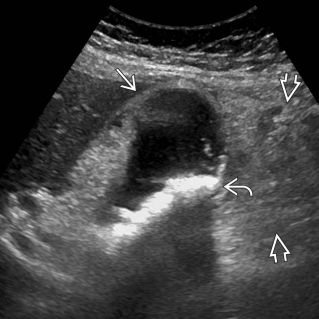
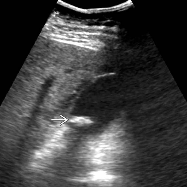
 with an acoustic shadow
  and a thickened gallbladder (GB) wall. These findings, along with a positive sonographic Murphy sign, suggested acute cholecystitis, confirmed at surgery.
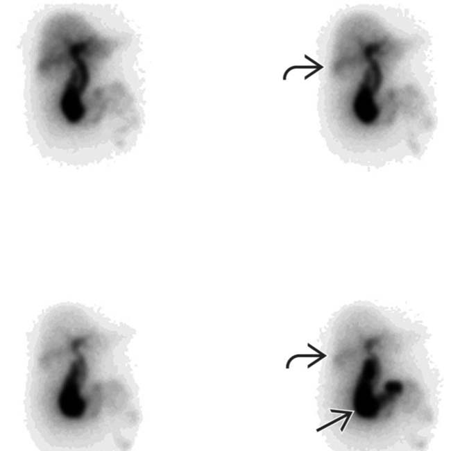
 , absence of GB activity, and a subtle GB fossa rim sign
  . Persistent pericholecystic activity may be due to tissue edema and biliary stasis.
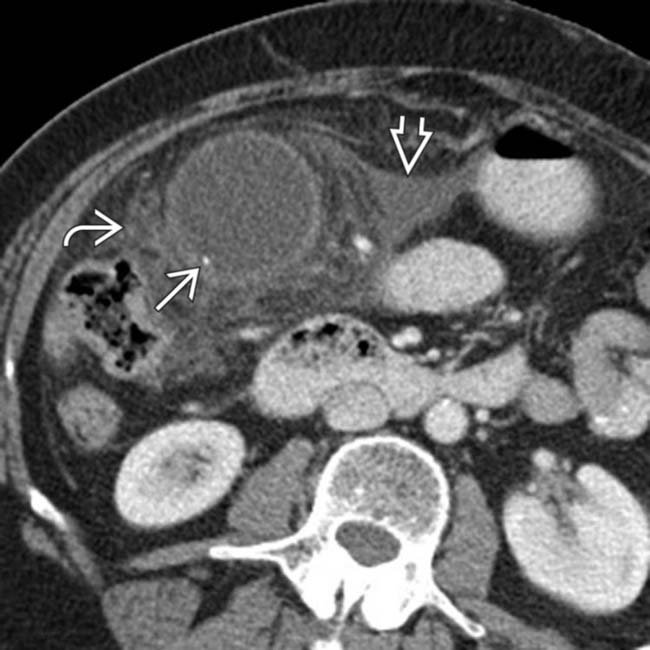
 within the gallbladder, with thickening of the GB wall, pericholecystic free fluid
  , and fat stranding
  , a constellation of findings diagnostic of acute cholecystitis.
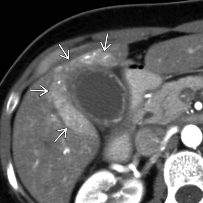
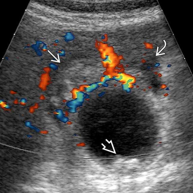
 . This surrounding hepatic parenchymal hyperemia is analogous to the rim sign of hepatobiliary scintigraphy.
IMAGING
General Features


CT Findings
• Uncomplicated cholecystitis
 GB wall thickening (> 3 mm) with mural and mucosal hyperenhancement and pericholecystic fat stranding
 GB wall thickening (> 3 mm) with mural and mucosal hyperenhancement and pericholecystic fat stranding


Ultrasonographic Findings
• Grayscale ultrasound
 Uncomplicated cholecystitis
 Complicated cholecystitis
Uncomplicated cholecystitis
Complicated cholecystitis
– Perforated cholecystitis: Pericholecystic abscess with collapsed GB


– Gangrenous cholecystitis: Asymmetric wall thickening, intraluminal linear membranes, and irregularity and ulcerations of GB wall


Nuclear Medicine Findings
• Hepatobiliary scintigraphy
 Nonvisualization of GB 4 hours after radiotracer injection (or 30 minutes after morphine administration)
 Nonvisualization of GB 4 hours after radiotracer injection (or 30 minutes after morphine administration)


PATHOLOGY
General Features


CLINICAL ISSUES
Presentation


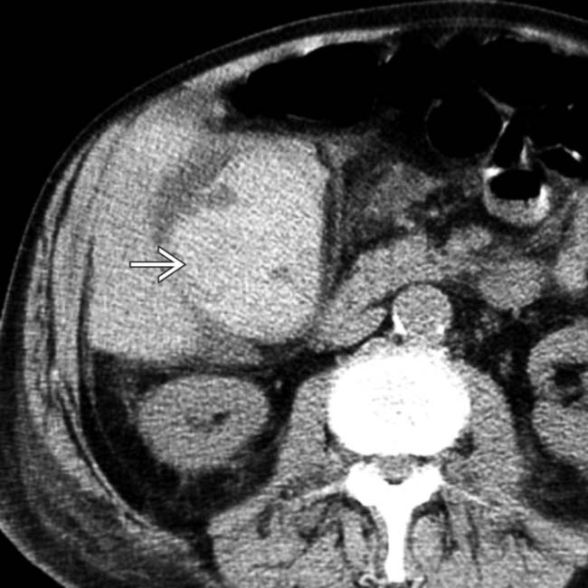
 with adjacent fat stranding
  , suggesting acute cholecystitis.
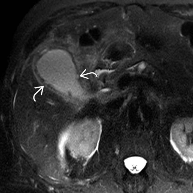
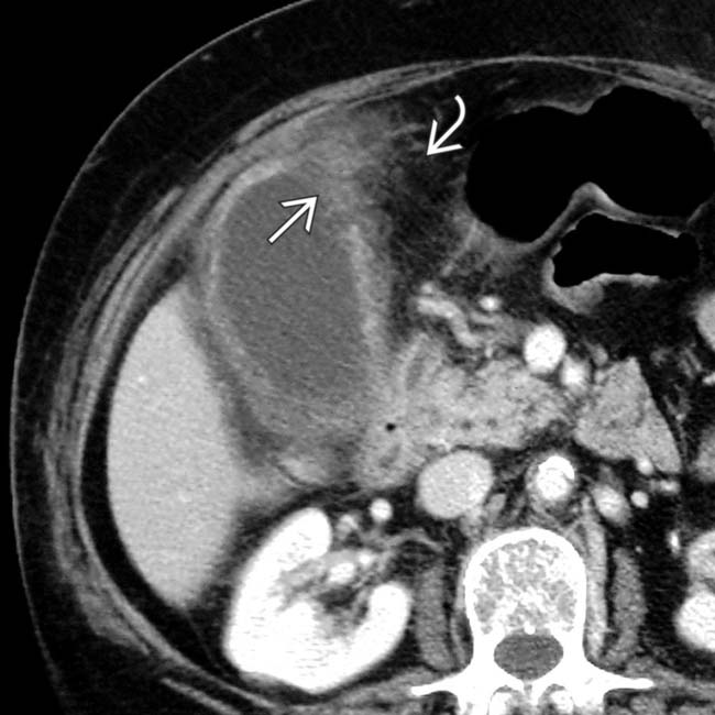
 . This was confirmed to represent gangrenous cholecystitis at surgery.
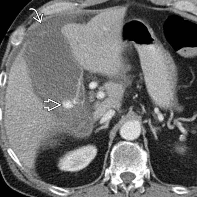
 near the fundus. Most importantly, there is active extravasation
  within the GB lumen, in keeping with this patient’s surgically confirmed hemorrhagic cholecystitis.
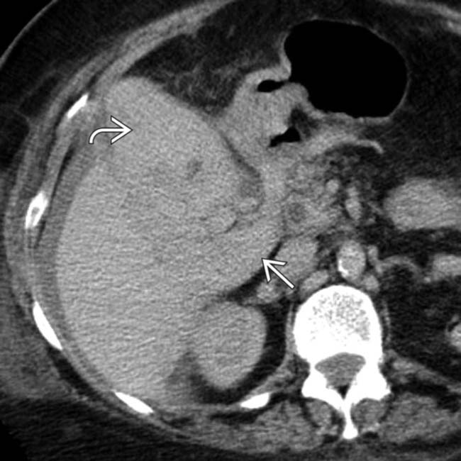
 , with similar density blood tracking in Morison pouch and medial to the GB
  . At surgery, the GB was gangrenous and contained hemorrhagic bile.
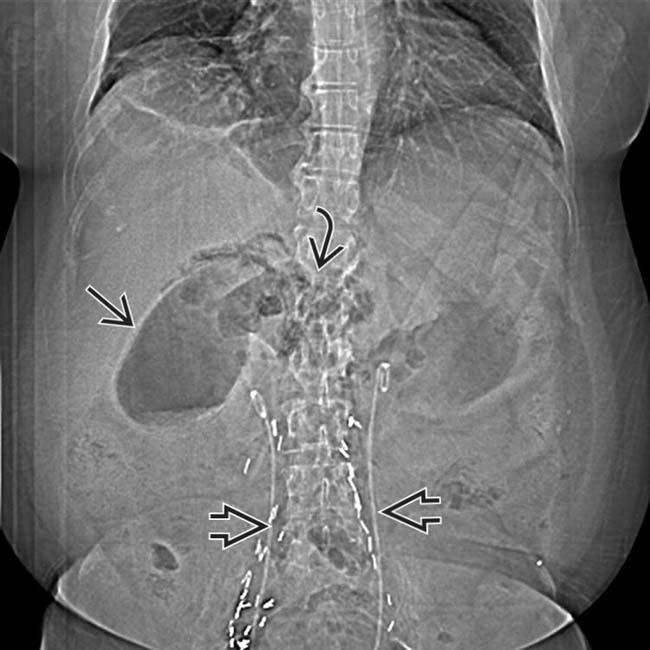
 . There also appears to be a collection of gas more medially
  . Ureteral stents
  and surgical clips are noted from the patient’s recent surgery.
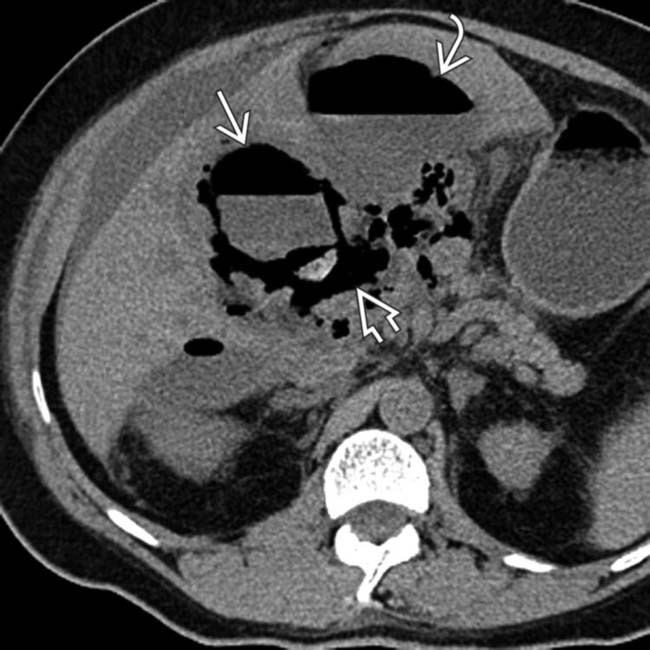
 in the GB lumen and wall. The fluid/gas
  tracking outward from the GB, as well as the adjacent gas and fluid collection
  , are consistent with perforation.
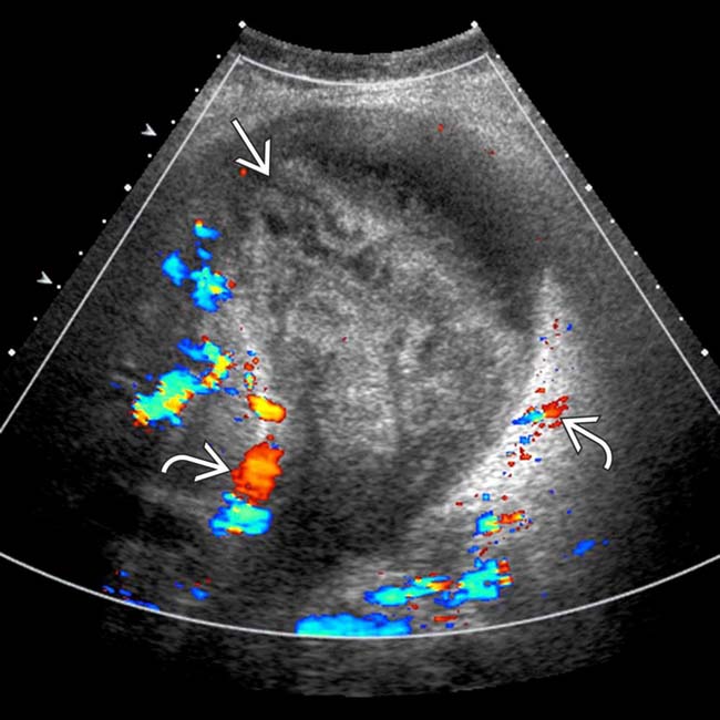
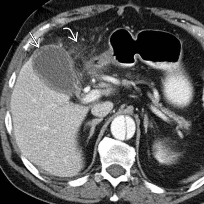
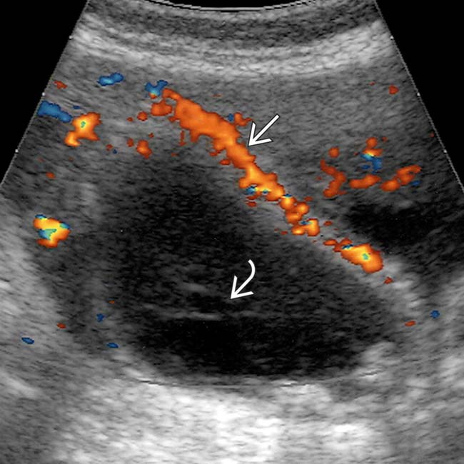
 , suggesting acute cholecystitis. However, the GB is distended and filled with high-density, heterogeneous blood products
  , suggesting hemorrhagic cholecystitis.
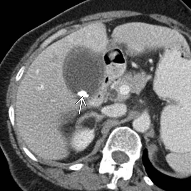
 within the GB, which appears thickened and inflamed, in keeping with acute cholecystitis.
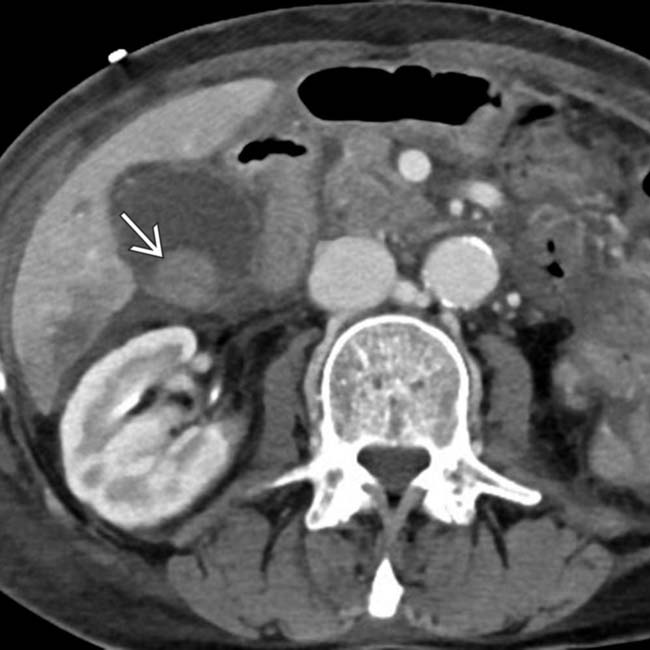
 within the GB lumen, suggesting acute hemorrhagic cholecystitis.
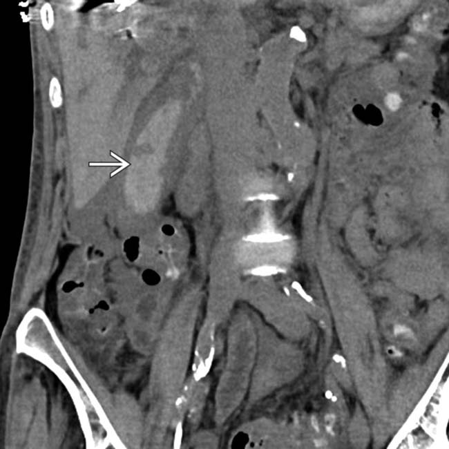
 has grown and now fills much of the GB lumen.
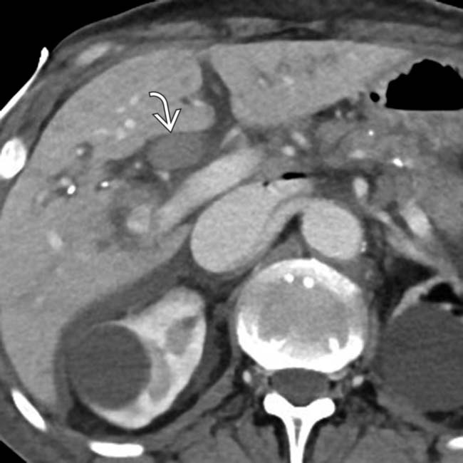
 distending and filling the common bile duct. Hemorrhagic cholecystitis in this patient was thought to be secondary to a recent embolization procedure.
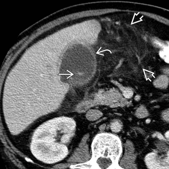
 , omental infiltration
  , and subtle layering of calcified stones
  .
.
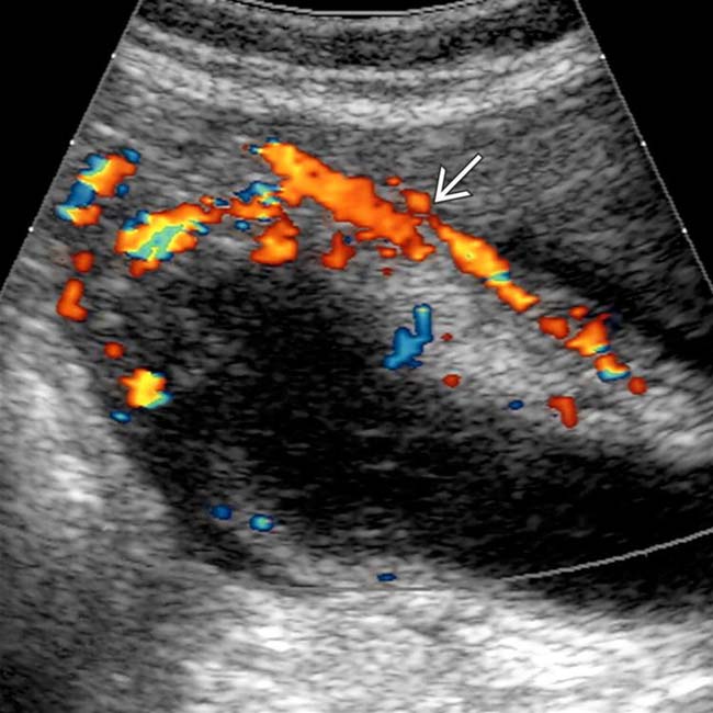
 , mild GB thickening
  , and sludge. Omentum is infiltrated
  , and an unequivocal sonographic Murphy sign was easily elicited to make the diagnosis of acute cholecystitis.
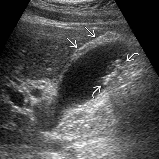
 and multiple small, weakly shadowing stones
  . A sonographic Murphy sign was confidently elicited.
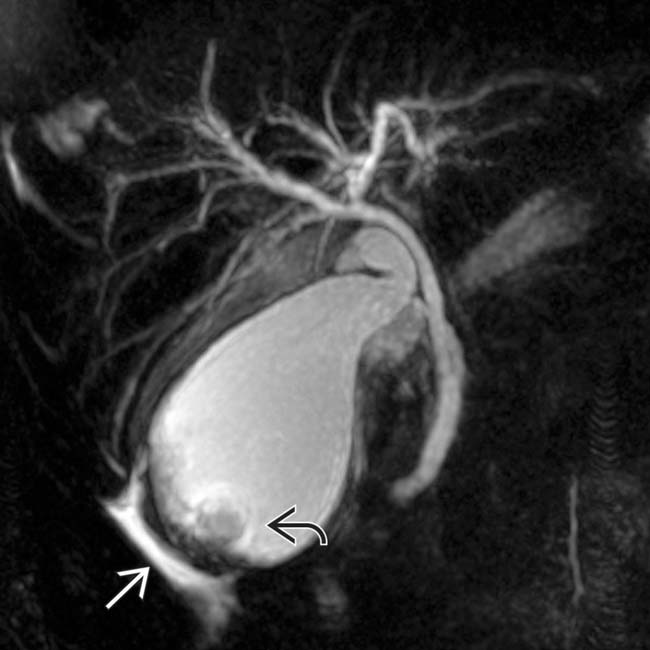
 , and pericholecystic fluid
  .
.
 , a small amount of pericholecystic fluid
  , and small gallstones
  .
.
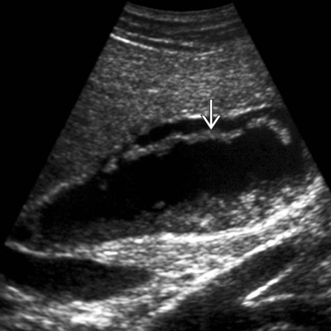
 and linear membranes in the GB lumen
  from fibrous strands of pus.
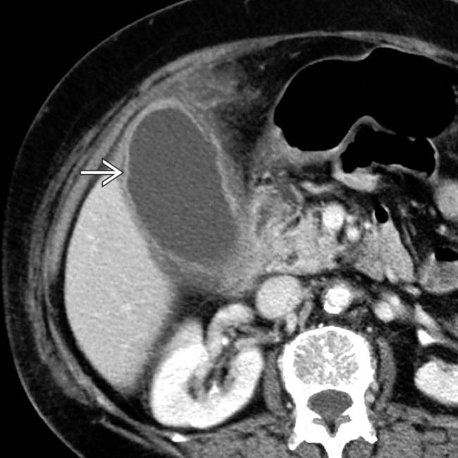
 .
.
 . Note the adjacent inflammatory changes in the omental fat
  .
.
 , but the GB wall was judged to be of normal thickness, and the area above it was not very tender.
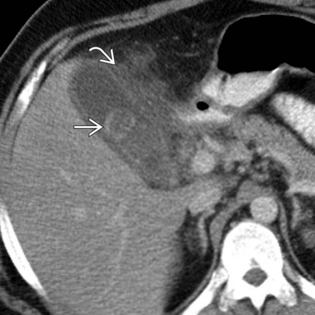
 as well as mural thickening and pericholecystic inflammation
  . Acute cholecystitis was confirmed at surgery.
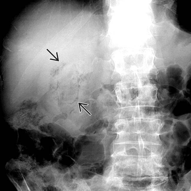
 .
.
 .
.
 of a thickened GB wall.
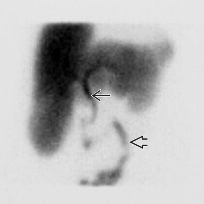
 and the small bowel
  without filling of the GB.
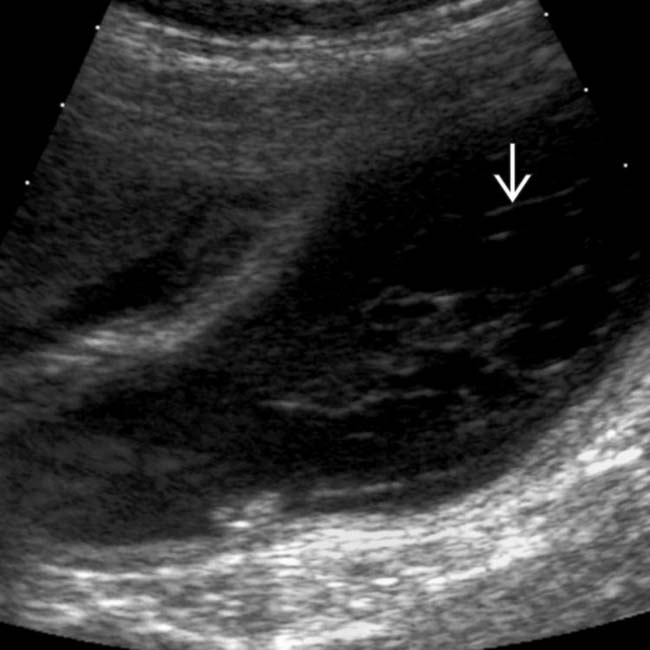
 .
.
 .
.

