Acid–base disorders: diagnosis and management

Published on 01/03/2015 by admin

Filed under Basic Science

Last modified 22/04/2025

Print this page

rate 1 star rate 2 star rate 3 star rate 4 star rate 5 star
Your rating: none, Average: 0 (0 votes)

This article have been viewed 1442 times

24

Acid–base disorders

diagnosis and management

Specimens for blood gas analysis

[H+] and PCO2 are measured directly in an arterial blood sample. This is usually taken from the brachial or radial arteries into a syringe that contains a small volume of heparin as an anticoagulant. It is important to exclude air from the syringe before and after the blood is collected. When the sample has been taken, any air bubbles in the sample should be expelled before the syringe is capped for immediate transport to the laboratory. Ideally, the syringe and its contents should be placed in ice during transit.

Acid–base problems may be discussed by referring to the three ‘components’ of the bicarbonate buffer system. In practice, blood gas analysers measure the [H+] of the sample and its PCO2. There is no need to measure the third variable, the bicarbonate. By the law of mass action:

image

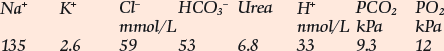
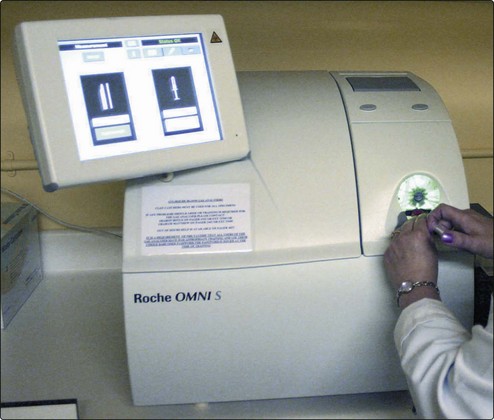
If the [H+] and the PCO2 are known, the bicarbonate can be calculated. Indeed, blood gas analysers (Fig 24.1) are programmed to provide this on all samples, as the ‘standard bicarbonate’ i.e. under standard conditions. Other parameters usually included are the PO2, and the base excess, another way of assessing the metabolic component.

In many laboratories, bicarbonate concentration is also determined directly as part of the electrolyte profile of tests on the laboratory’s main analyser, usually on a serum specimen obtained from a venous blood sample. These results (described as ‘total CO2’) are not identical to the printout from the blood gas analyser nor should this be expected since they include dissolved carbon dioxide, carbonic acid and other carbamino compounds. However, the results should not differ by more than 3 mmol/L. They may, therefore, be interpreted in the same way. A low bicarbonate in an electrolyte profile will usually indicate the presence of a metabolic acidosis.

Interpreting results

The most important information available for the interpretation and classification of an acid-base disorder is provided by the patient’s clinical history. The predicted compensatory responses in [HCO3] or PCO2 when [H+] changes as a result of primary acid–base disorders are shown in Table 24.1.

Table 24.1

Primary acid–base disorders and compensatory responses

Primary disorder Compensatory response
↑PCO2 (Respiratory acidosis) ↑HCO3
↓PCO2 (Respiratory alkalosis) ↓HCO3
↓HCO3 (Metabolic acidosis) ↓PCO2
↑HCO3 (Metabolic alkalosis) ↑PCO2

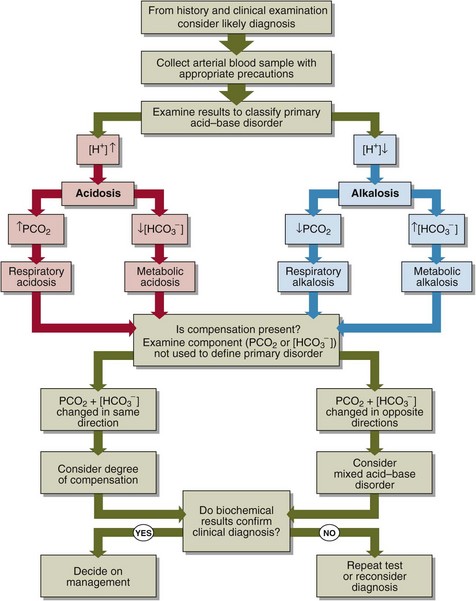
A practical approach to the interpretation of blood gas results is shown in Figure 24.2. The steps in classifying the acid–base disorder are:

image Look first at the [H+]. Decide if an acidosis or an alkalosis is present.

image If the [H+] is elevated, decide what is the primary cause of the acidosis. Look at the PCO2. If this is elevated, then there is a respiratory acidosis. Look at the bicarbonate. If this is decreased, there is a metabolic acidosis.

image If the [H+] is decreased, decide what is the primary cause of the alkalosis. Look at the PCO2. If low, then there is a respiratory alkalosis. Look at the bicarbonate. If this is high, then there is a metabolic alkalosis.

image Having decided on the primary acid–base disorder, look to see if there is compensation. If there is, there will be a change in the other component (the one which was not used to determine the primary disorder), in the direction which ‘compensates’ for the primary disorder, i.e. returns the ratio, and hence the [H+], towards normal. If there is not, the acid–base disorder may be uncompensated. If the change is in the opposite direction then a second acid–base disorder may be present. Even if there is compensation consider the possibility that there is a second acid–base problem that mimics the compensatory response.

image If there is compensation, decide if the disorder is fully compensated or partially compensated. If fully compensated, the resulting [H+] will be within the reference limits.

Clinical cases

The above practical advice is best illustrated by four case examples.

image A patient with chronic bronchitis. Blood gas results are: [H+] = 44 nmol/L; PCO2 = 9.5 kPa; [HCO3] = 39 mmol/L. A compensated respiratory acidosis is present.

image A patient who has had an acute asthmatic attack. Blood gas results are: [H+] = 24 nmol/L; PCO2 = 2.5 kPa; [HCO3] = 20 mmol/L. An acute, and hence uncompensated, respiratory alkalosis is present.

image A young man with a history of dyspepsia and excessive alcohol intake who gives a 24-hour history of vomiting. Blood gas results are [H+] = 28 nmol/L; PCO2 = 7.2 kPa; [HCO3] = 48 mmol/L. This is a partially compensated metabolic alkalosis.

image A 50-year-old man with a 2-week history of vomiting and diarrhoea. On examination he is dehydrated and his breathing is deep and noisy. Blood gas results are: [H+] = 64 nmol/L; PCO2 = 2.8 kPa; [HCO3] = 8 mmol/L. These results show a partially compensated metabolic acidosis.

Management of acid–base disorders

Many acid–base disorders are secondary to some other disorder. In most cases the management of an acid–base disorder is to treat the underlying illness. This may involve:

In cases where there is a life-threatening acidosis (e.g. [H+] >100 nmol/L), the infusion of sodium bicarbonate may be considered. The circumstances when this might be appropriate include severe diabetic ketoacidosis. Sodium bicarbonate must always be used with caution. Careful monitoring of the patient by repeatedly measuring the blood gases may be necessary. It should be noted, however, that once sodium bicarbonate has been administered, arterial blood gas results can be very difficult to interpret.