2. Achondroplasia (Dwarfism)
Definition
Achondroplasia is a hereditary, congenital autosomal dominant disorder that produces abnormalities in the growth and/or remodeling of cartilage and bone affecting the skull, spine, and extremities. The disorder, commonly known as dwarfism, frequently causes a person to be disproportionately short.
Incidence
The recent estimate for the incidence of achondroplasia in the United States is approximately 1:29,000; internationally, the estimate is approximately 1:40,000.
Etiology
Achondroplasia is an autosomal dominant inherited trait. The defect occurs on the short arm of chromosome 4—specifically, band 4p16.3.
Signs and Symptoms
• Abnormal odontoid
• Atlantoaxial instability
• Cleft lip
• Cleft palate
• Clubfoot
• Congenital heart disease
• Congenital odontoid absence
• Genu varum
• Hydrocephalus
• Kyphosis
• Laryngomalacia
• Micrognathia
• Obstructive sleep apnea
• Pulmonary hypertension
• Scoliosis
• Seizure disorder
• Spinal stenosis
• Tracheomalacia
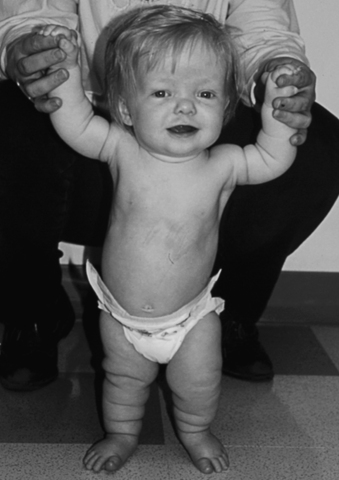 |
| Achrondroplasia (Dwarfism). This girl has short limbs relative to trunk length. She also has a prominent forehead, low nasal root, and redundant skin folds in the arms and legs. |
Medical Management
The patient’s short stature is frequently treated by administration of human growth hormone (somatotropin). The maximum benefits are realized when this treatment is initiated when the patient is in the age range of 1 to 6 years.
Surgical procedures may be necessary for a wide variety of occurrences, similar to any other patient. However, most procedures undertaken for patients with achondroplasia are orthopedic, with a significant portion of those involving the spine. Surgical correction of craniocervical stenosis, thoracolumbar kyphosis, spinal stenosis, lower extremities angular deformity (such as genu varum), and lengthening short extremities are frequently performed on these patients. Foramen magnum decompression and ventriculoperitoneal shunt insertion are also carried out when indicated.
Complications
• Apnea
• Brainstem compression
• Cervicomedullary compression
• Cyanosis
• Dysesthesias
• Dysphagia
• Genu varum
• Hydrocephalus
• Hypotonia
• Incontinence
• Kyphosis
• Lordosis
• Neurogenic claudication
• Obesity
• Paraparesis
• Paresthesias
• Pneumonia
• Quadriparesis
• Recurrent otitis media
• Respiratory insufficiency
• Scoliosis
• Spinal cord stenosis
• Sudden death
Anesthesia Implications
The patient with achondroplasia typically has cardiovascular and respiratory system abnormalities. Therefore the patient should have a chest x-ray, electrocardiogram, and ideally, transthoracic echocardiogram.
The patient with achondroplasia frequently has one or more craniofacial anomalies. The patient’s airway must be thoroughly assessed preoperatively. The anesthetist should prepare for encountering a difficult airway. The patient with achondroplasia frequently has laryngomalacia and subglottic stenosis; thus a smaller than “calculated” endotracheal tube should be within easy reach. The urgent need to surgically secure this patient’s airway should be anticipated.
Another possible impediment to securing the patient’s airway is an extremely limited range of motion of the neck as the result of atlantoaxial instability. The patient’s range of motion should be observed preoperatively and prominently documented before surgery and anesthesia. The anesthetist must not attempt to hyperextend the patient’s neck because the atlantoaxial joint may be easily dislocated with high compromise of the spinal cord, possibly resulting in quadriplegia.
Pulmonary volumes may be altered significantly by kyphosis. The patient’s respiratory function should be evaluated extensively preoperatively via chest x-ray, arterial blood gas analysis, and pulmonary function testing.
Laryngeal mask airways (LMAs) may be utilized on the patient with achondroplasia. The use of such an airway device should be appropriate for the proposed surgical intervention and may facilitate tracheal intubation in a difficult situation.
Because of associated restrictive pulmonary disease, higher respiratory rates with reduced ventilatory volume may be required. This may be associated with high ventilatory pressures. As a result, pressure-controlled ventilation methods may provide better respiratory exchange and reduce the risk of air trapping and barotrauma.
Regional anesthesia is acceptable for the patient with achondroplasia, but the patient’s degree of kyphoscoliosis may make instillation difficult. The anesthetist must be aware that even a relatively small volume of anesthetic may spread more than expected and cause a higher subarachnoid block than originally intended.
Positioning is critical particularly because of the patient’s atlantoaxial joint instability. The patient’s head and neck must be maintained in a neutral position at all times. Movement of the patient must be slow and deliberate, especially when the patient must be placed in either the lateral decubitus or the prone position.
Postoperative pain control may be a challenge. The patient with achondroplasia may have obstructive sleep apnea, and the effects of opioid analgesics on the respiratory system may be magnified by the obstructive tendencies.