Abdominal pain in a young woman

Published on 10/04/2015 by admin

Filed under Surgery

Last modified 22/04/2025

Print this page

rate 1 star rate 2 star rate 3 star rate 4 star rate 5 star
Your rating: none, Average: 0 (0 votes)

This article have been viewed 832 times

Problem 13 Abdominal pain in a young woman

The patient is otherwise in good health with an unremarkable medical history. She is considerably distressed by the pain, but the physical examination is normal apart from some tenderness in the right upper quadrant. She is overweight with a BMI of 34. Arrangements are made for her admission.

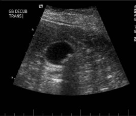
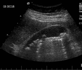
An ultrasound examination of the upper abdomen is performed (Figures 13.1 and 13.2).

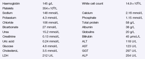
There is bilirubin + on urinalysis. Her serum biochemistry is shown below.

The day after admission the patient is feeling much better and is pain-free.

Her liver function tests are improving. She listens to your advice and agrees to a plan of ERCP followed by elective cholecystectomy. Her weight has not previously been mentioned as a problem. Should it be added to the introduction if it is used her? She is discharged home with arrangments for an ERCP the following week.

Five days later you are asked to see the patient in the emergency department. She has had further bouts of pain on and off for 2 days and has vomited a number of times. She also reports that her urine has darkened and that her stools are pale. She has not slept well the past 2 nights because of drenching sweats and she reports two violent shivering attacks this morning. She is uncertain of the date or day.

On examination she looks ill, distracted and has a temperature of 39.5°C. She is flushed and has yellow sclera. Her pulse is 110 bpm and her blood pressure is 90/50 mmHg. She has dry mucous membranes. Examination of her cardiorespiratory system is unremarkable. Her abdomen is soft and there is no localized area of tenderness.

With prompt resuscitation the patient improves and an ERCP is performed once she is stable. In preparation for this procedure, informed consent must be obtained.

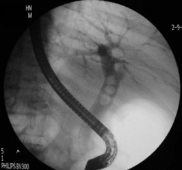
The ERCP is performed and the image shown in Figure 13.3 is obtained.

The patient makes a rapid recovery from her illness and her liver function tests return to normal. She is discharged home, manages to lose 20 kg and subsequently undergoes an elective laparoscopic cholecystectomy.

Answers

A.1 Further information must be sought on:

These severe episodes of right upper quadrant pain requiring opiate analgesia are characteristic of biliary colic (stones in the gallbladder or bile duct). The pain will often radiate around the costal margin to the back. Biliary colic may result from obstruction of the cystic duct, which typically occurs when a gallstone becomes impacted in Hartmann’s pouch. The pain – which is typically unremitting and constant – lasts until the gallstone falls back into the gallbladder and the obstruction is relieved.

Other diagnoses to consider include:

A.2 The following investigations are required:

A.3 This is an image from an ultrasound of the gallbladder showing a thin-walled gallbladder with several small echogenic foci which cast acoustic shadows beyond. These are gallstones. The biliary system does not appear to be dilated and no stones can be seen outside the gallbladder. The serum biochemistry results show deranged liver function with moderate elevation of the transaminases, alkaline phosphatase and bilirubin. All these would be in keeping with the patient’s current problem being related to duct stones.

A.4 This patient needs to be advised that her current episode of pain is almost certainly biliary in origin, as were the previous ones. Left untreated she will most likely have further problems.

She needs a cholecystectomy. The frequency and severity of her symptoms suggests that this should be done relatively soon. The standard of care would be to offer her a laparoscopic cholecystectomy during the current admission. Two things mitigate against this: her obesity and the high likelihood of duct stones. These two factors would increase the risk of conversion from a laparoscopic to an open operation. Opinion is divided on optimum treatment. Some surgeons might elect to proceed to laparoscopic cholecystectomy, cholangiography and intraoperative duct clearance – knowing that this might involve conversion to open surgery. An alternative approach would be endoscopic retrograde cholangiopancreatography (ERCP), duct clearance and weight reduction prior to planned laparoscopic cholecystectomy.

A.5 She has the classical triad that describes cholangitis: fever, right upper quadrant pain and jaundice. Of greater concern, she is hypotensive and is becoming confused. In this life-threatening situation of biliary sepsis the patient requires rapid resuscitation. She must be given intravenous fluids, supplemental oxygen and broad-spectrum antibiotics. Once intravenous access has been established blood samples must be sent for laboratory investigations, including blood cultures and coagulation studies. One litre of isotonic saline can be run in rapidly and further infusion judged on the response. Suitable antibiotic regimens would include gentamicin/amoxicillin/metronidazole or a second-generation cephalosporin such as cefoxitin. Initial management should be in a high-dependency unit with pulse oximetry, blood pressure and cardiac monitoring.

The ultrasound should be repeated, looking for biliary obstruction. She will likely need an urgent ERCP to relieve the obstruction and achieve biliary drainage so you should contact the gastroenterologists or biliary surgeons.

A.6 In obtaining informed consent for ERCP, you need to explain the rationale for the procedure and its potential risks and benefits. The aim of the procedure is to decompress the common bile duct and get sepsis under control. The endoscopist may place a stent into the common bile duct and/or may be able to retrieve the stone. The benefit of the ERCP in the present circumstances is that, if successful, the patient may be able to avoid major and hazardous open surgery to her common bile duct. The main risks of ERCP are pancreatitis (1–5%) and haemorrhage (1–2%). Both of these complications can be life-threatening and are more common when there is prolonged instrumentation of the biliary tree as in complex cases like these. There is a also a small risk of a retroperitoneal perforation following sphincterotomy and the patient must be informed of these risks in addition to the risks of sedation or anaesthesia needed to carry out the ERCP. In the present circumstances, the potential benefits of ERCP far outweigh the risks.

A.7 The image shows opacification of the biliary tree with contrast in the common bile duct and the intrahepatic system. There are three filling defects in the common bile duct. These are gallstones. A guidewire has been passed up the duct above the stones. This will allow a catheter with a balloon tip to be introduced in order that the stones may be extracted. In preparation to removing the calculi, the endoscopist will have made a cut through the sphincter of Oddi using diathermy. This procedure is known as a sphincterotomy. A stent will be placed after the stones have been removed to facilitate drainage.

Revision Points

Gallstones

Further Information

, http://www.quackwatch.com/. One practitioner’s efforts to expose many of the fraudulent claims made by the proponents of alternative medicine. Search for ‘gallstones’

, http://www.gastro.org/patient-center/digestive-conditions/gallstones. Up-to-date information on gallstones with a patient slant, from the American Gastroenterological Society