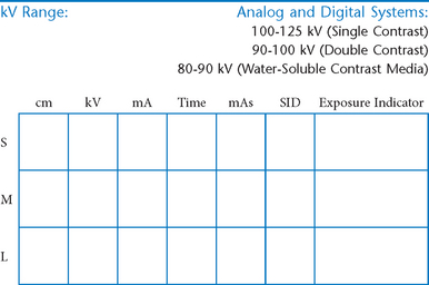
Abdomen and Common Contrast Media Procedures
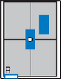
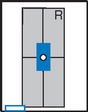
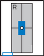
• Shielding and positioning landmarks
• Barium distribution and body positions
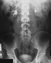
 AP supine (KUB) (R)
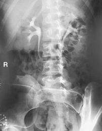
 AP erect (S)
 AP supine and AP erect critique
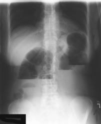
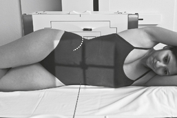
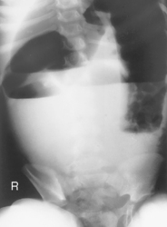
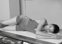
 Lateral decubitus (S)
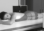
 Dorsal decubitus (S)
 Lateral and dorsal decubitus critique
 AP supine (R)
 AP erect (R)
 Lateral decubitus (S)
 AP supine (R)
 AP erect (R)
 Lateral decubitus (S)
 AP supine (KUB) (R)
 AP erect (S)
 AP supine and erect abdomen critique
 RAO
RAO Lateral
 RAO and lateral critique
 AP (PA)
 PA
PA RAO
RAO PA and RAO critique
 Lateral
 AP
AP Lateral and AP critique
 LPO
LPO LPO critique
 PA
PA PA (AP)
 PA (AP) critique
 Anterior/Posterior oblique
 Oblique critique
 Lateral rectum
 R and L lateral decubitus (double contrast)
 AP/PA axial (butterfly position)
 Lateral decubitus and AP/PA axial critique
 AP/PA (scout and series)
 Posterior obliques
 AP and posterior oblique critique
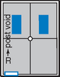
 AP erect (postvoid)
 AP
AP Posterior obliques and lateral
 AP and posterior oblique critique
Abdomen and Common Contrast Media Procedures
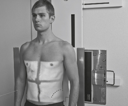
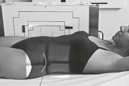
Shielding and Positioning Landmarks
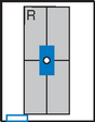
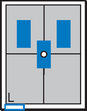
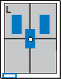
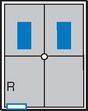
Gonadal Shielding

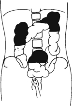
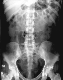
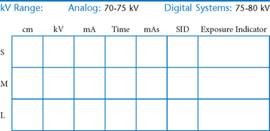
AP Abdomen (KUB)*
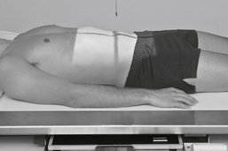
Position
• Supine, legs extended, arms at sides
• Midsagittal plane aligned and centered to centerline
• Ensure no rotation (ASISs equal distance from tabletop)
• Center of IR to level of iliac crests, ensuring that upper margin of symphysis pubis is included on lower IR margin. (A large hypersthenic patient may require that the IR be placed crosswise with a second IR centered higher.)
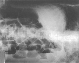
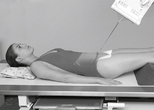
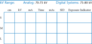
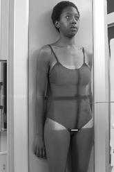
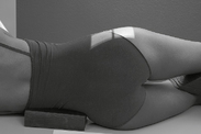
Erect AP Abdomen*
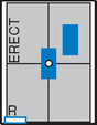
AP Supine and AP Erect Abdomen


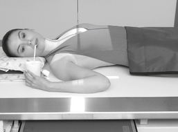
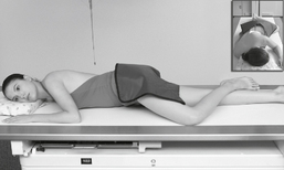
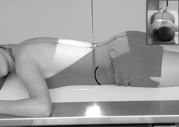
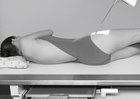
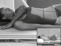
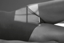
Abdomen*
• Arrow marker to include upside
• Patient should be on side a minimum of 5 minutes before exposure; 10 to 20 minutes is preferred.
Position
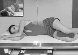
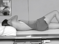
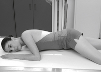
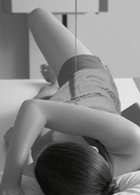
• Patient on side (on decubitus board or support to elevate downside abdomen), knees partially flexed, arms up near head
• Adjust patient and stretcher so center of IR and table (and CR) is approximately 2″ (5 cm) above level of iliac crest (to include diaphragm)
• Adjust height of IR to ensure that upside of abdomen is included for possible free air

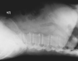
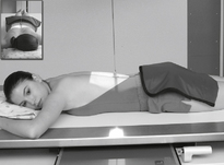
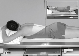
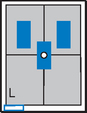
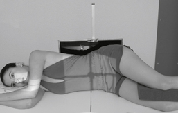
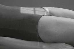
Abdomen*
Position
• Patient supine (on decubitus board or support to elevate posterior abdomen), side against table, arms above head
• Secure stretcher (lock wheels)
• Center of IR and table (and CR) at level of iliac crest (2″ above iliac crest to include diaphragm)
• Adjust height of IR to align midcoronal plane to centerline of IR
Lateral and Dorsal Decubitus Abdomen


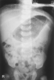
AP Pediatric Abdomen (KUB)*

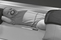
AP Erect Pediatric Abdomen*


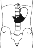
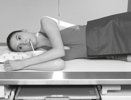
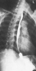
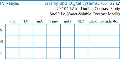
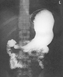
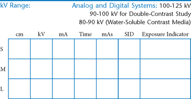
Esophagogram—RAO*

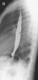
Esophagogram—Lateral*


Esophagogram—AP (PA)*

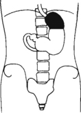
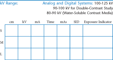
Upper GI—PA*


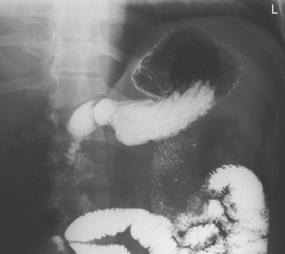
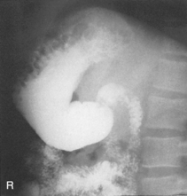
Upper GI—RAO*


Upper GI—Lateral*

Upper GI—AP*

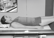
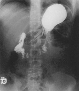
Fig. 9-31 AP supine Trendelenburg, upper GI (stomach) (Trendelenburg position best demonstrates hiatal hernia).
Lateral and AP Upper GI


Upper GI—LPO*

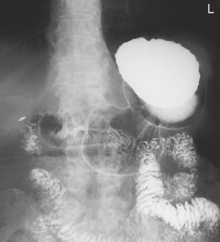

Small Bowel Series—PA*
A common routine includes images at 15- or 30-minute intervals until barium reaches ileocecal valve.

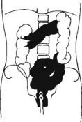
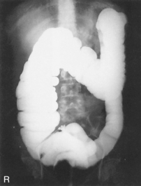
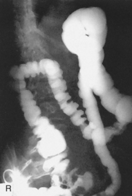
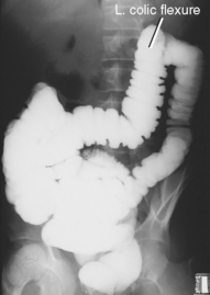
Barium Enema—PA or AP*
PA (AP) Barium Enema

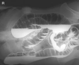
Barium Enema—RAO and LAO*
Oblique Barium Enema


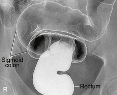
Barium Enema—Lateral Rectum (Ventral Decubitus)*

Alternative ventral decubitus projection is often performed for double-contrast studies.
Barium Enema—Lateral Decubitus*
Both right and left lateral decubitus are commonly taken as part of a double-contrast series.

Lateral Decubitus and AP/PA Axial Barium Enema

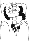
Intravenous Urogram*


Intravenous Urogram*
AP and Posterior Oblique IVU
Evaluation Criteria


Intravenous Urogram*

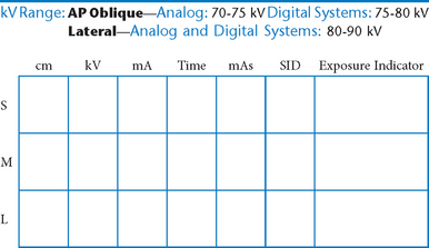
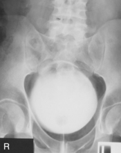
Cystogram—AP*

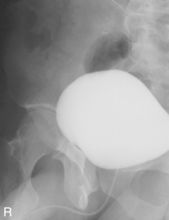
Cystogram—Posterior Obliques*
(RPO, LPO, and Optional Lateral)

Note: Cystogram routine may not include a lateral because of high gonadal dose.


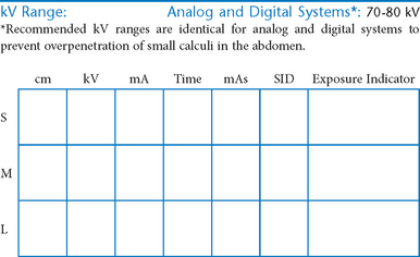
*Bontrager Textbook, 8th ed, p. 116.
*Bontrager Textbook, 8th ed, p. 119.
*Bontrager Textbook, 8th ed, p. 118.
*Bontrager Textbook, 8th ed, p. 120.
*Bontrager Textbook, 8th ed, p. 644.
*Bontrager Textbook, 8th ed, p. 645.
*Bontrager Textbook, 8th ed, p. 478.
*Bontrager Textbook, 8th ed, p. 479.
*Bontrager Textbook, 8th ed, p. 480.
*Bontrager Textbook, 8th ed, p. 483.
*Bontrager Textbook, 8th ed, p. 482.
*Bontrager Textbook, 8th ed, p. 484.
*Bontrager Textbook, 8th ed, p. 486.
*Bontrager Textbook, 8th ed, p. 485.
*More rotation for hypersthenic patients
*Bontrager Textbook, 8th ed, p. 513.
*Bontrager Textbook, 8th ed, p. 515.
*Bontrager Textbook, 8th ed, pp. 516 and 517.
*Bontrager Textbook, 8th ed, p. 519.
*Bontrager Textbook, 8th ed, p. 520.
*Bontrager Textbook, 8th ed, p. 523.
*Bontrager Textbook, 8th ed, p. 554.
*Bontrager Textbook, 8th ed, p. 556.
*Bontrager Textbook, 8th ed, p. 557.