Tubo-Ovarian Abcess and Pelvic Inflammatory Disease

Published on 10/03/2015 by admin

Filed under Obstetrics & Gynecology

Last modified 22/04/2025

Print this page

rate 1 star rate 2 star rate 3 star rate 4 star rate 5 star
Your rating: none, Average: 0 (0 votes)

This article have been viewed 2586 times

Tubo-Ovarian Abcess and Pelvic Inflammatory Disease

Synonyms/Description

Pelvic infection
Pelvic abcess
Pyosalpinx

Etiology

Pelvic inflammatory disease (PID) is an ascending pelvic infection causing inflammation of the upper genital tract, including cervicitis, endometritis, salpingitis, pelvic peritonitis, and occasionally resulting in development of a tubo-ovarian abscess (TOA). Inflammation damages the fallopian tubes, leading to infertility, ectopic pregnancy, and chronic pelvic pain. Acutely, the fallopian tubes swell and become congested, leading to salpingitis and potentially pyosalpinx if the tube fills with pus. Untreated PID may progress from inflammation of the tubo-ovarian complex to the formation of a tubo-ovarian abscess (TOA).
Most cases of PID are sexually transmitted and caused by Chlamydia trachomatis, Neisseria gonorrhoeae, or other similar causative agents. Pelvic inflammatory disease can also occur as a result of a gynecologic or abdominal procedure or surgery, as well as tuberculosis and appendicitis.

Ultrasound Findings

Ultrasound is very sensitive for detecting ovarian and tubal involvement of PID (sensitivity of 90% and 93%, respectively). When infected, the uterus enlarges and the echogenicity of the endometrium becomes heterogeneous and blotchy. The endometrial cavity often contains small amounts of echogenic fluid (exudate). The outer border of the uterus is often indistinct, with loss of clear separation between the uterus and adnexa.
Inflamed fallopian tubes are typically thick walled and dilated, filled with echogenic fluid and debris. The walls are irregular, and the tubes are elongated and tortuous with abundant blood flow on color Doppler examination. Milder infections may result in thickening of the tube without the presence of fluid (salpingitis). These thickened tubes are very tender during the transvaginal ultrasound examination.
A TOA is usually a solid, cystic or complex multiseptate mass, most often hypoechoic with areas of mixed echogenicity and thick septa.
Adhesions form within the pelvis, causing the tubo-ovarian complex to be adherent to nearby bowel, giving the appearance of a large, complex, adnexal mass with indistinct borders. Abundant blood flow is typical on Doppler studies.

Differential Diagnosis

The sonographic appearance of PID and TOA is nonspecific, and the differential diagnosis includes diseases that can cause complex hydrosalpinges and pain. These include ectopic pregnancy (there must be a positive pregnancy test), extensive endometriosis (typically a patient with chronic, cyclic pelvic pain), and tubal carcinoma (typically asymptomatic). Patients with PID and TOA are usually febrile and appear quite ill; therefore the clinical setting is an important part of evaluating a patient with a complex multiseptate tubo-ovarian mass.

Clinical Aspects and Recommendations

PID and TOAs are infections of the upper reproductive tract, usually sexually transmitted but occasionally polymicrobial; therefore, treatment is with antibiotics. The choice of antibiotics depends on the source of infection. The diagnosis of PID is more of a clinical diagnosis than one based on ultrasound findings. Clinicians are taught to maintain a low threshold for suspecting, diagnosing, and treating PID because long-term complications are more common if treatment is delayed. Easily obtainable sensitive pregnancy tests have helped quickly exclude possible ectopic pregnancy in patients who present with lower abdominal pain. Appendicitis and ovarian torsion are part of a clinical differential and may be assisted by ultrasound findings. TOAs may be more chronic and the diagnosis better assisted by the presence of complex adnexal masses as well as clinical signs, symptoms, and laboratory findings. Antibiotics or surgical intervention may be indicated, but specific recommendations are beyond the scope of this book.

Figures

image

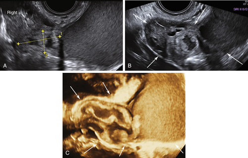
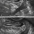
Figure T5-1 Very large pyosalpinx in a very sick patient with severe PID. A and B show the 2-D view of the very large pyosalpinx. The distal end of the tube is filled with a large amount of echogenic fluid. The rest of tube is more narrow and folded upon itself (calipers and arrows). C is a 3-D rendering of the entire dilated tube (arrows).


image

Figure T5-2 A and B, Acute PID with a very dilated tube in a patient later found to have a TOA. Note that the tubal wall is thick and edematous with multiple cystic spaces (pockets of pus) inside.

image

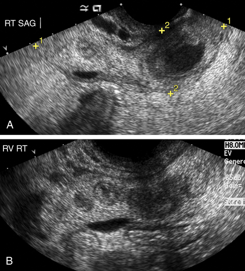
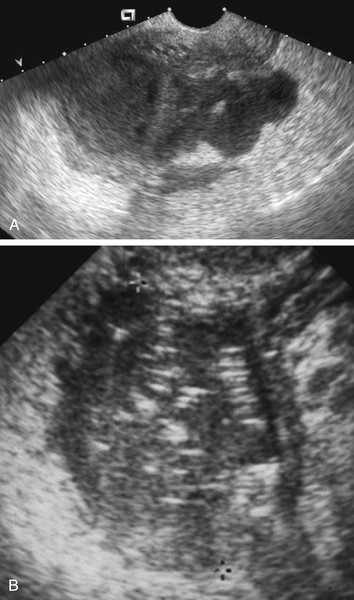
Figure T5-3 Tubo-ovarian abscess in two different patients. A shows a large complex cystic mass with irregular borders and septations in a patient with severe PID. Note that the borders of the mass are indistinct and blurry because of the surrounding edema. Although the sonographic appearance of the mass is nonspecific, the setting of a septic patient helps to make the diagnosis more definitive. B shows a completely solid adnexal mass containing small linear echoes throughout, consistent with an air-containing abscess.


image

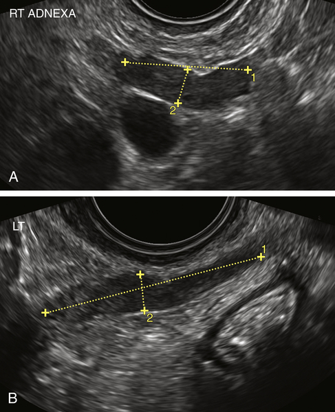
Figure T5-4 A and B, Salpingitis in two different patients. Note the elongated and straight swollen-appearing tubes (calipers). Both patients had localized pain in the area of the tube. For the patient in A, the diagnosis of pyosalpinx was confirmed at laparoscopy. The second patient improved with antibiotics.


Suggested Reading

Chappell C.A., Wiesenfeld H.C. Pathogenesis, diagnosis, and management of severe pelvic inflammatory disease and tuboovarian abscess. Clin Obstet Gynecol. 2012;55:893–903.

Cicchiello L.A., Hamper U.M., Scoutt L.M. Ultrasound evaluation of gynecologic causes of pelvic pain. Obstet Gynecol Clin North Am.. 2011;38:85–114.

Crossman S.H. The challenge of pelvic inflammatory disease. Am Fam Physician. 2006;73:859–864.

Ghiatas A.A. The spectrum of pelvic inflammatory disease. Eur Radiol. 2004;14(suppl):E184–E192.

Kamaya A., Shin L., Chen B., Desser T.S. Emergency gynecologic imaging. Semin Ultrasound CT MRI. 2008;29:353–368.

Kim M.Y., Rha S.E., Oh S.N., Jung S.E., Lee Y.J., Kim Y.S., Byun J.Y., Lee A., Kim M.R. MR imaging findings of hydrosalpinx: a comprehensive review. Radiographics. 2009;29:495–507.

Soper D.E. Pelvic inflammatory disease. Obstet Gynecol. 2010;116:419–428.

Varras M., Polyzos D., Perouli E., Noti P., Pantazis I., Akrivis C.H. Tubo-ovarian abscesses: spectrum of sonographic findings with surgical and pathological correlations. Clin Exp Obstet Gynecol. 2003;30:117–121.