A woman with acute upper abdominal pain

Published on 10/04/2015 by admin

Filed under Surgery

Last modified 22/04/2025

Print this page

rate 1 star rate 2 star rate 3 star rate 4 star rate 5 star
Your rating: none, Average: 0 (0 votes)

This article have been viewed 1064 times

Problem 14 A woman with acute upper abdominal pain

To refine your list of differential diagnoses you order some investigations.

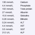
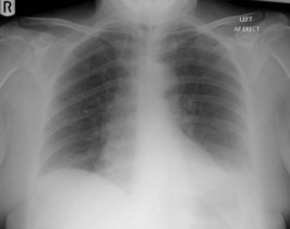
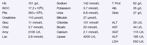
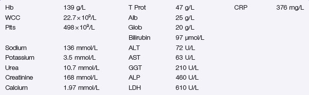
The patient’s ECG reveals a sinus tachycardia and no other changes. Her chest X-ray is shown in Figure 14.1 and her blood test results are shown in Investigation 14.1. Her BMI is calculated to be 34 kg/m2.

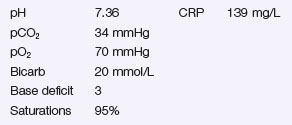
The results of the arterial blood gas analysis on inspired room air and the C-reactive protein (CRP) are shown in Investigation 14.2.

Overnight, she remains stable and begins to feel better.

An ultrasound scan shows gallstones in a distended gallbladder, with dilatation of the intrahepatic bile ducts. The extrahepatic biliary tree and the pancreas are obscured by bowel gas.

Over the next 24 hours the patient deteriorates. She develops a pyrexia of 38.8°C and becomes jaundiced. She is transferred to the high dependency unit and her repeat blood results are shown in Investigation 14.3.

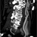
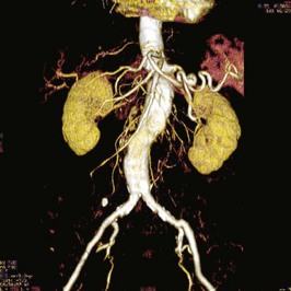
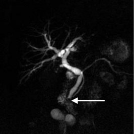
The magnetic resonance cholangiopancreatogram is shown in Figure 14.2. The MRCP shows dilated intra- and extrahepatic ducts and a solitary stone (arrow) impacted at the bottom of the common bile duct. The pancreatic duct is also visible passing to the right of the picture.

She has a successful sphincterotomy, duct clearance and stent insertion at ERCP and makes a prolonged, but otherwise uneventful recovery over the next 10 days.

Answers

A.1 The diagnoses to consider include acute pancreatitis, perforated peptic ulcer and acute cholecystitis. It is possible that she has been suffering with attacks of biliary colic for some time and, given the progressive nature of her pain and its radiation through to the back, acute pancreatitis would be the most likely clinical diagnosis. Although not always the case, you may expect the patient to have a rigid abdomen if the cause were a perforated peptic ulcer. It is important to consider the possibility of a myocardial infarction or a lower lobe pneumonia giving rise to similar symptoms, especially considering the findings on respiratory examination.

Other diagnoses to consider are ischaemic gut and intestinal obstruction. Ischaemic gut is a difficult diagnosis to make but may be more likely in those with pre-existing vascular disease or cardiac dysrythmias, particularly atrial fibrillation. There are few, if any, features to suggest the possibility of intestinal obstruction.

A.2

A.3 The chest X-ray shows a small left-sided pleural effusion. The white cell count is elevated, as are the serum lipase and amylase estimations. Given the degree of hyperamylasaemia and hyperlipasaemia this patient almost certainly has acute pancreatitis. The clinical picture and the raised white cell count and blood glucose are certainly in keeping with this. A rise in the serum amylase to at least three times the upper limit of normal would be expected. Hyperamylasaemia to a lesser degree can, however, be associated with alternative intra-abdominal conditions such as acute cholecystitis and intestinal ischaemia. In situations of diagnostic difficulty, measurement of the serum lipase has a higher sensitivity and specificity than serum amylase.

She has deranged liver function tests, with an elevated alkaline phosphatase and a slightly raised bilirubin, although this is unlikely to be detectable clinically. It would be prudent to check the clotting profile in this circumstance.

She needs arterial blood gas analysis and a C-reactive protein measurement.

A.4 This patient needs careful management. Initially, she should be fluid resuscitated with intravenous crystalloids. She may require a considerable volume of fluid over the next few days because of the acute inflammatory response leading to fluid sequestration around the inflamed pancreas, within bowel loops and in the interstitial fluid compartment. She needs close monitoring of fluid input and output and will need a urinary catheter connected to an hourly drainage bag.

If vomiting is a problem, she may benefit from a nasogastric tube, although patients with acute pancreatitis may, if tolerated, benefit from oral intake and nasogastric feeding in an attempt to reduce the incidence of septic complications.

She will require opiate analgesia.

Given the presence of a small pleural effusion at presentation and oxygen saturations of 95% on air, she should have supplemental oxygen, although at this stage nasal cannulae with 2–4 L/minute will probably suffice. She will require regular measurement of oxygen saturations by pulse oximetry.

She will need regular monitoring of serum electrolytes, calcium and blood sugar. She may require supplemental calcium and intravenous sliding scale insulin therapy.

The Atlanta classification (1992) is the most widely used clinically based scoring system that stratifies patients as having either mild or severe disease depending largely on the severity of associated organ dysfunction, combined with the presence or absence of local and systemic complications. However, numerous attempts have been made to develop scoring systems with the aim of predicting which patients will go on to develop severe pancreatitis to allow early intensive monitoring and therapy. On admission, a BMI of >30 and the presence of a pleural effusion are predictive of severe disease. The acute physiology and chronic health evaluation (APACHE) II scoring system has been used in pancreatitis and can be applied to patients with acute pancreatitis from the time of admission. A score of >8 predicts severe disease. The modified Glasgow score consists of eight variables applied after 48 hours of admission. Each positive variable scores 1, with a total of ≥3 predicting severe pancreatitis (see revision points). A CRP of >150 IU after the first 24 hours is indicative of severe disease. However, much still depends on clinical impression of severity, especially if there is persistent or deteriorating organ failure extending beyond 48 hours after admission.

A.5 The most common cause of acute pancreatitis in Western medicine is gallstones. Every patient diagnosed with acute pancreatitis should have an abdominal ultrasound scan within 24 hours.

A.6 Given the presence of gallstones and dilated intrahepatic bile ducts on ultrasound, in conjunction with a rising serum bilirubin, a rising WCC and pyrexia, the most useful investigation is magnetic resonance cholangiopancreatography (MRCP) to confirm the presence of a stone within the bile duct.

Your patient is at risk of developing severe pancreatitis. With a BMI >30, she had a left pleural effusion at presentation, her CRP has risen to >150 mg/L and her modified Glasgow score is 5 (age > 55, WCC >15 × 109/L, calcium <2 mmol/L, LDH >600 U/L and albumin <32 g/L).

A.7 She needs urgent endoscopic retrograde cholangiopancreatography (ERCP), with sphincterotomy and stone extraction +/− removable stent insertion to ensure relief of biliary obstruction, ideally with 72 hours.

A.8 This patient needs a cholecystectomy. This should ideally be arranged within the same hospital admission and as a laparoscopic procedure. The biliary stent should be removed endoscopically once her gallbladder has been removed.

Revision Points