A 68-year-old woman with breathlessness and yellow sputum

Published on 10/04/2015 by admin

Filed under Surgery

Last modified 22/04/2025

Print this page

rate 1 star rate 2 star rate 3 star rate 4 star rate 5 star
Your rating: none, Average: 0 (0 votes)

This article have been viewed 1018 times

Problem 31 A 68-year-old woman with breathlessness and yellow sputum

You ponder the differentials, and move on to examining the patient.

Her temperature is 38°C, her pulse 140 bpm, regular and bounding, and her blood pressure is 150/100 mmHg with a paradox of 25 mmHg. The patient has a barrel chest with markedly reduced chest expansion and a tracheal tug with use of her sternocleidomastoid muscles and intercostal recession. Precordial dullness is lost. There is no finger clubbing or flap.

Her jugular venous pulse is raised 4 cm. Her apex is not palpable and her heart sounds are inaudible. Her chest is hyper-resonant with globally reduced breath sounds, a prolonged expiratory phase and a loud wheeze. There is no cervical lymphadenopathy and the abdomen is not examined formally due to the patient’s distress. There is mild ankle oedema. She is unable to perform simple spirometry due to the severity of her breathlessness.

The following result is obtained:

Investigation 31.1 Arterial blood gas analysis on air

pO2 41 mmHg
pCO2 75 mmHg
pH 7.25
HCO3 32 mmol/L

Investigation 31.2 Arterial blood gas analysis on 3 L/min O2

pO2 58 mmHg
pCO2 89 mmHg
pH 7.21
HCO3 32 mmol/L

The electrolytes are normal other than bicarbonate.

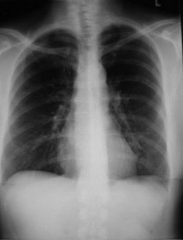
The chest X-ray is shown in Figure 31.1.

An ECG shows a sinus tachycardia with a right axis deviation and a prominent R wave in lead V1.

The patient is managed in the specialist respiratory ward. Her blood gases are maintained within a satisfactory range using low-flow nasal cannulae at 1 L/min oxygen. Salbutamol nebulizers are given hourly.

After blood cultures have been taken, she is started on oral amoxicillin/clavulanic acid and intravenous clarithromycin. Oral steroids are given starting at 50 mg prednisolone.

Twenty-four hours later you are called to see the patient because she has an obvious tremor and a temperature of 37.6°C. Her pulse is 140 bpm and her blood pressure is 140/90 mmHg.

Her chest findings are unchanged and abdominal examination is unremarkable. A repeat chest X-ray shows no change and an ECG shows a sinus tachycardia with a rate of 146 bpm.

Her nebulized salbutamol is decreased to 4 hourly and the tachycardia slowly improves. The prednisolone dose is decreased and then ceased over 7 days. Tiotropium is instituted as a once-daily inhaler.

Seven days after admission she has improved to the extent where she can walk slowly around the ward. Formal pulmonary function tests are done prior to discharge. The results are as follows:

Investigation 31.5 Arterial blood gases on air

pO2 50 mmHg
pCO2 61 mmHg
pH 7.40
HCO3 34 mmol/L

The patient is told firmly that she must stop smoking immediately.

The patient ignores your advice on smoking. She qualifies for home oxygen but is not issued the treatment due to her current smoking. She survived another 18 months, dying of another episode of acute on chronic respiratory failure.

Answers

A.1 She has at least a 100 pack/year history of smoking and chronic obstructive pulmonary disease (COPD) secondary to cigarette smoking is the most likely underlying diagnosis. This includes chronic bronchitis and emphysema and there may also be an element of reversible airways obstruction, i.e. asthma. Infection of the tracheobronchial tree causing an increase in airway inflammation and volume and viscosity of sputum is the most common precipitant of deterioration in these patients.

Cardiac failure, either diastolic failure secondary to ischaemia or decompensation of cor pulmonale secondary to increasing pulmonary artery pressures, are important factors to consider as precipitants or as confounders.

Other factors contributing to deterioration may include increased tobacco use, non-compliance with medication, use of sedative drugs, intercurrent pneumonia, left ventricular failure, pneumothorax, thromboembolism and development of a bronchogenic carcinoma or development of anaemia.

The differential diagnosis includes adult-onset asthma and bronchiectasis, and heart failure.

The occupational history is important as the woman may have pneumoconiosis in addition to her smoking-related disease. Exposure may be indirect, such as having laundered her husband’s clothes contaminated with asbestos material etc. Occupational history including exposure related to hobbies must always be asked about, even with an obvious smoking history. A history of childhood respiratory problems including exposure to pulmonary tuberculosis may also be relevant.

A.2 During the examination of this patient you will focus on:

A.3 Initial investigations (and reasons why):

A.4 The initial blood gas indicates acute on chronic respiratory failure. The bicarbonate is halfway between what is expected in acute respiratory acidosis (lower bicarbonate) and chronic respiratory acidosis (higher bicarbonate). This suggests renal (metabolic) compensation for chronic hypercapnia and acidosis by bicarbonate retention. This would normalize the pH in the chronic state. With an acute deterioration this compensation cannot occur, resulting in a moderate acidosis.

A.5 With the application of 3 L/min of oxygen there has been improvement in oxygenation but also further CO2 retention and exacerbation of the acidosis.

The patient has a raised haemoglobin, which is likely to reflect a secondary polycythaemia induced by chronic hypoxia. She has a neutrophilic leucocytosis, which indicates bacterial infection.

The chest X-ray reveals a cardiac size within normal limits and prominent pulmonary arteries. The lung fields are grossly inflated and there is some scarring at the lung apices. There is no evidence of consolidation. This suggests chronic obstructive pulmonary disease (COPD) with pulmonary hypertension. This diagnosis is supported by the ECG changes of right ventricular strain (right axis deviation and a prominent R wave in V1).

Overall, the clinical picture indicates that the patient has COPD with probable cor pulmonale. There has been an acute deterioration precipitated by infection but there is no evidence of pneumonia.

A.6 Oxygenation is the key. Patients need oxygen if they are hypoxic, but excessive supplemental oxygen can result in dangerous hypercapnia with mental stupor or coma and respiratory arrest. A compromise is needed. Improvement of the pO2 towards 50–60 mmHg should be attempted using the smallest amount of oxygen possible. At this level pulmonary vasodilatation is achieved. This is when Venturi masks are useful: low-dose oxygen via 24% Venturi mask improves oxygen without causing dangerous respiratory depression. If these cannot be tolerated, nasal cannulae can be substituted at 1 L/min. A modest rise in pCO2 is acceptable provided that the patient remains alert. If a 24% Venturi mask is tolerated, particularly if the patient improves, a 28% mask (or nasal cannulae at 1.5–2 L/min) may be tried. The blood gases will need to be checked to exclude deterioration in ventilation.

In addition to oxygen, the treatment of any reversible airways obstruction by removing secretions and treating infection will help. If these measures to improve ventilation and ventilation–perfusion mismatch fail non-invasive ventilation (NIV) will have to be considered, including with continuous positive airways pressure (CPAP) or bi-level positive airway pressure (BiPAP). Usually endotracheal intubation and mechanical ventilation would be regarded as the last resort.

The patient may only have a small component of reversible airways obstruction but attempted bronchodilatation by frequent salbutamol nebulizers is accepted practice. Salbutamol intravenous infusion is hardly ever used.

Steroids are used in acute exacerbations of chronic airways obstruction, although they are more important in asthma, and their effectiveness in the management of COPD is not uniform. A similar dose is often used during the acute phase of the illness, but unless there is a significant component of bronchospasm in the obstruction, the chronic use of steroids is not recommended.

Clearance of airway secretions in this patient is important. In the acute stage she would not tolerate routine chest physiotherapy, but regular encouragement of coughing and possibly nasotracheal suction should be performed.

Although the patient has no consolidation she has a fever and a severe deterioration associated with purulent sputum; intravenous antibiotics such as amoxicillin (or amoxicillin/clavulanic acid), doxycycline or clarithromycin should be added. It must be remembered that the macrolide antibiotics (erythromycin, roxithromycin, clarithromycin) interact with many medications including warfarin, anticonvulsants, oral contraceptives and any drugs metabolized by hepatic cytochrome P450. Intravenous erythromycin is now avoided wherever possible, as it can be associated with a highly irritant superficial thrombophlebitis.

This patient is critically ill with respiratory failure, and should be admitted to a dedicated respiratory speciality ward or if that is not available to a high dependency unit. If repeated gas measurements are necessary, an arterial line can be considered.

A.7 She would need some form of respiratory support. This would probably be non-invasive in the form of non-invasive ventilation such as BiPAP, or CPAP. Both these forms of ventilatory support are supplied via well-fitting face or nasal masks. If her respiratory failure worsened further or her conscious level decreased then she would need endotracheal intubation.

A.8 The most likely explanation for the tachycardia is the combination of fever and the frequent administration of salbutamol. Potassium levels may have changed, especially if diuresis has occurred. In addition, she may have cor pulmonale which makes her prone to atrial tachyarrhythmias, further exacerbated by hypoxaemia. You should arrange an ECG to rule out ischaemia, given her history, repeat electrolytes and a chest X-ray.

A.9 Her lung function tests show an obstructive pattern with significant reversibility to salbutamol. According to GOLD criteria this lady has severe COPD with respiratory failure, stage IV. The reduced DLCO demonstrates markedly impaired gas exchange and is typical of severe emphysema.

Her gases show a return to her chronic state. She is no longer acidotic and her bicarbonate has risen back to its chronically elevated level.

In the short term she should finish her course of antibiotics and tail off her steroids over the following week.

She should have an assessment of her inhaler technique and if required receive more education on device use. She should be discharged with inhalers of tiotropium and a corticosteroid/long-acting beta agonist combination as well as salbutamol if needed.

She must stop smoking. Tobacco smoke inhibits mucociliary function and hence clearance of airway secretions. Smoking also stimulates increased airway mucus production so cessation of smoking will decrease sputum production and bronchospasm. In addition home oxygen cannot be prescribed if the patient continues to smoke, because of the fire hazard.

This patient should have the pneumococcal vaccine every 5 years and an annual influenza vaccine. Compliance with medication should be ensured and inhaler technique should be checked regularly. Infective exacerbations should be treated early with oral antibiotics, and intensive chest physiotherapy. If manifestations of cor pulmonale (such as ankle swelling) worsen, they may have to be treated with diuretics.

Referral to a respiratory rehabilitation programme is helpful, and provision of home improvements may improve the ability of the chronically breathless person to cope. This patient would fit the criteria for home oxygen provided she can become an established non-smoker and that there is reversal of hypoxaemia with domiciliary oxygen without progressive hypercapnia.

Revision Points

Chronic Obstructive Pulmonary Disease (COPD)

Cigarette smoking is by far the most important aetiological factor in this disease. Smoking cessation slows the progression of the disease and there may be improvement in airway obstruction. Marijuana smoking has been largely undervalued as a risk factor and it is estimated that one marijuana cigarette may be as damaging as 8–15 cigarettes.

Further Information

, www.COPDX.org.au. The COPD-X Plan, Australian and New Zealand guidelines for the management of chronic obstructive pulmonary disease

, www.GOLDCOPD.com. GOLD – Global Initiative for Chronic Obstructive Lung Disease

, www.lungfoundation.com.au. The Australian Lung Foundation is a key agent of change in Australia for promoting the understanding, management and relief of lung disease