A 63-year-old woman with a screen-detected abnormality

Published on 10/04/2015 by admin

Filed under Surgery

Last modified 22/04/2025

Print this page

rate 1 star rate 2 star rate 3 star rate 4 star rate 5 star
Your rating: none, Average: 0 (0 votes)

This article have been viewed 861 times

Problem 9 A 63-year-old woman with a screen-detected abnormality

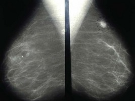
The mammogram is reviewed (Figure 9.1).

An ultrasound examination is performed and shows a hypoechoic lesion with irregular margins, distorting and invading the surrounding breast tissue.

A fine needle aspiration (FNA) is arranged. In the outpatient clinic and with ultrasound guidance a 23 gauge needle is passed several times through the lesion. The aspirate is spread on a microscope slide and examined immediately. Benign cells with atypia are seen, but there is no evidence of malignancy.

A core biopsy is arranged. This procedure requires the use of an automated biopsy gun and 12–18 G biopsy needle. The biopsy is performed under local anaesthesia using ultrasound guidance. A core of architecturally intact breast tissue is removed and invasive ductal carcinoma of intermediate nuclear grade is confirmed.

In this particular case, staging investigations (liver function tests, CT scan chest and abdomen, whole body bone scan) did not reveal any evidence of metastatic disease.

A surgical procedure is planned for the patient. The following investigation is performed immediately before the operation (Figure 9.2).

The patient undergoes wide local excision, specimen X-ray and sentinel node biopsy. Histopathology demonstrates a 22 mm grade II ductal carcinoma with focal vascular invasion. Tumour excision is complete with the closest resection margin being 5 mm from the inferior edge. Of the three sentinel lymph glands removed, two demonstrated metastatic tumour deposits. The patient subsequently underwent completion axillary dissection. Histopathology of the axillary fat demonstrated 1/13 further lymph glands with metastatic tumour. The cancer stained strongly for oestrogen receptor and progesterone receptor but was negative for HER-2 (human epidermal growth factor receptor-2).

The patient underwent chemotherapy followed by sequential breast radiotherapy and hormone treatment using an aromatase inhibitor. Regular follow-up was arranged to include clinical examination and annual mammography.

Answers

A.1 The two most obvious risk factors are that the patient is female and over the age of 50 years. Breast cancer is a disease almost exclusively affecting women although approximately 1% of all breast cancers affect men.

Family history is an important breast cancer risk factor. The nature of the family history is important whereby closely related relatives, bilaterality, age of onset and the number of affected relatives all play a role in determining breast cancer risk. The presence of other organ-specific cancers (i.e. ovarian, soft tissue malignancies) is also important and may indicate a hereditary form of the disease involving breast cancer-specific genes such as BRCA1 or BRCA2.

Hormonal factors also affect breast cancer risk. Women who start menstruating early in life, have a late menopause or have a late age of first pregnancy are at increased risk of developing breast cancer. Current users of hormone replacement therapy (especially combined preparations using both oestrogen and progesterone) are also at increased risk of breast cancer. The risk increases with longer duration of hormone replacement therapy use. The risks associated with HRT disappear 5 years after stopping this treatment.

A.2 The following features on the mammogram would suggest malignancy:

The following features are more suggestive of a lesion being benign:

This woman’s mammogram shows:

These features are suggestive of a malignant lesion and the patient will require further investigtion.

A.3 The woman should be formally assessed by a medical practitioner using the triple test. This includes:

In this particular clinical scenario, an ultrasound of the area is helpful and will add further information. High frequency (≥7.5 Mhz) ultrasound has a high sensitivity and distinguishes most solid and cystic lesions and can differentiate benign from malignant lesions with a high degree of accuracy. Ultrasound is indicated for focal breast lesions at all ages. For women under the age of 35 years, ultrasound is usually the only imaging technique required. Ultrasound is less sensitive than mammography in the early detection of breast cancer at a population-based screening level and is therefore not recommended for such purposes.

A.4 While cytological examination of a fine needle aspirate has a sensitivity for breast cancer of approximately 90% a negative finding does not exclude breast cancer. In some cases, the cytology may demonstrate atypical cells suspicious of malignancy, without confirming the diagnosis. In other cases, the fine needle aspiration may not be representative of the lesion. Under these circumstances, the fine needle aspiration could be repeated or a core biopsy may be performed.

A.5 The diagnosis needs to be given clearly and sympathetically. As with breaking any bad news, the following principles should be observed:

A.6 Current breast cancer treatment may involve a combination of surgery (breast conserving surgery or mastectomy), radiotherapy or systemic treatments including chemotherapy, hormonal therapy or targeted treatments such as Herceptin.

Breast-conserving treatment (involving wide local excision of the tumour with complete resection margins and the subsequent use of breast radiotherapy) is the most common procedure undertaken for breast cancer. This type of treatment is suitable for unifocal cancers (usually less than 3 cm in diameter) where the volume of breast tissue to be resected will still leave a satisfactory cosmetic outcome. Options for patients with tumours considered too large (relative to the size of the breast) for breast-conserving treatment include neoadjuvant systemic therapy (i.e. chemotherapy or hormone therapy) to shrink the tumour, an oncoplastic procedure which involves transfer of tissue into the breast defect from elsewhere (i.e. latissimus dorsi miniflap) or reconstructing the breast using various modifications of breast reduction techniques, to achieve a more satisfactory cosmetic outcome. Contraindications to breast-conserving treatment include large tumours, clinically evident multifocal/multicentric disease, the presence of an extensive duct carcinoma in-situ (DCIS) component or where the patient specifically prefers mastectomy. Patients with a previous history of collagen vascular disorders may not be suitable candidates for breast-conserving therapy due to the adverse tissue effects following radiotherapy. Under these circumstances, mastectomy is recommended.

For cancers which are not clinically palpable, preoperative localization is undertaken to help the surgeon identify the site of the cancer and facilitate its excision. Various techniques include hookwires or the use of carbon particles which are injected into the breast under ultrasound or mammographic guidance. At the time of surgery, the excised breast specimen is orientated using sutures and metallic marker clips and submitted for intraoperative specimen X-ray using mammography to demonstrate the presence of the lesion within the specimen. If the lesion is located close to a designated resection margin, the surgeon can then immediately re-excise breast tissue from that margin to help obtain tumour clearance. Approximately 1 in 6 women who undergo breast-conserving surgery may require a further operation to obtain tumour clearance due to the presence of tumour at one or more resection margins. In some circumstances, mastectomy may be required.

A.7 This is a lymphoscintigram, demonstrating two sentinel lymph glands in the axilla. The sentinel lymph glands represent the first glands that drain lymph from a tumour-bearing site. After injecting the breast with radioactive colloid (in this case, technetium-99-labelled antimony trisulphide colloid) around the tumour or beneath the areola, a lymphoscintigram is undertaken to identify the location of the sentinel lymph gland(s). These are usually located in the axilla but may be identified in other sites such as the internal mammary region or supraclavicular area. At the time of surgery, a lymphotropic blue dye is also injected into the breast to assist the surgeon in identifying the sentinel lymph gland(s). A handheld gamma probe is used by the surgeon to confirm the presence of radioactivity in the sentinel lymph gland(s) which are also stained blue. The premise of sentinel node biopsy means that in cases where there is no spread of cancer to the sentinel lymph gland(s), the likelihood of cancer in the remaining lymph glands is small (<3%), thus obviating the need for axillary dissection. Randomized controlled studies have demonstrated a significant reduction in arm morbidity and lymphoedema and an improvement in quality-of-life for patients undergoing sentinel node biopsy-based treatment in early breast cancer. It is currently offered as the preferred option in staging the axilla for women with clinically node-negative breast cancer. In women where there is clinical lymph node involvement, axillary dissection is recommended.

A.8 This patient has a relatively poor prognosis. The likelihood of breast cancer recurrence and death from breast cancer in this particular case is approximately 60% and 40% respectively.

The important pathological prognostic factors currently used to determine prognosis are:

These factors are often used in various prognostic calculators such as the Nottingham Prognostic Index (which defines four separate prognostic categories – excellent, good, moderate and poor) or ADJUVANT ONLINE (an online prognostic calculator that provides absolute disease-free survival or breast cancer survival specific for individual cases and the perceived benefit with various adjuvant treatments).

Several molecular predictive factors are important in predicting response to systemic treatments. These include oestrogen receptor and progesterone receptor (predict response to hormonal treatment) and HER-2 (human epidermal growth factor receptor-2, which predicts for a more aggressive tumour phenotype, less responsive to hormone treatment alone).

Hormonal treatments are usually recommended for oestrogen receptor-positive tumours. For postmenopausal women, anti-oestrogens (e.g. tamoxifen) or aromatase inhibitors are now offered. Recommendations about specific hormonal treatments depend on a number of tumour-related factors and co-morbidities of the patient. For example, tamoxifen is associated with certain side-effects including an increased risk of thromboembolic disease and stroke whereas aromatase inhibitors place women at increased risk of osteoporosis and fracture. Recent clinical trials suggest that aromatase inhibitors are associated with a reduction in breast cancer recurrence compared with tamoxifen, particularly for high-risk cancers. Adjuvant chemotherapy is recommended for moderate or high-risk breast cancers that do not express oestrogen receptor. Chemotherapy is sometimes recommended for women with moderate or high-risk oestrogen receptor-positive cancers where the prospects of disease control are improved with the addition of chemotherapy to hormone therapy. Cancers that over-express HER-2 respond to monoclonal antibody therapy using Herceptin.

Gene expression profiles are currently being assessed in various clinical trials. These techniques use fresh or paraffin-embedded breast cancer tissue using a number of PCR reactions for specific gene sequences to subdivide patients into various prognostic subgroups with the aim of individually tailoring prognosis and treatment. Although gene expression profiles are not routinely used in clinical practice, it is envisaged that in the future, these will guide decisions on the choice of hormonal or chemotherapy agents for each patient.

Treatment recommendations following breast cancer surgery are preferably undertaken at a multidisciplinary meeting attended by surgeons, medical oncologists, radiation oncologists, breast care nurses and geneticists. Recent studies have demonstrated benefits to both the patient and treating specialists in terms of breast cancer outcomes, entry into clinical trials and an understanding of the various treatments between specialties.

Revision Points

Further Information

, www.cancer.gov. An excellent website from the US National Cancer Institute covering many aspects of cancer including consensus statements on the management of breast cancer

, www.cancerscreening.nhs.uk. An interesting website covering various aspects of cancer screening including for breast cancer in the UK

, www.nbocc.com. The website of the National Breast and Ovarian Cancer Centre. Australia’s most significant breast cancer website providing consumer information, regular breast cancer updates and recommendations to medical practitioners about best clinical practice.

. Breast surgery – a companion to specialist surgical practice. Dixon J.M., editor, third ed. London: Elsevier Saunders. 2006.