A 54-year-old man with a high-voltage electrical conduction injury

Published on 10/04/2015 by admin

Filed under Surgery

Last modified 22/04/2025

Print this page

rate 1 star rate 2 star rate 3 star rate 4 star rate 5 star
Your rating: none, Average: 0 (0 votes)

This article have been viewed 815 times

Problem 10 A 54-year-old man with a high-voltage electrical conduction injury

The man was standing on the platform of a rising ‘scissor lift’. As the platform struck overhead cables, eye-witnesses reported a loud bang, sparks and arcing of electrical current. The patient was wearing steel toe-capped boots and was trapped for approximately 2 minutes, before co-workers pushed the platform away from the cables with a wooden pole. The incident occurred at 14.00. He was taken to his local hospital by ambulance before transfer to a level 1 trauma hospital by an airborne medical retrieval team. He arrived at the trauma centre at 20.45.

Q.2

Describe the important points of your initial assessment and calculate his initial intravenous fluid requirements.

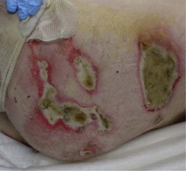
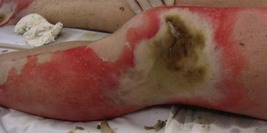
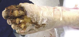
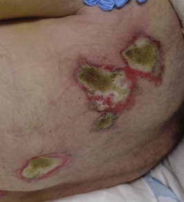
The patient was intubated and ventilated and a urinary catheter inserted in preparation for transport. His blood pressure was 125/80 mmHg with a pulse rate of 130/min. His clothes were removed to allow an accurate assessment to be made of the extent and severity of his injuries. The photographs show the burned areas of his buttocks, left knee and right foot (Figures 10.110.3). There was a 6 cm burn on the scalp with a similar appearance to those on his buttocks. His back had four similar burns. The changes to the left foot resembled those seen on the right foot. Five similar-sized burns were present on the upper limbs. Blood samples were collected for haemoglobin estimation, cross-matching, serum electrolytes and creatinine kinase.

The left calf was ‘rock hard’ on palpation; the left foot was white, cool to touch and displayed a capillary refill time of 5 seconds. His right foot was pink but there were severe burns to the digits with bone and joint exposure. There was a deep defect, an arcing ‘blow-out’ hole, lateral to the left knee. The compartments of the left forearm were ‘tight’. Some surgical interventions are required.

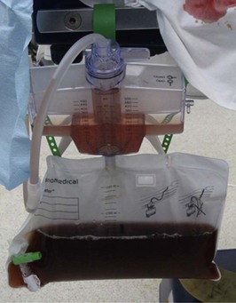
His urine output was 1 mL/kg/hour (appropriate for an adult); its appearance is shown in Figure 10.5.

The patient attended the operating theatre as soon as trauma clearance was completed (21.30). A full assessment of his burns was possible after an aggressive scrub debridement of all skin lesions. The surgeon confirmed left lower leg compartment syndrome and that the hole over the anterolateral aspect of the left knee communicated with the knee joint. The left 1st to 4th toes were non-viable, the sole was cold and white. The right calf compartments were soft. There was a full-thickness burn (~1% total body surface area (TBSA)) over the lateral right ankle. The right 1st to 3rd toes were deeply destroyed and ‘burst’ open to reveal damaged joints and bones. The distal right foot was deeply burned. The left forearm compartments were swollen and tight. Full-thickness burns were noted over the right lateral thigh, lateral to the right knee, left elbow, radial and ulnar left forearm and on the scalp. Fasciotomies were performed medially and laterally to the left lower leg, lateral left thigh and radial and ulnar left forearm. Clearly demarcated non-viable tissue was excised at its visible margins effectively requiring a left forefoot amputation and an equally radical debridement of the right distal foot. Fascial excision of burn over the anterolateral left knee defect was performed and the knee joint was washed out (arthroscopy two days later would show the cartilages to be undamaged). Tangential excision of all deep burns was performed. The scalp burns were debrided down to galea aponeurotica. Superficial burns were recorded at left thigh and right lateral calf. All wounds were dressed.

The terminology for burns has changed and ‘first’, ‘second’ and ‘third degree’ burns are no longer described. The term ‘fourth degree burns’ persists.

Two days later there had been a sharp declaration of the left calf and foot necessitating below-knee amputation – if the arthroscopy had shown severe damage to the left knee joint, an above-knee amputation would have been likely, since this joint was open and the defect required complex free flap repair. This surgery would not have been merited if the knee joint was unlikely to be functional later. Necrosis of deep foot muscles was noted on the right and further debridement was performed. All wounds were redressed. A VAC (vacuum assisted dressing) was applied to the right forefoot debridement.

The patient began to recover physiologically and 4 days after his injury he was extubated. He was transferred to the burns unit 2 days later. He underwent skin grafting to the scalp, left forearm, hip and back wounds which healed well, but the right foot tissues deteriorated and further deep necrosis was evident. He returned to theatre for right below-knee amputation. Prior to amputation, some of the lateral right calf tissue (including the peroneal artery as its blood supply) was used to create a free flap which was used to close the left knee defect (with the flap peroneal artery anastomosed to the artery to the vastus medialis of the left leg).

The patient continued to improve, aided by strong support from family and friends. Approximately 6 weeks after his injury, he was transferred to a rehabilitation facility where he remained for a further 6 weeks. The patient today walks on bilateral below-knee prostheses, is independent and happy. No late sequelae had developed at 6 months post-injury.

Answers

A.1 Since the patient arrived intubated and ventilated, no direct history is available. In any trauma case it is important to interview witnesses, ambulance officers, members of the retrieval team, etc. to obtain as much information as possible about the incident leading up to the injury and what has happened in the time between the injury and arrival at the hospital. The following information is important:

Some information may have to wait until family arrive, or may necessitate a phone call to the patient’s family doctor, such as:

A.2 The initial assessment (primary survey) must assess:

3. Circulation

A secondary survey can now be performed looking for joint disruptions or dislocations, bony fractures and to fully assess the burn size, burn sites and burn depth. The standard techniques for assessing burn size (Wallace rule of nines, Lund and Brouder charts, etc.), while good at assessing cutaneous injuries, tend to underestimate fluid requirements since electrical conduction causes deep tissue injury which cannot be measured with any accuracy. However, skin injury calculation can at least provide a ‘starting’ volume for fluid resuscitation using an approved formula. The modified Parkland formula suggests that the fluid volume (in Hartmann’s solution) needed in the first 24 hours can be calculated by multiplying the patient’s weight in kilograms by the percentage of the total body surface area which has been burnt by 3 or 4 mL. In this case it would be prudent to err on the 4 mL factor, to account in part for the underestimation mentioned above. Thus, with the 25% TBSA cutaneous burn in this 80 kg patient, the fluid requirement in the first 24 hours is 4 × 25 × 80 = 8000 mL, half (4000 mL) to be given in the first 8 hours after injury and the second half over the next 16 hours. All formulae are merely guidelines to be increased or decreased according to the patient’s physical response picked up by monitoring (particularly urine output).

A.3 The burns occupy around 25% of the patient’s total body surface area and are, as usual, heterogenous in depth. The estimation is not easy from the photographs because the burns on the trunk, head and upper limbs were caused by arcing at multiple sites separated by uninjured skin. The burns to the skin of the toes of both feet are ‘fourth degree’ (involving non-skin deep tissue such as vessels, tendons, bones, ligaments and joints). The burn over the lateral left knee is similarly fourth degree since deep tissue disruption to the knee joint capsule is evident). These injuries are devastating and are the commonest reason for amputation in burns practice. More proximally on the feet, the burns are white and dry – these are full thickness (although venous thrombosis is visible below the burn). The burns over the shins and anterior ankles are deep dermal. The only superficial/mid-dermal burns (which could be treated conservatively) are over the anterior thighs. The left buttock, hip, flank and scapular burns are deep full thickness (involving subcutaneous fat), as are the scalp burns.

A.4 Escharotomy is division or ‘opening’ of circumferential full-thickness burns of the limbs or trunk. An escharotomy does not extend through the subcutaneous fat. An escharotomy is performed through incisions running down the midaxial lines but avoiding important superficial structures (ulnar nerve at elbow, common peroneal nerve as it winds around the neck of the fibula, etc.). Fasciotomy is division of the muscular fascia of all affected compartments in a limb injured by severe crush or, in this case, electrical conduction. Skin incisions are designed to afford best exposure of the muscular fascia. The clinical findings mandate fasciotomy release of the left calf muscular compartments and those of the left forearm. Radical debridement of severely disrupted and non-viable tissue of both feet was also performed early (within a few hours). Close and frequent operative review of tissues obviously having conducted large current is necessary. The electrical injury has damaged the left foot to such an extent that the patient is likely to require a left below-knee amputation.

A.5 The urine has a maroon colour which is almost certainly due to the presence of haemochromogens – most likely to be myoglobin, although haemoglobin usually accompanies it. These large proteins are released by damaged muscle and vessels and can result in sluggish blood flow (especially where hypovolaemia is allowed to co-exist). The main problem is the challenge they pose to the kidneys leading to acute renal failure. Intravenous fluid administration should be increased to raise the urine output up to 2 mL/kg/hour rather than the 1 mL/kg/hour normally optimal in adult burn patients during resuscitation; 25 g of mannitol (an osmotic diuretic) can be administered immediately intravenously followed by the addition of 12.5 g to each litre of administered fluid. Finally, intravenous administration of bicarbonate (2 vials stat) alkalinizes the urine, making the pigments more water-soluble and facilitating their excretion. With these manoeuvres, the urine colour lightens from black/dark purple through purple, plum, maroon, dark red/brown, rose, pink, orange and dark yellow on its way to a normal straw colour.

A.6 ‘Fourth degree’ burns involve tissues deep to the subcutaneous fat such as deep vessels (including large arteries), tendons, bones, ligaments and joints. They are devastating and the commonest cause of amputation in burns practice.

A.7 A ‘free flap’ is a composite piece of tissue, which can be made up of skin, fat, fascia, tendon/muscle and bone (in any combination depending on the origin and the defect to be repaired), which is raised with the arterial inflow and venous drainage intact. The vascular pedicle is then divided (usually as far from the flap as possible, giving the greatest pedicle length possible), separating the tissue from its donor site. The artery and vein(s) within the vascular pedicle are then anastomosed to vessels at the recipient site (the defect to be repaired) and the flap inset to fill the defect. The tissue is thus technically a flap because it has an immediate blood supply but a ‘free’ flap because the blood supply has to be established by surgical anastomosis. In this case a composite piece of tissue was raised from tissue planned to be discarded as part of the right below-knee amputation, its blood supply was the peroneal artery which was anastomosed into the vastus medialis artery so the flap could repair the left knee defect.

A.8 The immediate complications of an electrical injury include cardiac arrhythmias (which can cause instant death), skin burns and compression fractures. Important early complications include hypovolaemia secondary to inaccurate assessment of burn size (failing to account for the hidden internal injury), vascular compromise, nerve destruction (muscular paralysis/loss of sensation), muscle breakdown and myoglobinuria leading to acute renal failure, other injuries (including compression fractures of the vertebrae), compartment syndrome and sepsis secondary to inadequate debridement of necrotic tissue.