A 42-year-old woman with hypertension

Published on 10/04/2015 by admin

Filed under Surgery

Last modified 22/04/2025

Print this page

rate 1 star rate 2 star rate 3 star rate 4 star rate 5 star
Your rating: none, Average: 0 (0 votes)

This article have been viewed 1570 times

Problem 6 A 42-year-old woman with hypertension

She was diagnosed with hypertension 8 years ago and this has never been well controlled. She has been on the current treatment regimen for the last 3 years and is compliant. She self-monitors her blood pressure at home and obtains readings between 140/90 mmHg and 200/100 mmHg. She has no explanation for this variation.

She was diagnosed with ‘anxiety’ around the same time the diagnosis of hypertension was made. This diagnosis was based on intermittent episodes of palpitations, shortness of breath, sweating, associated with a feeling of ‘impending doom’. She has had no headaches. She has been diagnosed and managed by her local general practitioner.

In the last 2 years she has experienced central weight gain (10 kg), which has occurred despite diet and exercise. She has never had depression and has not had any problems with concentration. She has not experienced increased facial hair or acne. Her muscle strength has always been good. She slipped and fell 2 months ago and sustained a fractured radius. Her periods are regular. She has two children – aged 15 and 10. Both pregnancies were uncomplicated. She is not on the oral contraceptive and does not take any other medications. She drinks 1–2 standard drinks per week and is a non-smoker. Her mother had diabetes; her father is alive and well. There is no other family history of hypertension or endocrine or renal disease.

On examination she appears slightly anxious, and is tremulous. She is overweight, but there are no clinical features of Cushing’s. Her blood pressure is 170/90 mmHg lying and 160/85 mmHg standing. Her pulse is 110 bpm and regular. She has a forceful, non-displaced apex beat; examination of the cardiovascular system is otherwise unremarkable. There are no abdominal masses or striae visible or bruits audible. She is able to stand from a squat without difficulty. On fundoscopy there is silver wiring.

The following results are available.

Investigation 6.1 Summary of results

Fasting glucose (3.8–5.5 mmol/L) 10
Urea (2.7–8.0 mmol/L) 7.3
Creatinine (50–120 µmol/L) 68
eGFR (mL/min/1.73 m2) >60
Ionized calcium (1.1–1.25 mmol/L) 1.31
Total calcium (2.10–2.55 mmol/L) 2.75
Phosphate (0.65–1.45 mmol/L) 1.27
Plasma metanephrine 18 100 pmol/L (<500 pmol/L)
Plasma normetanephrine 9260 pmol/L (<900 pmol/L)

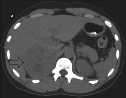
Some imaging studies are arranged and a CT scan of the abdomen performed. A representative slice is shown (Figure 6.1).

She is tolerating phenoxybenzamine well, but has been tachycardic (heart rate 110–120 bpm for the last 2 days).

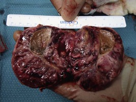
She undergoes a laparoscopic adrenalectomy and the large adrenal tumour is removed intact. As soon as the adrenal vein is ligated the blood pressure falls and the anaesthetist is able to reduce the alpha blockade. The opened surgical specimen is shown (Figure 6.2).

All medications are stopped immediately postoperatively.

Over the next week her blood pressure without medication varies between 100/70 and 125/80 mmHg. The histopathology is consistent with a phaeochromocytoma.

Answers

A.1 In this patient, secondary hypertension should be suspected because she has refractory/resistant hypertension – remaining hypertensive despite three antihypertensive drugs – and has no family history of hypertension. The possible causes of secondary hypertension in this patient include:

She must be investigated for a secondary cause of hypertension.

A.2 The following components of the history should be clarified:

Specific enquiry should be made regarding:

Table 6.1 Phaeochromocytoma – catecholamine excess

Symptoms Signs
‘Classic triad’ – episodic headache, sweating, tachycardia (palpitations) Hypertension (essential or paroxysmal)
Diaphoresis
Tremor
Tachycardia

Table 6.2 Cushing’s syndrome – hypercortisolism

Symptoms Signs

The physical examination should include inspection both for signs of a secondary cause of hypertension and for complications of longstanding hypertension.

A.3 She needs to be investigated for endocrine causes of hypertension – phaeochromocytoma, Cushing’s and Conn’s syndrome.

Investigations:

A.4 The elevated plasma metanephrine levels are diagnostic of phaeochromocytoma. Further biochemical testing (e.g. clonidine suppression testing) is not required. Elevated blood glucose may occur in phaeochromocytoma – a result of insulin resistance produced by the elevated catecholamines. This often resolves after treatment of the phaeochromocytoma. The elevated plasma calcium may be seen in phaeochromocytoma due to catecholamine-induced volume contraction/dehydration or to primary hyperparathyroidism – which may be sporadic (although unusual in this age group) or associated with the phaeochromocytoma as part of a multiple endocrine neoplasia syndrome.

The priority of management of this patient is treatment of the phaeochromocytoma.

With appropriate endocrine consultation the principles of management are:

A.5 The scan shows a well-circumscribed heterogeneous mass behind the liver in the region of the right adrenal gland. The lesion is 10 cm diameter.

A.6 She is consistently tachycardic so a beta-blocker should be introduced (e.g. metoprolol). The definitive management of this patient is adrenalectomy and she should be referred to an experienced adrenal surgeon and anaesthetist.

A.7 Postoperative hypotension may occur which can be managed with fluid loading. Hypoglycaemia may sometimes occur because of the loss of catecholamine-induced suppression of insulin secretion. Other complications include the surgically related problems of atelectasis, infection and deep vein thrombosis.

A.8 Serum metanephrines should be checked to determine whether she has been cured. There is a risk of recurrence – which is highest in younger patients, those with extra-adrenal or familial disease, or bilateral and/or large tumours. Recommendations are increasingly being made that patients with phaeochromocytoma should undergo long-term surveillance (10 years in sporadic phaeochromocytoma and indefinitely in familial disease) (Lenders et al. 2005). This patient will also need a fasting blood glucose and serum calcium levels checked, as both were elevated preoperatively. If she remains hypercalcaemic then she will need investigation for primary hyperparathyroidism. If a repeat fasting blood glucose level is elevated, then this is diagnostic of diabetes mellitus.

Revision Points

Phaeochromocytomas are catecholamine-producing neuroendocrine tumours arising from chromaffin cells of the adrenal medulla (80–85%) or extra-adrenal paraganglia (10–15%; paraganglioma). Phaeochromocytomas are rare, accounting for 0.1–0.6% of all hypertension. Approximately 25% are diagnosed during the investigation of the ‘adrenal incidentaloma’ (an adrenal tumour >1 cm in diameter discovered incidentally by radiological investigations performed for another purpose).

Although phaeochromocytomas are usually sporadic, they may occur as a component of a familial syndrome (neurofibromatosis 1 (NF1), von Hippel–Lindau (VHL), multiple endocrine neoplasia 2 (MEN2), succinate dehydrogenase (SDH) syndromes). Features suggestive of familial disease include young age at diagnosis (age <30), bilateral phaeochromocytomas, family history, associated manifestations of NF1, VHL, MEN2, SDH.

The clinical presentation of phaeochromocytoma is variable. The classic presentation is with episodic headache, sweating and tachycardia. Hypertension may be sustained or paroxysmal. Other symptoms include: palpitations, dyspnoea, anxiety, pallor, orthostatic hypotension, weight loss, polyuria, polydipsia. The initial presentation of a phaeochromocytoma may be with an adrenal incidentaloma – such patients are frequently normotensive and asymptomatic – because the phaeochromocytoma is either non-secretory or secreting only low levels of catecholamines.

Measurements of either plasma-free or 24-hour urine metanephrines are increasingly thought to be among the most sensitive of tests for the diagnosis of phaeochromocytoma. Metanephrines are formed as a result of catecholamine metabolism (most of which occurs within the tumour) – and hence increased sensitivity of metanephrine testing is due to the continuous production of these metabolites. Conversely, the production of catecholamines from phaeochromocytomas is highly variable.

Interpretation of investigations can be made difficult because of medications which alter metanephrine or catecholamine levels (e.g. tricyclic antidepressants, phenoxybenzamine, stimulants – caffeine, nicotine, amphetamines). Where possible medications should be stopped prior to repeat testing.

In the clonidine suppression test clonidine is administered to suppress catecholamine release; plasma catecholamines or metanephrines are measured. This should be able to distinguish increased catecholamine levels due to a phaeochromocytoma from increased levels due to sympathetic activation.

Once a biochemical diagnosis is made, imaging (CT abdomen and pelvis) is needed to localize the tumour. If the imaging is negative then there are two possibilities: (a) the diagnosis of phaeochromocytoma is incorrect or (b) the tumour is extra-adrenal (paraganglioma). Nuclear imaging using a tracer compound, MIBG, can help localize the tumour. MIBG is a compound resembling noradrenaline and is taken up by adrenergic tissue. MIBG scanning can also be useful in detecting metastatic disease.

Once the diagnosis has been established and the tumour localized there are several steps in management: