A 41-year-old man involved in a car crash

Published on 10/04/2015 by admin

Filed under Surgery

Last modified 22/04/2025

Print this page

rate 1 star rate 2 star rate 3 star rate 4 star rate 5 star
Your rating: none, Average: 0 (0 votes)

This article have been viewed 1106 times

Problem 26 A 41-year-old man involved in a car crash

Your examination reveals the following:

Airway (with cervical spine control): the patient’s airway is patent, he is speaking to you and has no evidence of facial trauma or airway foreign body. His cervical spine is immobilized with a hard collar.

Breathing: he has a respiratory rate of 30 bpm and his pattern of breathing is shallow. He is not centrally cyanosed. Examination of his chest reveals superficial abrasions and contusion to his left anterior hemithorax and shoulder. There is no evidence of paradoxical chest movement or open wound. He is tender over the anterior aspects of the left 6th–9th ribs and crepitus is present. There is a paucity of breath sounds on the left side and the percussion note is increased on that side. His trachea is central. Pulse oximetry measurement reveals his oxygen saturation is 99% on 15 litres of O2 by non-rebreather mask.

Circulation: he has a pulse rate of 130/min and a blood pressure of 105/85 mmHg. His jugular venous pressure is not elevated and he has a peripheral capillary return time of 3–4 seconds. He has no obvious external haemorrhage or major long bone fractures. The examination of his abdomen reveals bruising consistent with a seatbelt injury and generalized tenderness of both upper quadrants, maximally on the left, with marked guarding.

Disability: a rapid skeletal and neurological survey shows that the patient has no obvious deformity or tenderness of his limbs, has normal power in all limbs and has normal sensation and reflexes. His Glasgow Coma Scale score (GCS) is 14/15, with his verbal score being confused (4/5). His pupils are equal and react normally to light. You also note the odour of alcohol. Breath alcohol analyser shows a breath alcohol level (BAL) of 0.210 g/dL. He has no neck pain or midline cervical tenderness.

Exposure/environmental control: the patient’s temperature is 36.1°C. In order to facilitate examination and X-rays his shirt and trousers’ are cut off. He complains that he is cold in the resuscitation room.

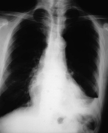
A second wide-bore intravenous cannula is inserted into a peripheral vein and 1 L of a crystalloid solution administered rapidly. Blood samples are collected for cross-matching, complete blood picture and biochemistry. In addition forensic blood alcohol samples are obtained. The chest X-ray is shown in Figure 26.1.

The chest X-ray confirms your clinical suspicion of a haemopneumothorax. A chest drain is inserted successfully and connected to an underwater seal. Air and approximately 500 mL blood drain out. The patient confirms that his breathing is much improved.

A log roll and inspection of the patient’s back is subsequently performed which is unremarkable. FAST scan done on arrival is equivocal.

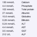
The initial laboratory results are also unremarkable and his haemoglobin is 131 g/L.

One hour after admission and initial resuscitation you reassess the patient and note that although he has received a total of 2 L intravenous fluid and his pain is adequately controlled with parenteral opiates, his pulse has risen to 145/min and his blood pressure is 90/75 mmHg. His JVP is not visible. His airway and breathing are stable. Examination of his chest shows reasonable air entry to all areas, but there is still dullness at the left base. There is a further 100 mL fresh blood in the chest drainage bottle. His abdomen is markedly tender in the left upper quadrant.

A further haemoglobin estimation is performed. It is now 102 g/L. He is rapidly transfused 2 units blood and a further 1 L crystalloid. His urine dipstick is negative for blood and a urinary catheter is inserted with hourly urine output monitored. His blood pressure stabilizes at 105/80 mmHg with a pulse of 120 bpm.

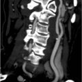
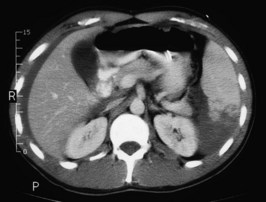
A CT scan of his head, cervical spine, chest, abdomen and pelvis is performed. A slice from the upper abdomen is shown in Figure 26.2.

The patient is transfused a further 2 units of blood. He remains stable, his tachycardia settles and so it is decided to trial treating his splenic injuries conservatively.

He is admitted to an intensive care unit where he is monitored closely and has an uneventful course. His chest drain is removed at day 5. His splenic injury is monitored with a further CT, which showed gradual reabsorption of the haematoma. He is discharged home 2 weeks after the accident and advised to avoid strenuous exertion for a further month.

Answers

A.1 This patient has been involved in a high-speed crash and is likely to be suffering from multiple traumatic injuries. In order to avoid overlooking significant injuries and to treat the most life-threatening conditions first it is wise to take a structured approach. Advanced Trauma Life Support® (also known as Early Management of Severe Trauma®) is one such system. This system relies on:

The mechanism of injury is especially important. You should establish the speed and type of vehicle and the mode of impact, i.e. direction and whether the car hit a mobile or immovable object. Also, the condition of the vehicle after the accident, the provision and use of safety features, the position of the patient, the ability to self-extricate or the time taken to release trapped victims are all important features and may give valuable information as to the likely nature and severity of injuries. Head injuries and alcohol intoxication may result in an altered mental state which may preclude the patient from being able to give a precise history or report pain, e.g. from a cervical vertebrae fracture.

A.2 As always in any resuscitation your priorities are Airway, Breathing and Circulation, with Disability and Exposure/environmental control of additional importance in trauma.

The assessment of this patient reveals no imminent problems with A and the cervical spine is being appropriately managed with hard collar immobilization. Further investigation of the cervical spine beginning with cross-table lateral X-ray should be arranged.

The assessment of B demonstrates problems that need to be addressed rapidly. The chest wall shows obvious signs of trauma with probable rib fractures of at least three ribs, the underlying lung is probably damaged and the increase in percussion note on the left side suggests a pneumothorax. The patient does not appear to be deteriorating rapidly, and the pneumothorax does not appear to be under tension. Therefore it is reasonable, in this case, to proceed to imaging of the chest to rule out diaphragmatic rupture and intrathoracic abdominal contents before proceeding to chest drain insertion. Whilst the X-ray is performed you ask for a chest drain set and underwater seal to be prepared and you proceed to assess C.

C: this is a relatively young patient who will have a good cardiovascular reserve. This means that even when bleeding and shocked, he can initially maintain his blood pressure and central perfusion. As he has a marked tachycardia and is diverting his blood from his peripheries (prolonging his capillary return), restoration of his circulating volume should be started immediately. Two large-bore peripheral cannulae should be adequate at this stage. After determining a baseline haemoglobin via a point-of-care haemoglobin analyser, blood samples should be sent for haematology, biochemistry and urgent six-unit cross-match. Coagulation studies may be of benefit if massive transfusion (the loss of one blood volume in 24 hours or 50% of blood volume in 3 hours) is thought likely. Many jurisdictions require all patients involved in motor vehicle accidents to have forensic blood alcohol samples taken. A large volume (1–3 L) of intravenous crystalloid (e.g. normal saline) should be infused over the first hour, titrated to physiologic response.

The history of injury to the left side of the torso must raise the possibility of damage to the rib cage and the abdominal wall, and the contents in the immediate vicinity. Any trauma of sufficient force to break ribs, particularly if associated with shock, may well have produced substantial internal injury.

Disability: a rapid skeletal and neurological survey shows that the patient has no obvious deformity or tenderness of his limbs and has normal sensation. Assessment of D may not show any immediate acute problems, but should be repeated later.

Exposure/environmental control: it is important to adequately and rapidly expose the patient so as to not miss injuries or other examination findings and to allow full access for investigations and interventions. This must be tempered with ensuring the patient is not allowed to become hypothermic so use of passive warming devices, e.g. blankets, or active warming, e.g. warm intravenous fluids, may need consideration.

Apart from X-rays of the chest and cervical spine, a pelvic X-ray should be performed, especially in view of his haemodynamic instability. An ECG should also be performed to look for evidence of cardiac contusion. Provided it can be arranged without compromising immediate patient care, a CT scan of the neck, chest and abdomen (including pelvis) will yield more information than plain radiology.

A.3 The X-ray shows collapse of the upper part of the left lung with air in the pleural space from the mid-zone to the apex. There is blunting and flattening of the left costophrenic angle, suggesting a haemopneumothorax. There are fractures of the 6th–8th ribs.

In this situation the following potential complications must be remembered:

Flail Segment

Multiple ribs fractured in more than one place prevent that section of the chest moving normally. The affected section of the chest moves paradoxically and can limit normal ventilation. It is not this that causes hypoxia, but the associated severe contusion to the underlying lung. Contused pulmonary tissue is very sensitive to both under- and over-perfusion. Very careful fluid balance is needed in these cases. A short period of assisted ventilation (which may be non-invasive or invasive) may be required to optimize ventilation and perfusion of the affected lung.

A.4 Despite your initial resuscitation this patient has become hypotensive and this is likely to be due to hypovolaemia. While other causes of hypotension must be considered, in this setting the cause of the problem is almost certainly blood loss. The patient requires:

Apart from a further clinical examination, imaging studies must be considered. The two main imaging methods used in these circumstances are CT scan and ultrasound.

If available, ultrasound examination in the emergency department (FAST scan – focused assessment with sonography of trauma) by the emergency physician or trauma surgeon is useful in the unstable patient, and can be performed in the resuscitation cubicle. It is particularly useful for rapidly detecting free fluid (e.g. haemorrhage) in the abdomen and has largely replaced the more invasive diagnostic peritoneal lavage.

A CT scan with intravenous contrast is the imaging investigation of choice, and will aid in defining internal injuries including splenic injury. A CT scan showing a contained splenic bleed with acceptable patient haemodynamics may allow non-operative management and spleen preservation. This might include splenic embolization. If the patient is significantly haemodynamically unstable moving the patient out of the emergency department or resuscitation area for an imaging investigation may be dangerous. In this case a diagnosis of ruptured internal organ may be made solely on clinical grounds and the patient moved straight to theatre from the emergency department.

When scanning this patient it is likely that he would receive a ‘full body scan’, i.e. a scan of his head to look for cerebral contusions or bleeds, of the cervical spine to clear his neck (see Issues to Consider), of the chest to look at the extent of chest injury, and of the abdomen and pelvis to assess for intra-abdominal injury.

A.5 This is a section through the upper abdomen. Free blood can be seen between the right lobe of the liver and the abdominal wall. There is a large haematoma within the spleen, particularly involving the lower pole. The laceration goes towards the splenic hilum and most of the splenic capsule appears to be intact.