A 31-year-old man with sudden onset headache and vomiting

Published on 10/04/2015 by admin

Filed under Surgery

Last modified 22/04/2025

Print this page

rate 1 star rate 2 star rate 3 star rate 4 star rate 5 star
Your rating: none, Average: 0 (0 votes)

This article have been viewed 869 times

Problem 46 A 31-year-old man with sudden onset headache and vomiting

On arrival he is alert but agitated, disorientated to time and obeying commands. He is afebrile, but has marked neck stiffness. His heart rate is 52/min and blood pressure 130/80 mmHg. Neurological examination shows no cranial nerve or focal neurological deficit. On investigation his haemoglobin is 127 g/L, white cell count 8.4 and platelets 111 000. His serum biochemistry (including calcium and glucose) is within normal limits and a serum troponin is negative. His chest X-ray is normal and an ECG shows sinus bradycardia with T-wave inversion.

The patient required intravenous morphine and midazolam for the radiological investigation.

On the advice of the consulting neurosurgeon, the patient was retrieved to the nearest neurosurgery centre and had further investigations to determine the cause of the subarachnoid haemorrhage.

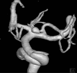
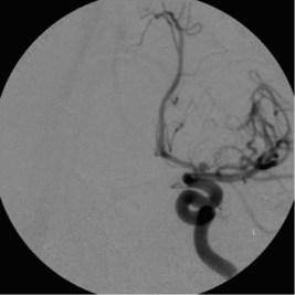
Endovascular coiling was attempted in the patient but was technically unsuccessful.

The patient underwent craniotomy and clipping of the aneurysm which was successful. The patient was making good progress until the third postoperative day when he became progressively confused, verbally abusive and drowsy.

The patient’s blood pressure was 140/80 mmHg, temperature 38.6°C and oxygen saturation 100% on room air. A CT head showed no new intracerebral haemorrhage, no hypodensity consistent with infarction or hydrocephalus. The angiogram did not show any evidence of vasospasm. The serum sodium was 121 mmol/L, potassium 4.6 mmol/L, osmolality 266 mmol/kg (270–300), urine osmolality 116, urine sodium 177 mmol/L (40–100).

The patient was transferred to the high dependency unit for administration of hypertonic saline, monitoring of arterial blood pressure and fluid balance monitoring and oral sodium replacement. The patient progressively improved and was transferred back to his local hospital 3 weeks following his haemorrhage. He was reviewed 3 months following surgery and was doing well. He was smoking again.

Answers

A.1 This is a young man who has suddenly collapsed with vomiting and headache and therefore subarachnoid haemorrhage (SAH) should be considered the diagnosis of exclusion in this patient. Primary intracerebral haemorrhage, secondary to an arteriovenous malformation or hypertensive haemorrhage should also be considered, given the young age. The possibility of a traumatic intracerebral haemorrhage, e.g. subdural haematoma, particularly in a patient with unclear history and a history of alcohol abuse, needs to be considered. Intracranial infection, e.g. meningitis, would be less likely to present with such an acute ictus.

A.2 The neurological examination of any patient has two key aims – to establish the diagnosis and to determine the site of the lesion.

Focus on:

A.3 A CT head scan with and without contrast needs to be performed urgently in any patient in whom subarachnoid haemorrhage is in the differential diagnosis. If the CT cannot be performed at the receiving facility then the patient should be retrieved to an appropriate facility as soon as possible. Patients who are agitated may require intravenous sedation and even intubation to facilitate or even safely perform this test. It is preferable to avoid intubation to allow careful neurological assessment to be made.

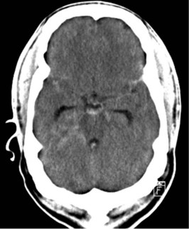
A.4 The CT head shows diffuse subarachnoid haemorrhage around the brainstem.

A.5 The first is a CT angiogram and shows a left terminal internal carotid artery aneurysm. The second test is a cerebral angiogram (digital subtraction angiogram) which shows the aneurysm at the termination of the left internal carotid artery – at the point where it bifurcates into the middle and anterior cerebral arteries.

A.6 Endovascular coiling and craniotomy and surgical clipping of the aneurysm are the two choices. Both techniques can be effective in securing the aneurysm and preventing rebleeding and death. Important variables that may favour one technique over the other include the relative neck to dome ratio (coiling more favourable with a narrow neck), ease of access to the aneurysm location (posterior circulation aneurysms are more difficult surgically), and the presence of active vasospasm and brain swelling (surgical retraction may be more hazardous).

A.7 For 3 weeks following aneurysmal SAH the patient is at potential risk for a rebleed (if the aneurysm is not secured), cerebral vasospasm, fluid and electrolyte disturbance and postoperative surgical and medical complications, including seizures, wound or systemic infection. The history should determine when the deterioration occurred (vasospasm unlikely before 3 days, peaking at 7–10 days and less likely after 2 weeks), whether the deterioration occurred gradually (electrolyte disturbance or progressive ischaemia from vasospasm) or suddenly (acute vascular events or seizures). Vital signs, wound review and a detailed neurological exam will establish important changes from prior baseline. Electrolytes, cardiac enzymes, full blood count, ECG, CXR would be appropriate. An urgent CT with angiography is essential.

A.8 The patient has hyponatraemia. Following SAH hyponatraemia is most commonly due to either cerebral salt wasting (CSW) or the syndrome of inappropriate antidiuretic syndrome (SIADH). The former is currently believed to be due to release of atrial natriuretic (ANF) following SAH which results in the loss of urinary sodium and also water through the kidneys. The distinction can be made by examining the urinary sodium excretion which is well above normal in CSW as well as there being clinical and biochemical evidence of hypovolaemia (reduced CVP, elevated haematocrit). The treatment of CSW is sodium and volume replacement together and for SIADH, volume restriction. A potential hazardous situation may arise if an erroneous diagnosis of SIADH is made, the patient is fluid restricted and hypovolaemia is exacerbated resulting in inadequate cerebral perfusion and ischaemia. If in doubt treatment for CSW with volume and sodium replacement is safer.

A.9 Risk factors for the formation of aneurysms include a prior aneurysm, smoking, cocaine use, hypertension, a family history of aneurysms and some inherited connective tissue disorders, e.g. Marfan’s syndrome. Up to 20% of patients who have one aneurysm may have another at some point in the future. The most modifiable of all the patient’s risk factors will be to cease smoking.