A 25-year-old woman with chest pain and breathlessness

Published on 10/04/2015 by admin

Filed under Surgery

Last modified 22/04/2025

Print this page

rate 1 star rate 2 star rate 3 star rate 4 star rate 5 star
Your rating: none, Average: 0 (0 votes)

This article have been viewed 1328 times

Problem 29 A 25-year-old woman with chest pain and breathlessness

On further questioning, the patient reports having had left calf pain for 1 month but she thought it was a ‘cramp’. She has had no episodes of haemoptysis or syncope. She has not had recent surgery. There is no known family history of heart or lung problems. She is compliant with her oral contraceptive pill and she is not pregnant. She has not embarked on any recent long distance travel. She has had a recent papsmear which was negative for malignancy.

Complete blood picture and baseline electrolytes are normal.

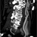
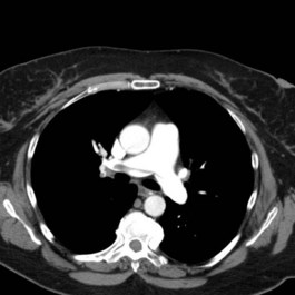
An imaging study is undertaken and two images from the sequence are shown (Figure 29.2). Her left Doppler showed an extensive clot into proximal femoral veins.

The nurse asks you to review the patient again because she is concerned that her BP is now 85/60 mmHg. Her pulse has increased to 128 beats/min with no central or peripheral cyanosis. She appears distressed and her oxygen saturations are 90% on 2 L/min O2 with use of nasal prong. In addition to her raised JVP, you palpate a left parasternal heave. A third heart sound is now heard (gallop rhythm). You order an urgent bedside echocardiogram and the sonographer tells you the right ventricle is moderately dilated with moderate RV dysfunction and RVSP of 40 mmHg.

Intravenous unfractionated heparin loading dose is given calculated to your institution’s weight-based protocol. O2 is increased to 10 L with non-rebreather mask. The patient is taken to ICU. Thrombolysis is given with alteplase. Within 4 hours the patient is normotensive, pulse rate 82 and O2 requirements have reduced and her JVP is not visible. The next day the patient is transferred to your ward with IV heparin running.

Oral warfarin is commenced early, while on IV heparin. Once her INR has been within the therapeutic range for 2 days, the IV heparin is ceased. An echocardiogram is repeated and shows normalization of right ventricular size and function. Prior to discharge, her thrombophilic screen of tests is reported as showing no abnormalities.

Advice is provided about her condition and taking warfarin tablets. The hospital’s outpatient warfarin clinic is going to monitor her INR, and an alert bracelet is arranged for her to wear oral contraceptive pill is ceased and referal to gynaecologist regarding progestrone only contraception.

The woman returns to see you in the outpatient clinic after 3 months. She has been well and no longer experiences chest pain or breathlessness on exertion. Her INR has been well maintained within the therapeutic range. She has quit smoking.

Answers

A.1 This young lady has pleuritic chest pain, dyspnoea and hypoxia (based on pulse oximetry) unexplained by her chest X-ray.

Further specific questions should be asked to determine her pre-test probability of DVT/PE:

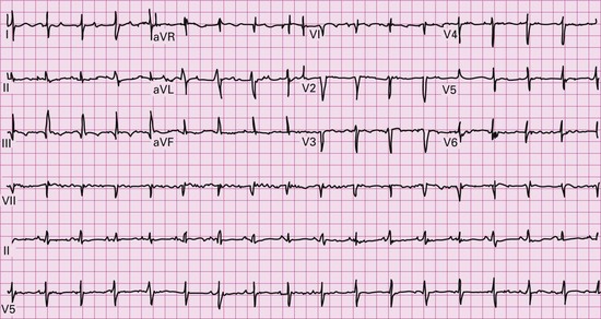
A.2 The following investigations must be undertaken:

A D-dimer blood test is not appropriate in this case as it is only useful in excluding a diagnosis of PE when there is a low pre-test clinical probability. This lady has known risk factors for and a clinical picture consistent with PE; therefore, specific imaging should be performed. A CTPA should be chosen in this setting because there is atelectasis on her chest X-ray which makes a V/Q scan difficult to interpret.

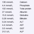
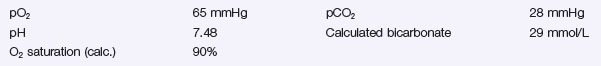
A.3 The arterial blood gases show the following:

This is type 1 respiratory failure, and suggests an acute event, causing hypoxia. The patient then hyperventilates to compensate, and releases carbon dioxide leading to hypocapnia and an alkalosis. A similar pattern may be seen in acute asthma, pneumothorax and pulmonary oedema.

This ECG shows the classically quoted changes seen with PE of S-wave in lead I with a Q-wave and T-wave inversion in lead III (S1Q3T3). However, this is a rare occurrence and the most common abnormality seen is sinus tachycardia. More extensive emboli often result in new-onset atrial fibrillation.

A.4 This high-definition image confirms the diagnosis of PE showing clot at the bifurcation of the pulmonary artery into its right and left main branches (saddle PE).

The signs of PE will generally depend on the size of the embolus and underlying co-morbidities. Large, haemodynamically significant PE, as in this case, can be associated with:

A.5 This patient requires emergency resuscitation. You need to increase the oxygen requirements, anticoagulate the patient with IV heparin and call for help from senior colleagues. Blood should be sent for thrombophilic screen to detect underlying abnormalities of coagulation prior to commencing heparin. These include:

Testing for Prothrombotic Disorders

ANA, antinuclear antigen; CRP, C reactive protein; ENA, extractable nuclear antigen; MTHFR, methylenetetrahydrofolate reductase.

Immediate anticoagulation is the usual treatment for PE. In this case, unfractionated IV heparin should be given to achieve rapid anticoagulation.

Thrombolyis is indicated for life threatening PE when associated with one or more of the following features:

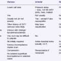
Thrombolysis should be performed in consultation with a major centre experienced in PE management.

This young woman is in impending cardiogenic shock (as defined above). Provided there are no contraindications, IV thrombolysis should be given and the patient should be transferred to HDU/ICU. Rarer treatment options that are only available in highly specialized centres include surgical embolectomy (where the clot is surgically removed) and catheter fragmentation (where the clot is broken up into smaller pieces with a special device) in patients with contraindications to thrombolysis.

Contraindications to Thrombolysis

Relative

A.6 Intravenous (systemic) alteplase was chosen for thrombolysis as this has been shown in clinical trials to be effective in treatment of acute massive PE with right ventricular dysfunction.

Baseline coagulation studies are important (INR and APTT). A prolonged APTT prior to heparin is a common presentation of a lupus anticoagulant, and may indicate an underlying thrombophilic disorder. Thrombophlia is a very important diagnosis as it will mean the APTT cannot be used to monitor the heparin therapy if unfractionated heparin is to be used.

She requires ongoing anticoagulation both in the short and long terms.

A.7 The patient should be issued with an information card about warfarin, a medical alert bracelet, and advised not to commence any new medications without first talking to a doctor. She must understand that warfarin can interact with many drugs. Alcohol, antibiotics, and non-steroidal anti-inflammatory drugs (NSAIDs) are common culprits in these instances. Also, contraception while on warfarin is essential as it is teratogenic in the first trimester. The oral contraceptive pill has been associated with increased risk of venous thrombosis, and expert opinion of alternative contraception methods should be sought (e.g. intra-uterine device, barrier methods, etc. depending on patient preference).

The patient should also be encouraged to lose weight through regular exercise and a good diet. She should have regular breast examinations and pap smears to ensure that an underlying malignancy does not become apparent. Furthermore, she should be helped to stop smoking.

A.8 The minimum treatment duration of warfarin therapy is 3 months which is appropriate in situations where there has been a short-lived risk factor such as recent surgery or orthopaedic injury.

A V/Q should be performed between 3 and 6 months after acute PE to reassure you that there has been resolution of thrombus as evidenced by normalization of perfusion.

A proportion of patients with life threatening PE go on to develop high blood pressure in the pulmonary circulation, known as chronic thromboembolic pulmonary hypertension (CTEPH). This is a rare but serious complication that can occur after acute PE and is often missed. CTEPH should be considered in patients who present with progressive breathlessness who have a distant history of PE or DVT. CTEPH is life-threatening but potentially curable with surgery called pulmonary thromboarterectomy.

Revision Points

Pulmonary Embolism