[level-membership-for-anesthesiology-category]
Neonatal cardiovascular physiology
Fetal circulation
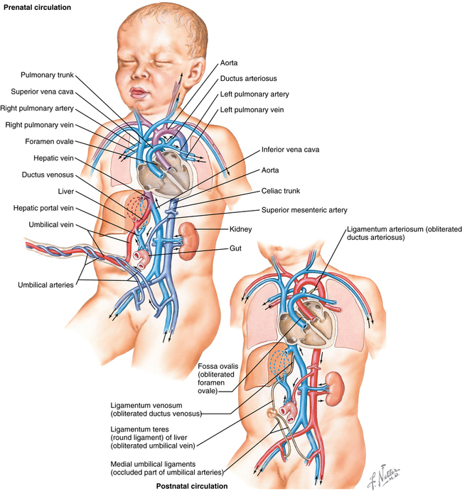
From the placenta, blood with a partial pressure of oxygen (PO2) of 30 to 35 mm Hg flows to the fetus via the umbilical vein (UV) (Figure 191-1), which, in the liver of the fetus, separates into two branches, with one branch joining the portal vein and the other becoming the DV, which joins the inferior vena cava (IVC). Approximately 30% to 50% of the oxygenated blood flowing through the UV will bypass the liver and flow directly through the DV into the IVC, flowing along its posterior wall. As this oxygenated blood enters the right atrium, it is directed across the FO into the left atrium by the eustachian valve, flowing through the left ventricle (∼35% of fetal circulation) into the aorta to supply the head and upper torso.
[/level-membership-for-anesthesiology-category][not-level-membership-for-anesthesiology-category]
Neonatal cardiovascular physiology
Fetal circulation
From the placenta, blood with a partial pressure of oxygen (PO2) of 30 to 35 mm Hg flows to the fetus via the umbilical vein (UV) (Figure 191-1), which, in the liver of the fetus, separates into two branches, with one branch joining the portal vein and the other becoming the DV, which joins the inferior vena cava (IVC). Approximately 30% to 50% of the oxygenated blood flowing through the UV will bypass the liver and flow directly through the DV into the IVC, flowing along its posterior wall. As this oxygenated blood enters the right atrium, it is directed across the FO into the left atrium by the eustachian valve, flowing through the left ventricle (∼35% of fetal circulation) into the aorta to supply the head and upper torso.
[/not-level-membership-for-anesthesiology-category]
