[level-membership-for-anesthesiology-category]
CHAPTER 2 Respiratory and Pulmonary Physiology


2 What is closing capacity? What factors affect the closing capacity? What is the relationship between closing capacity and functional residual capacity?
5 Discuss the factors that affect the resistance to gas flow. What is laminar and turbulent gas flow?
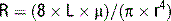
where R is resistance, L is the length of the tube, μ is the viscosity, and r is the radius of the tube. At higher flow rate (in obstructed airways and heavy breathing), the flow is turbulent. At these flows the major determinants of resistance to flow are the density of the gas (ρ) and the radius of the tube, r.

6 Suppose a patient has an indwelling 7-mm endotracheal tube and cannot be weaned because of the increased work of breathing. What would be of greater benefit, cutting off 4 cm of endotracheal tube or replacing the tube with one of greater internal diameter?

10 Review the different zones (of West) in the lung with regard to perfusion and ventilation


11 What are the alveolar gas equation and the normal alveolar pressure at sea level on room air?
The alveolar gas equation is used to calculate the alveolar oxygen partial pressure:

where PAO2 is the alveolar oxygen partial pressure, FiO2 is the fraction of inspired oxygen, Pb is the barometric pressure, PH2O is the partial pressure of water (47 mm Hg), PaCO2 is the partial pressure of carbon dioxide, and RQ is the respiratory quotient, dependent on metabolic activity and diet and is considered to be about 0.825. At sea level the alveolar partial pressure (PAO2) is:

Knowing the PaO2 allows us to calculate the alveolar-arterial O2 gradient (A-a gradient). Furthermore, by understanding the alveolar gas equation we can see how hypoventilation (resulting in hypercapnia) lowers PaO2, and therefore PaO2.
12 What is the A-a gradient and what is a normal value for this gradient?

A normal A-a gradient is estimated as follows:

14 What are the causes of hypoxemia?


19 Define absolute shunt. How is the shunt fraction calculated?

where Qs is the physiologic shunt blood flow per minute, Qt is the cardiac output per minute, CiO2 is the ideal arterial oxygen concentration when V/Q = 1, CaO2 is arterial oxygen content, and CvO2 is mixed venous oxygen content. It is estimated that 2% to 5% of cardiac output is normally shunted through postpulmonary shunts, thus accounting for the normal alveolar-arterial oxygen gradient (A-a gradient). Postpulmonary shunts include the thebesian, bronchial, mediastinal, and pleural veins.
21 Calculate arterial and venous oxygen content (CaO2 and CvO2)

where 1.34 is the O2 content per gram hemoglobin, SaO2 is the hemoglobin saturation, [Hgb] is the hemoglobin concentration, and PaO2 is the arterial oxygen concentration.

and

 ) (70%), and combined with hemoglobin (23%).
23 How is PCO2 related to alveolar ventilation?

where VCO2 is total CO2 production and Valveolar is the alveolar ventilation (minute ventilation less the dead space ventilation). In general, minute ventilation and PCO2 are inversely related.

27 What are the causes of hypercarbia?


1. Barash P.G., Cullen B.F., Stoelting R.K. Clinical anesthesia. Philadelphia: Lippincott Williams & Wilkins, 2006;790-812.
2. Wilson W.C., Benumof J.L. Respiratory physiology and respiratory function during anesthesia. In: Miller R.D., editor. Miller’s anesthesia. Philadelphia: Churchill Livingstone; 2005:679-722.
[/level-membership-for-anesthesiology-category][not-level-membership-for-anesthesiology-category]
CHAPTER 2 Respiratory and Pulmonary Physiology
2 What is closing capacity? What factors affect the closing capacity? What is the relationship between closing capacity and functional residual capacity?
5 Discuss the factors that affect the resistance to gas flow. What is laminar and turbulent gas flow?
where R is resistance, L is the length of the tube, μ is the viscosity, and r is the radius of the tube. At higher flow rate (in obstructed airways and heavy breathing), the flow is turbulent. At these flows the major determinants of resistance to flow are the density of the gas (ρ) and the radius of the tube, r.
6 Suppose a patient has an indwelling 7-mm endotracheal tube and cannot be weaned because of the increased work of breathing. What would be of greater benefit, cutting off 4 cm of endotracheal tube or replacing the tube with one of greater internal diameter?
10 Review the different zones (of West) in the lung with regard to perfusion and ventilation
 Zone 1: Alveolar pressure (PAlv) exceeds pulmonary artery pressure (Ppa) and pulmonary venous pressure (Ppv), leading to ventilation without perfusion (alveolar dead space) (PAlv > Ppa > Ppv).
[/not-level-membership-for-anesthesiology-category]
