[level-membership-for-internal-medicine-category]
208 Old choroiditis
Salient features
Examination
• Old or inactive retinochoroiditis appears as white, well-defined areas of chorioretinal atrophy with pigmented edges (caused by proliferation of retinal pigment epithelium) (Fig. 208.1B)
• The retinal blood vessels pass over the lesions undisturbed.
Questions
[/level-membership-for-internal-medicine-category][not-level-membership-for-internal-medicine-category]
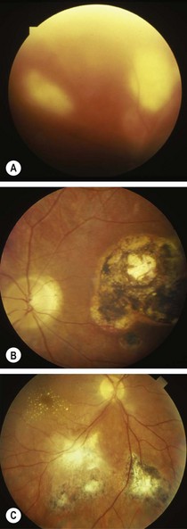
208 Old choroiditis
Salient features
Examination
• Old or inactive retinochoroiditis appears as white, well-defined areas of chorioretinal atrophy with pigmented edges (caused by proliferation of retinal pigment epithelium) (Fig. 208.1B)
• The retinal blood vessels pass over the lesions undisturbed.
Buy Membership for Internal Medicine Category to continue reading. Learn more here
[/not-level-membership-for-internal-medicine-category]
