[level-membership-for-obstetrics-gynecology-category]
Uterine Sarcoma
Synonyms/Description
Etiology
Ultrasound Findings
Differential Diagnosis
Clinical Aspects and Recommendations
Figures
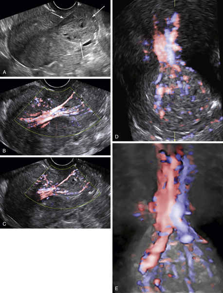
Figure U2-1 A, Adenosarcoma presenting as a mass (arrows) protruding through the cervical canal. Note the small cystic areas within the solid matrix of the mass. B and C show abundant color flow with a large stalk originating from the uterine fundus. D and E show a 3-D image of the vasculature of the mass as it attempts to pass through the cervix.
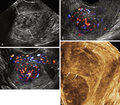
Figure U2-2 Endometrial stromal sarcoma. A, Note that the solid mass (arrows) is largely intraluminal; however, the color flow images (B and C) show abundant blood flow coming from the uterine fundus where the borders of the mass are ill-defined. D is a 3-D rendered image of the endometrial cavity showing a sharp border on one side but no real border (arrows) at the right fundus where the tumor appears to reach the serosa.
Suggested Reading
Seddon B.M., Davda R. Uterine sarcomas—recent progress and future challenges. Eur J Radiol. 2011;78(1):30–40.
Shah S.H., Jagannathan J.P., Krajewski K., O’Regan K.N., George S., Ramaiya N.H. Uterine sarcomas: then and now. AJR Am J Roentgenol. 2012;199(1):213–223.
Wu T.I., Yen T.C., Lai C.H. Clinical presentation and diagnosis of uterine sarcoma, including imaging. Best Pract Res Clin Obstet Gynaecol. 2011;25(6):681–689.
Xue W.C., Cheung A.N. Endometrial stromal sarcoma of uterus. Best Pract Res Clin Obstet Gynaecol. 2011;25(6):719–732.
[/level-membership-for-obstetrics-gynecology-category][not-level-membership-for-obstetrics-gynecology-category]
Uterine Sarcoma
Synonyms/Description
Etiology
Ultrasound Findings
[/not-level-membership-for-obstetrics-gynecology-category]
