[level-membership-for-physical-medicine-and-rehabilitation-category]CHAPTER 99
Complex Regional Pain Syndrome
Jiaxin Tran, MD; V.S. Ramachandran, MD, PhD; Eric L. Altschuler, MD, PhD
Definition
Complex regional pain syndrome (CRPS) is a perplexing medical condition. CRPS begins when a patient has an injury—to a nerve, bone, soft tissue, or connective tissue. The injury heals as assessed by both clinical examination and imaging, yet the patient still has pain significantly out of proportion not only to the healed injury but at times even to the injury itself. In addition to pain, the patient can have hyperesthesia or allodynia at the site of the injury. Curiously, the temperature of the affected limb can be different from that of the other side, and there can be hair, nail, and skin changes. In some cases, these changes can spread beyond the injury site to the entire limb, and in the worst case, in addition to excruciating pain and hyperesthesia, the skin becomes pachydermic.
Before 1994, this bizarre disease and constellation of symptoms was known, among other names, as reflex sympathetic dystrophy. Despite this moniker, it was never proved that the sympathetic nervous system mediates all the components of the disease, nor was a peripheral reflex or feedback loop causing the disease found. In 1994, a conference was held to clarify the classification of the disease [1]. It was renamed CRPS with two classifications, CRPS II for apparent major nerve injury and CRPS I for no major nerve injury. The disease is complex, and it can often be regional rather than confined to a dermatome, myotome, or territory of a single peripheral nerve. Pain is the hallmark of the disease and, for a patient with history of an injury, the sine qua non of diagnosis. It is multifaceted.
The new name, interestingly, is less specific than the former name, expressing the current lack of a full understanding of the disease. More than a decade ago, one of us (V.S.R.) suggested that central remapping of sensory neurons in the cerebral cortex in response to a peripheral injury to the corresponding body area might be a root cause of pain and ultimately other problems in this condition [2]. This hypothesis has not only collected evidence since but led to new and potentially most useful treatments [3].
CRPS is relatively rare. Even with fracture, the most common inciting event, only 1% of patients develop CRPS [4]. The incidence has been estimated to be up to 26 per 100,000 person-years [5]. The incidence of CRPS type I is generally higher than that of CRPS type II. The lower extremity is more often affected than the upper extremity. Female sex, adult age, postmenopausal status, and smoking are all risk factors for its development [6]. In the pediatric population, the incidence of CRPS increases just before puberty [7].
The pathophysiologic mechanism of the disease is, again, poorly understood. There is emerging evidence that CRPS does not only affect the peripheral nervous system. Functional and structural reorganization [8] of the central nervous system has been demonstrated with functional magnetic resonance imaging [9], single-photon emission computed tomography, electroencephalography, and transcranial magnetic stimulation mapping. Neuroplasticity of both the cortical somatosensory [10] and motor [11] systems is implicated. Distorted sensory and motor mapping is hypothesized to promote spontaneous pain and neglect symptoms in CRPS.
Symptoms
CRPS is a clinical diagnosis. The diagnosis can be suggested by the history of present illness alone. An individual experiences an injury. The injury can come in a variety of forms, whether traumatic (e.g., crush injury, gunshot wound, burn, or protracted labor [12]) or nontraumatic (e.g., leprosy [13]). The “injury” can also be an otherwise uncomplicated surgery. While healing takes place, the individual develops pain variably progressive in intensity, area involvement, and duration. Patients with both CRPS type I and type II may report neuropathic pain that is often intense, constant, burning, and present even without stimulation or movement. Symptoms are not confined to a particular nerve or anatomic territory; CRPS can spread [14] to the contralateral limb and even progress to all four limbs. In addition to pain, motor complaints are common and include weakness, cramps, and stiffness. There may also be signs of neglect [15], or more subtly, some patients may refer to the affected limb in the third person (e.g., “it is not moving”). It is important to ask about abnormal sweating patterns, localized edema, and skin flushing as clues of autonomic dysfunction in the history.
Symptoms and signs of CRPS are historically grouped into three stages. Stage 1 consists of severe pain and inflammatory signs (e.g., pitting edema, rubor, increased hair and nail growth). Stage 2 is marked by more intense pain, brawny edema, pallor, ridged nails, and osteoporosis. Unyielding pain and irreversible skin and bone changes (e.g., dystrophy, contracture, and extensive osteoporosis) are the primary features of stage 3. Although CRPS does not always follow a stepwise progression, these stages are useful descriptively. Classifying the syndrome solely on the basis of the severity of pain (i.e., mild, moderate, and severe) may be appropriate to follow its clinical course over time. We also classify CRPS as irreversible (i.e., presence of pachydermic skin changes) or potentially fully reversible (i.e., absence of pachydermic skin changes).
Mood disturbance is common in the chronic stages of the illness. Patients often report anxiety as the disease progresses beyond the second month; and by the sixth month, all patients will exhibit varying degrees of depression, sleep disturbance, and anxiety [16].
Physical Examination
Begin the examination by inspecting for signs of injury at or near the affected site. The injury may have been long ago and only a scar remains. If possible, compare all findings with the contralateral side to identify subtle asymmetry. Look for evidence of autonomic disturbances that are manifested as localized edema, skin color changes, and abnormal sweating pattern; note that skin color is best observed under natural light. Late-stage CRPS is characterized by trophic changes, such as abnormal hair and nail growth, thin and shiny skin, and fibrosis. At rest, disuse atrophy is the most apparent motor disturbance, but spontaneous muscle fasciculation, tremor, and cramp in the supporting muscles of the limb may also be observed.
Proceed to gentle palpation of the affected area. Make note of marked temperature asymmetry. Studies have shown that a clinician can adequately identify temperature and limb circumference asymmetry without special instruments [17]. Nonetheless, an infrared thermometer or a surface-probe thermometer can give more precise measurements. A temperature difference of more than 1.1° C between the affected area and a nonaffected, homologous body part is significant [1], but clinicians should have a low index of suspicion for CRPS such that even a small temperature change may be clinically relevant. Next, estimate the extent of involvement by sensory disturbances, such as increased sensitivity (hyperesthesia), exaggerated pain response to a painful stimulus (hyperalgesia), pain to an innocuous stimulus (allodynia), and paradoxically in some patients, hypoesthesia [18]. Ascertain abnormal findings with repeated testing. The sensory examination includes tests for light touch, pinprick, temperature, vibration, and proprioception. Consider testing the forehead because sensory disturbances on the ipsilateral side have been described [19].
Have the patient actively range the affected joints. Make note of difficulty with initiation of motion and range of motion deficits, which in turn require passive range to assess for contracture. The clinician can then perform a thorough musculoskeletal and neurologic examination including strength, stability, and reflexes to assess for deficits, be they related or unrelated to the CRPS.
Functional Limitations
The most immediate effects are dysfunction in activities of daily living from disuse of the affected limb. Lower limb involvement results in gait impairment. Even properly treated patients may continue to experience disability and decreased quality of life in the long term [20]. Permanent disability may ensue if contracture develops over time. Chronic pain also leads to the well-known syndrome of deconditioning, sleep disturbance, anxiety, and depression. With inadequate treatment, quality of life can be severely affected, often with devastating social, recreational, financial, and vocational consequences.
Diagnostic Studies
Several sets of diagnostic criteria (e.g., Bruehl and Veldman) exist, and none has been shown to be superior [21]. The International Association for the Study of Pain (IASP) criteria are most widely referenced in the literature and made up of the following four components: (1) presence of an identifiable noxious event or cause of immobilization; (2) persistent pain, allodynia, or hyperalgesia, disproportionate to any inciting event; (3) edema, changes in skin blood flow, or abnormal sudomotor activity; and (4) exclusion of other diagnoses as the cause of these symptoms [1]. IASP criteria are notably limited by poor inter-rater reliability and a specificity of only 36% [22]. They were subsequently revised in 2003 and published in 2007 as the Budapest criteria, which boast an increased specificity [23]. The Budapest criteria replace the second and third components of IASP criteria with at least one symptom and one sign in three of the following four categories: sensory, vasomotor, sudomotor or edema, and motor or trophic changes.
There is no “gold standard” for diagnosis of CRPS. It is a diagnosis of exclusion. Electromyography and nerve conduction studies can help identify nerve injury, if there is clinical suspicion. Doppler flowmeter, vascular scintigraphy, and vital capillaroscopy evaluate for vasomotor changes. Quantitative sudomotor axon reflex testing measures sweat output after mild electrical stimulation.
Plain films are often normal early in the course. Demineralization may become evident by the second month. Magnetic resonance imaging demonstrates nonspecific marrow edema, soft tissue swelling, and joint effusion. Even bone scintigraphy, in which the most suggestive finding is increased periarticular activity in the affected limb, has variable sensitivity and specificity [24]. A recent meta-analysis supports the use of triple-phase bone scan for assessment owing to its high sensitivity and negative predictive value [25].
Workup should focus on excluding other diagnoses. Frequently ordered tests include routine blood tests to screen for infection and acute inflammation, plain radiographs to screen for fracture, and electrodiagnostics to screen for coexisting nerve injury and muscle fiber loss.
Treatment
The old saying that prevention is the best treatment may hold true for CRPS. Since 1999 [26], a series of studies have suggested that a 50-day regimen of daily vitamin C of at least 500 mg, started immediately after upper extremity trauma [27] or extremity surgery [28]—including foot, ankle, distal radius, hand, and wrist—may prevent the development of CRPS. This recommendation has been adopted into the 2010 American Academy of Orthopaedic Surgeons clinical guideline [29].
When prevention is ineffective, aggressive management of CRPS early in the course of disease may minimize long-term impairment. Pharmacotherapy, physical therapy, interventional pain procedures, and neuromodulation are common management options. The treatment of CRPS, in reality, is highly controversial because quality evidence is lacking.
Anti-inflammatory Medications
Inflammation is implicated at least in the early development of CRPS; nonsteroidal anti-inflammatory drugs are thus often employed as first-line agents. There is no definitive evidence supporting this practice. Topical formulations of dimethyl sulfoxide and N-acetylcysteine, both free radical scavengers thought to buffer the excess byproducts from inflammatory processes, are viable options to consider. Three randomized controlled studies (RCTs) found topical dimethyl sulfoxide 50% and N-acetylcysteine formulations somewhat effective at reducing pain and improving function in CRPS type I [30]. Corticosteroids have also been used to manage inflammation with mixed success. Two RCTs showed that oral prednisone may improve inflammatory symptoms [31]. Conversely, an RCT of intrathecal methylprednisolone was stopped prematurely because there was no benefit at midpoint analysis [32].
Neuropathic Medications
Antidepressants and anticonvulsants have been extensively studied, and there is strong evidence demonstrating their effectiveness in treating neuropathic pain. There is, however, not yet a clinical study of antidepressants specifically on the treatment of CRPS. One RCT [33] demonstrated some efficacy of gabapentin, yet no statistical significance was found in another study [34]. The sedating side effects of both classes of medication can provide the added benefit of treating sleep disturbance.
Opioids and NMDA Receptor Antagonists
Opioids may be useful in the acute stages of CRPS for control of pain. Still, their use in chronic pain conditions remains controversial. Methadone may be considered for opioid-tolerant patients with severe neuropathic pain because of its N-methyl-D-aspartate (NMDA) receptor antagonist activity, which attenuates pain transmission through dorsal horn cells to the central nervous system.
Intravenous and 10% topical formulations of ketamine, a noncompetitive NMDA receptor antagonist, have been shown to significantly improve pain [35]. However, intravenous administration of ketamine warrants close monitoring, given a risk of psychedelic crisis, and repeated administrations have been associated with acute transaminitis [36]. Memantine is another noncompetitive NMDA receptor antagonist; its effectiveness when it is used alone has to be validated [37].
Bisphosphonates
Focal demineralization is a notable radiographic feature of CRPS, with a small contribution from disuse. Bisphosphonates are used to treat pathologic bone metabolism. Four RCTs have suggested that both oral and intravenous bisphosphonate formulations can improve pain in the acute inflammatory phase [32]. More research is necessary to determine the optimum formulation, dosage, and duration.
Novel Medications
A handful of novel medications were studied in the past decade. These studies are small and report only short-term outcomes, but they represent the continued effort of the medical community to find a better pharmacologic agent to treat CRPS. Tadalafil, a phosphodiesterase inhibitor aimed at reversal of the vasoconstrictive effect of CRPS, is one such medication under investigation and may provide pain reduction for patients with cold CRPS in the lower limbs [38]. Another promising RCT finds intravenous magnesium to improve pain and quality of life at 12 weeks after administration [39]. Last but not least is intravenous immune globulin [40], which significantly improved pain for three patients in an RCT with a sample size of 13.
Despite the early success described, many pharmacologic agents have proved ineffective. For more details on various medications that have been examined—including baclofen, lidocaine, and intranasal calcitonin—refer to a most comprehensive systematic review by Van Zundert et al [41].
Rehabilitation
Physical and Occupational Therapy
Existing RCTs find both physical and occupational therapies effective at improving pain and functional level. Relative to occupational therapy, physical therapy may provide quicker pain reduction at a lower cost [42]. Studies are unable to detect greater benefit with varying of the frequency of physical therapy or use of specific aspects of therapy.
Complementary Therapies
Three large RCTs suggest that acupuncture, opposing needling, and electroacupuncture for post-stroke CRPS may improve pain, edema, and function [43]. Similar benefits were not apparent in a separate RCT studying post-traumatic CRPS [44]. Hyperbaric oxygen therapy may improve pain and edema on the basis of the result of one medium-sized RCT [45]. Last but not least, Qigong may provide long-term anxiety relief for chronic, refractory CRPS [46].
Mirror Therapy
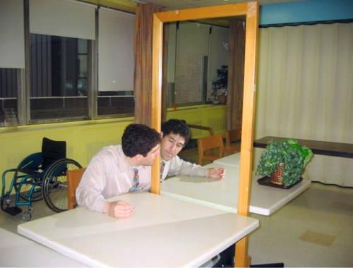
Mirror therapy was first devised as a therapeutic tool to relieve pain in amputees from a poorly mobile or spasmodic phantom limb [47]. In mirror therapy, a patient moves both limbs—the affected limb as best as possible—while watching the reflection of the good limb (Fig. 99.1). Visual feedback from the reflection of the good limb—which looks like the affected limb moving normally—feeds into and improves the motor control loop of the affected limb, ideally improving movement of the affected limb [3]. One of us (V.S.R.) was the first to suggest that mirror therapy might be helpful in reflex sympathetic dystrophy/CRPS [2]. RCTs [48,49] and case reports [3,50,51] have since affirmed the benefit of mirror therapy in CRPS.
Mirror therapy can be useful by visual feedback, providing an “active assist” to promote and to improve movement. It can also reduce hyperesthesia and allodynia through progressive contact exercises of both limbs while the reflection of the unaffected limb is watched in the mirror.
In general, we urge patients to use the limb as much as is safely possible and to take back “ownership” of the limb (e.g., not to speak of the limb in the third person). The hope is that with early intervention for CRPS with mirror therapy and rehabilitation, pharmacologic, and other methods, it may be possible to prevent patients from progressing to the pachydermic, irreversible stage of CRPS.
Procedures
Neuromodulation
Repetitive transcranial magnetic stimulation, in contrast to spinal cord stimulation, is approved by the Food and Drug Administration only for resistant major depressive disorder. It employs electromagnetic currents to stimulate cortical targets, which has previously been shown effective in chronic pain management. Two RTCs [52,53] for CRPS found repetitive transcranial magnetic stimulation to improve pain, possibly in the sensory discrimination and emotional dimensions of pain. An early attempt to use direct motor cortex electrical stimulation has had some success, but further study is needed [54].
Regional Procedures
Two types of regional sympathetic blocks are commonly used for CRPS: stellate ganglion block and lumbar sympathetic block. Stellate ganglion block, also known as cervicothoracic ganglion block, targets ganglion cells along the pharynx. It theoretically interrupts afferent painful signals from the upper limbs. Lumbar sympathetic block targets ganglion cells along the spine at L2-L4 levels and attempts to interrupt afferent painful signals from the lower limbs. The effectiveness of stellate ganglion and lumbar sympathetic blocks is variable in existing RCTs and on average shows only transient analgesic benefits [55]. Of note, compared with lumbar sympathetic block using bupivacaine alone, one small RCT found that the addition of botulinum toxin A may prolong the effects [56]. Longer lasting effects from stellate ganglion and lumbar sympathetic blocks may be achieved with radiofrequency lumbar sympathectomy and phenol neurolysis, but no further studies have been published [57].
Bier block can be used in the upper or lower limbs, and it involves use of a tourniquet to restrain blood flow into the affected limb while intravenous local anesthetic is administered. A small RCT in the 1980s found serial Bier blocks with guanethidine to be as effective as serial Bier block with lidocaine in management of upper limb reflex sympathetic dystrophy pain. However, recent RCTs did not find intravenous guanethidine, bupivacaine, or methylprednisolone effective for most patients [58].
Surgery
Spinal cord stimulation is approved by the Food and Drug Administration for treatment of refractory CRPS and neuropathic pain. Its action is attributed to central excitability suppression. Three medium-sized RCTs found the combination of spinal cord stimulation with physical therapy to be more effective than physical therapy alone for pain control in the short term, but this result does not appear to carry over in the long term [59]. Percutaneous cervicothoracic or lumbar sympathectomy is reserved for patients with severe CRPS, but effectiveness is questionable [60].
Potential Disease Complications
Functional disability from pain and hyperesthesia of CRPS is a significant concern. The most feared complication is progression of an affected limb to pachydermia, an irreversible condition. Patients will then face severe, chronic pain. Limb contracture and muscle atrophy can result in loss of extremity function and potentially permanent disability.
Potential Treatment Complications
Adverse effects of pharmacotherapy vary according to the medication selected. For example, long courses of corticosteroids can produce significant endocrinologic disturbances and should be avoided; and nonsteroidal anti-inflammatory drugs have well-known adverse effects on the gastric, hepatic, and renal systems. Because polypharmacy may be necessary, care should be used in selection of interacting drugs that do not result in untoward side effects. Potential complications of stellate ganglion block are inadvertent arterial injection and seizures or recurrent laryngeal nerve injury. Perforation of the aorta, vena cava, or kidney can occur during lumbar sympathetic block. The most common complications of spinal cord stimulation include hardware failure, lead migration, infection, and failure to provide pain relief. Even for seemingly benign rehabilitation such as mirror therapy, one should be aware that patients can be at an increased risk for fall when they have increased mobility.
[/level-membership-for-physical-medicine-and-rehabilitation-category][not-level-membership-for-physical-medicine-and-rehabilitation-category]CHAPTER 99
Complex Regional Pain Syndrome
Jiaxin Tran, MD; V.S. Ramachandran, MD, PhD; Eric L. Altschuler, MD, PhD
Definition
Complex regional pain syndrome (CRPS) is a perplexing medical condition. CRPS begins when a patient has an injury—to a nerve, bone, soft tissue, or connective tissue. The injury heals as assessed by both clinical examination and imaging, yet the patient still has pain significantly out of proportion not only to the healed injury but at times even to the injury itself. In addition to pain, the patient can have hyperesthesia or allodynia at the site of the injury. Curiously, the temperature of the affected limb can be different from that of the other side, and there can be hair, nail, and skin changes. In some cases, these changes can spread beyond the injury site to the entire limb, and in the worst case, in addition to excruciating pain and hyperesthesia, the skin becomes pachydermic.
Before 1994, this bizarre disease and constellation of symptoms was known, among other names, as reflex sympathetic dystrophy. Despite this moniker, it was never proved that the sympathetic nervous system mediates all the components of the disease, nor was a peripheral reflex or feedback loop causing the disease found. In 1994, a conference was held to clarify the classification of the disease [1]. It was renamed CRPS with two classifications, CRPS II for apparent major nerve injury and CRPS I for no major nerve injury. The disease is complex, and it can often be regional rather than confined to a dermatome, myotome, or territory of a single peripheral nerve. Pain is the hallmark of the disease and, for a patient with history of an injury, the sine qua non of diagnosis. It is multifaceted.
The new name, interestingly, is less specific than the former name, expressing the current lack of a full understanding of the disease. More than a decade ago, one of us (V.S.R.) suggested that central remapping of sensory neurons in the cerebral cortex in response to a peripheral injury to the corresponding body area might be a root cause of pain and ultimately other problems in this condition [2]. This hypothesis has not only collected evidence since but led to new and potentially most useful treatments [3].
CRPS is relatively rare. Even with fracture, the most common inciting event, only 1% of patients develop CRPS [4]. The incidence has been estimated to be up to 26 per 100,000 person-years [5]. The incidence of CRPS type I is generally higher than that of CRPS type II. The lower extremity is more often affected than the upper extremity. Female sex, adult age, postmenopausal status, and smoking are all risk factors for its development [6]. In the pediatric population, the incidence of CRPS increases just before puberty [7].
The pathophysiologic mechanism of the disease is, again, poorly understood. There is emerging evidence that CRPS does not only affect the peripheral nervous system. Functional and structural reorganization [8] of the central nervous system has been demonstrated with functional magnetic resonance imaging [9], single-photon emission computed tomography, electroencephalography, and transcranial magnetic stimulation mapping. Neuroplasticity of both the cortical somatosensory [10] and motor [11] systems is implicated. Distorted sensory and motor mapping is hypothesized to promote spontaneous pain and neglect symptoms in CRPS.
Symptoms
CRPS is a clinical diagnosis. The diagnosis can be suggested by the history of present illness alone. An individual experiences an injury. The injury can come in a variety of forms, whether traumatic (e.g., crush injury, gunshot wound, burn, or protracted labor [12]) or nontraumatic (e.g., leprosy [13]). The “injury” can also be an otherwise uncomplicated surgery. While healing takes place, the individual develops pain variably progressive in intensity, area involvement, and duration. Patients with both CRPS type I and type II may report neuropathic pain that is often intense, constant, burning, and present even without stimulation or movement. Symptoms are not confined to a particular nerve or anatomic territory; CRPS can spread [14] to the contralateral limb and even progress to all four limbs. In addition to pain, motor complaints are common and include weakness, cramps, and stiffness. There may also be signs of neglect [15], or more subtly, some patients may refer to the affected limb in the third person (e.g., “it is not moving”). It is important to ask about abnormal sweating patterns, localized edema, and skin flushing as clues of autonomic dysfunction in the history.
Symptoms and signs of CRPS are historically grouped into three stages. Stage 1 consists of severe pain and inflammatory signs (e.g., pitting edema, rubor, increased hair and nail growth). Stage 2 is marked by more intense pain, brawny edema, pallor, ridged nails, and osteoporosis. Unyielding pain and irreversible skin and bone changes (e.g., dystrophy, contracture, and extensive osteoporosis) are the primary features of stage 3. Although CRPS does not always follow a stepwise progression, these stages are useful descriptively. Classifying the syndrome solely on the basis of the severity of pain (i.e., mild, moderate, and severe) may be appropriate to follow its clinical course over time. We also classify CRPS as irreversible (i.e., presence of pachydermic skin changes) or potentially fully reversible (i.e., absence of pachydermic skin changes).
Mood disturbance is common in the chronic stages of the illness. Patients often report anxiety as the disease progresses beyond the second month; and by the sixth month, all patients will exhibit varying degrees of depression, sleep disturbance, and anxiety [16].
Physical Examination
Begin the examination by inspecting for signs of injury at or near the affected site. The injury may have been long ago and only a scar remains. If possible, compare all findings with the contralateral side to identify subtle asymmetry. Look for evidence of autonomic disturbances that are manifested as localized edema, skin color changes, and abnormal sweating pattern; note that skin color is best observed under natural light. Late-stage CRPS is characterized by trophic changes, such as abnormal hair and nail growth, thin and shiny skin, and fibrosis. At rest, disuse atrophy is the most apparent motor disturbance, but spontaneous muscle fasciculation, tremor, and cramp in the supporting muscles of the limb may also be observed.
Proceed to gentle palpation of the affected area. Make note of marked temperature asymmetry. Studies have shown that a clinician can adequately identify temperature and limb circumference asymmetry without special instruments [17]. Nonetheless, an infrared thermometer or a surface-probe thermometer can give more precise measurements. A temperature difference of more than 1.1° C between the affected area and a nonaffected, homologous body part is significant [1], but clinicians should have a low index of suspicion for CRPS such that even a small temperature change may be clinically relevant. Next, estimate the extent of involvement by sensory disturbances, such as increased sensitivity (hyperesthesia), exaggerated pain response to a painful stimulus (hyperalgesia), pain to an innocuous stimulus (allodynia), and paradoxically in some patients, hypoesthesia [18]. Ascertain abnormal findings with repeated testing. The sensory examination includes tests for light touch, pinprick, temperature, vibration, and proprioception. Consider testing the forehead because sensory disturbances on the ipsilateral side have been described [19].
Have the patient actively range the affected joints. Make note of difficulty with initiation of motion and range of motion deficits, which in turn require passive range to assess for contracture. The clinician can then perform a thorough musculoskeletal and neurologic examination including strength, stability, and reflexes to assess for deficits, be they related or unrelated to the CRPS.
Functional Limitations
The most immediate effects are dysfunction in activities of daily living from disuse of the affected limb. Lower limb involvement results in gait impairment. Even properly treated patients may continue to experience disability and decreased quality of life in the long term [20]. Permanent disability may ensue if contracture develops over time. Chronic pain also leads to the well-known syndrome of deconditioning, sleep disturbance, anxiety, and depression. With inadequate treatment, quality of life can be severely affected, often with devastating social, recreational, financial, and vocational consequences.
Diagnostic Studies
Several sets of diagnostic criteria (e.g., Bruehl and Veldman) exist, and none has been shown to be superior [21]. The International Association for the Study of Pain (IASP) criteria are most widely referenced in the literature and made up of the following four components: (1) presence of an identifiable noxious event or cause of immobilization; (2) persistent pain, allodynia, or hyperalgesia, disproportionate to any inciting event; (3) edema, changes in skin blood flow, or abnormal sudomotor activity; and (4) exclusion of other diagnoses as the cause of these symptoms [1]. IASP criteria are notably limited by poor inter-rater reliability and a specificity of only 36% [22]. They were subsequently revised in 2003 and published in 2007 as the Budapest criteria, which boast an increased specificity [23]. The Budapest criteria replace the second and third components of IASP criteria with at least one symptom and one sign in three of the following four categories: sensory, vasomotor, sudomotor or edema, and motor or trophic changes.
There is no “gold standard” for diagnosis of CRPS. It is a diagnosis of exclusion. Electromyography and nerve conduction studies can help identify nerve injury, if there is clinical suspicion. Doppler flowmeter, vascular scintigraphy, and vital capillaroscopy evaluate for vasomotor changes. Quantitative sudomotor axon reflex testing measures sweat output after mild electrical stimulation.
Plain films are often normal early in the course. Demineralization may become evident by the second month. Magnetic resonance imaging demonstrates nonspecific marrow edema, soft tissue swelling, and joint effusion. Even bone scintigraphy, in which the most suggestive finding is increased periarticular activity in the affected limb, has variable sensitivity and specificity [24]. A recent meta-analysis supports the use of triple-phase bone scan for assessment owing to its high sensitivity and negative predictive value [25].
[/not-level-membership-for-physical-medicine-and-rehabilitation-category]
