CASE 99
History: A patient presents with cyanosis.
1. What lesions characteristically manifest with decreased vascularity on radiography? (Choose all that apply.)
D. Pulmonary atresia with ventricular septal defect
2. What is the most likely diagnosis in this patient?
D. Pulmonary atresia with ventricular septal defect
3. Which valve is most likely abnormal?
A. Mitral
B. Aortic
C. Tricuspid
D. Pulmonary
4. Which imaging modality best assesses the valve leaflets?
B. CT
C. MRI
D. Angiography
ANSWERS
Reference
Higgins CB. Radiography of congenital heart disease. In: Webb WR, Higgins CB, eds. Thoracic imaging: pulmonary and cardiovascular radiology. ed 2 Philadelphia: Lippincott Williams & Wilkins; 2010.
Cross-Reference
Cardiac Imaging: The REQUISITES, ed 3, pp 206–207.
Comment
Etiology and Physiology
Ebstein anomaly is a congenital lesion characterized by an abnormality of the tricuspid valve; the septal and posterior leaflets are displaced apically, causing tricuspid regurgitation. A portion of the right ventricle is functionally incorporated into the atrium, which is known as “atrialization of the right ventricle,” although the atrialized portion of the ventricle contracts during ventricular systole. Tricuspid regurgitation causes marked enlargement of the right ventricle and atrium. Tricuspid regurgitation with a coexisting atrial septal defect results in a right-to-left shunt. Pulmonary vascularity is decreased, and cyanosis can develop. Ebstein anomaly is commonly associated with either an atrial septal defect or a patent foramen ovale. Less common associations include supraventricular tachycardia (25% to 50%), pulmonary atresia or stenosis (25%), and Wolff-Parkinson-White syndrome (10%). Maternal lithium use during the first trimester may result in Ebstein anomaly.
Imaging
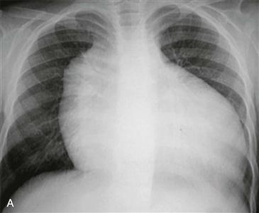
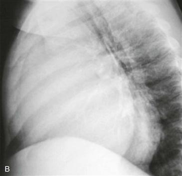
Chest radiography characteristically demonstrates marked enlargement of the heart, particularly the right atrium and ventricle (Figs. A and B). Echocardiography is used for further evaluation. MRI can also be performed to assess the anatomy and to quantify the severity of regurgitation. MRI provides the same information as echocardiography and gives more reliable and reproducible information in patients with limited acoustic windows. It can assess right ventricular size and function and accurately quantitate the volume of tricuspid regurgitation, which has a direct impact on the timing of valvular surgery in these patients.
Differential Diagnosis
A few other rare conditions may manifest with cyanosis, cardiomegaly, and reduced pulmonary vascularity, including (1) severe pulmonic stenosis with a restrictive atrial septal defect, (2) tricuspid atresia with a restrictive atrial septal defect, and (3) pulmonary atresia with a restrictive ventricular septal defect.


