CASE 98
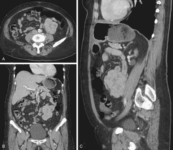
History: An 82-year-old woman presents with right flank pain and suspected renal colic.
1. Which of the following should be included in the differential diagnosis of the imaging finding shown in Figure A? (Choose all that apply.)
D. Asymptomatic and can be ignored
E. Retroperitoneal or mesenteric mass
2. What is the most common incidental finding of moderate importance on CT?
3. In which viscus is the finding of an incidental lesion containing fat most commonly found?
4. Which of the following incidental findings should not be ignored?
B. Adrenal nodule with washout
C. Liver lesion with nodular enhancement
D. Vertebral body lucency with polka-dot sclerotic foci
ANSWERS
CASE 98
Incidental Finding
1. A, B, and E
2. B
3. D
4. A
References
Berland LL, Silverman SG, Gore RM, et al: Managing incidental findings on abdominal CT: white paper of the ACR incidental findings committee. J Am Coll Radiol. 2010;7:754–773.
Cross-Reference
Gastrointestinal Imaging: THE REQUISITES, 3rd ed, p 138.
Comment
This patient presented with symptoms suggesting right renal colic. A low-dose CT scan without contrast enhancement was performed to evaluate for renal tract calculi and obstruction. An unusual appearance was observed by the radiologist on the left asymptomatic side. This then led to the standard contrast-enhanced CT scan presented here (see figures). The patient eventually went to surgery for resection of the mass, which was diagnosed histologically as a retroperitoneal leiomyosarcoma.
Improvements in imaging, especially CT, have seen an increase in the incidence of detecting incidental findings. Sometimes given the name incidentalomas, they are defined as findings that are unrelated to the clinical indication for the imaging examination performed. Most of these incidental findings are benign and have little or no clinical significance, but a small but significant proportion are serious. A careful decision needs to be made between performing appropriate further investigation and follow-up versus considered reassurance. Subjecting a patient with an incidentaloma to unnecessary testing and treatment can result in a potentially injurious and expensive cascade of tests and procedures.


