CASE 98
1. What should be included in the differential diagnosis? (Choose all that apply.)
A. Metastasis
C. Tamponade
D. Lymphoma
2. Which of the following is most specific for a malignant effusion?
B. Nodularity
C. Thickening
D. Enhancement
3. What is the most likely diagnosis?
A. Metastasis
C. Tamponade
D. Lymphoma
4. What does pulsus paradoxus indicate?
A. Tamponade
B. Tumor
C. Constriction
D. Hemorrhage
ANSWERS
References
Wang ZJ, Reddy GP, Gotway MB, et al. Cardiovascular shunts: MR imaging evaluation. Radiographics. 2003;23(Spec No):S181–S194.
Yared K, Baggish AL, Picard MH, et al. Multimodality imaging of pericardial diseases. JACC Cardiovasc Imaging. 2010;3(6):650–660.
Cross-Reference
Cardiac Imaging: The REQUISITES, ed 3, pp 277–278.
Comment
Pathogenesis and Etiology
Pericardial metastases are much more common than primary neoplasms of the pericardium. Tumor can seed the pericardium via lymphatic or hematogenous dissemination or invade the pericardium directly from the lung or mediastinum. Carcinomas of the breast and lung are the most common neoplasms to involve the pericardium, followed by lymphomas and melanomas.
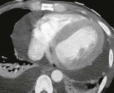
Imaging and Diagnostic Features
Pericardial metastasis is suggested on CT or MRI by the presence of effusion with an irregularly thickened, nodular pericardium or the demonstration of a pericardial mass (Figure). The pericardial effusion may be hemorrhagic, which produces high signal intensity on spin echo MRI. Malignant disease usually enhances after the administration of contrast agent.


