CASE 97
1. What should be included in the differential diagnosis? (Choose all that apply.)
B. Ventricular septal defect (VSD)
C. Patent ductus arteriosus (PDA)
D. Partial anomalous pulmonary venous connection (PAPVC)
2. What is the most likely diagnosis?
A. ASD
B. VSD
C. PDA
D. PAPVC
3. Which of these lesions commonly causes enlargement of the left atrium?
A. PDA
B. ASD
C. PAPVC
D. Pulmonary arteriovenous malformation
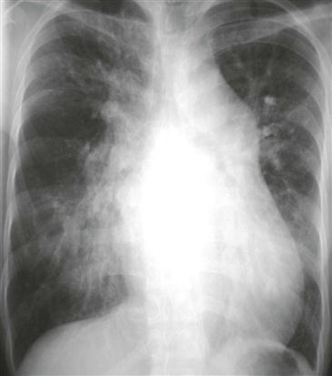
4. Which feature of left atrial enlargement is present in this patient’s chest radiograph?
ANSWERS
References
Higgins CB. Radiography of congenital heart disease. In: Webb WR, Higgins CB, eds. Thoracic imaging: pulmonary and cardiovascular radiology. ed 2 Philadelphia: Lippincott Williams & Wilkins; 2010.
Kilner PJ. Imaging congenital heart disease in adults. Br J Radiol. 2011;84(Spec No 3):S258–S268.
Wang ZJ, Reddy GP, Gotway MB, et al. Cardiovascular shunts: MR imaging evaluation. Radiographics. 2003;23(Spec No):S181–S194.
Cross-Reference
Cardiac Imaging: The REQUISITES, ed 3, pp 345–349.
Comment
Embryology, Clinical Features, and Treatment
In the fetus, the ductus arteriosus is a widely patent vessel that connects the proximal descending aorta to the main or left pulmonary artery, allowing the output of the right ventricle to bypass the lungs. The ductus usually closes shortly after birth as a result of the increase in partial pressure of oxygen in the circulation. If the ductus does not close, indomethacin can be administered in the first week of life, especially in premature infants. If the ductus remains patent, pulmonary hypertension can develop. In the absence of a coexisting anomaly, the ductus should be closed by surgical ligation or by transcatheter coil embolization.
Imaging
Radiography can demonstrate increased pulmonary vascularity and enlargement of the left atrium and aortic arch (Figure). Echocardiography usually depicts the location and size of the lesion. MRI may be performed in certain patients for better delineation of the lesion or for quantification of the pulmonary-to-systemic flow ratio (Qp/Qs), which is an indicator of the severity of the shunt.


