CASE 92
History: A 32-year-old woman presents with chest pain.
1. Which of the following should be included in the differential diagnosis of the imaging finding shown in the figures? (Choose all that apply.)
2. Why do focal nodular hyperplasia (FNH) lesions take up sulfur colloid?
A. The lesion contains hepatocytes.
B. The lesion contains Kupffer cells.
C. The lesion contains blood pools.
D. The lesion is hypervascular.
3. In what demographic group is FNH most commonly seen?
4. What is the vascular supply usually serving FNH?
ANSWERS
CASE 92
Focal Nodular Hyperplasia
1. A, B, D, and E
2. B
3. B
4. B
References
Brancatelli G, Federle MP, Grazioli L, et al: Focal nodular hyperplasia: CT findings with emphasis on multiphasic helical CT in 78 patients. Radiology. 2001;219:61–68.
Cross-Reference
Gastrointestinal Imaging: THE REQUISITES, 3rd ed, p 198.
Comment
Focal nodular hyperplasia (FNH) of the liver is the second most common benign tumor of the liver after hemangiomas. The tumor is a hyperplasia of normal, non-neoplastic liver tissue that has an abnormal arrangement and is similar to a hamartomatous type of lesion. It is believed to develop as some type of response to a congenital vascular abnormality in the region, and there is a slight association between hemangiomas and FNH. Histologically, the liver tissue is arranged in small nodules, with septa in between. Arterial vessels feed the nodules from a centrally located artery that is often within a central scar or septum. They do not have a portal venous supply. FNH contains Kupffer cells in most instances.
Adenomas and FNH are similar in that they both occur in young women, although FNH also occurs in children and older patients. Unlike adenomas, FNH is not associated with use of oral contraceptives. This tumor is rarely found in men. FNH must be differentiated from other hepatic tumors, such as adenomas, fibrolamellar hepatomas, and giant cavernous hemangiomas. All these tumors are large vascular tumors that affect young women and often have a central scar.
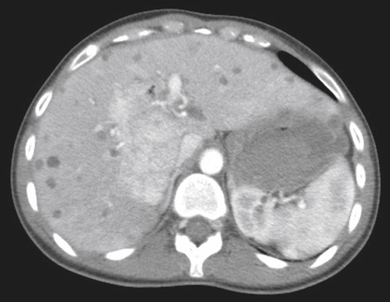
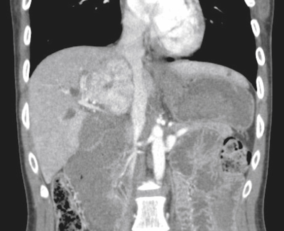
Focal nodular hyperplasia is sometimes detected on ultrasound as a well-defined lesion with echogenicity different from that of the normal liver. A large central vessel may be detectable on Doppler scanning. On CT scanning, the lesion’s density may be similar to that of the liver and therefore may be difficult to distinguish from the normal liver parenchyma (see figures). A central scar or low-density area may be visible (see figures).
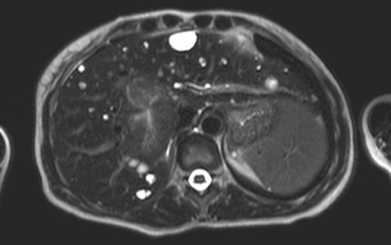
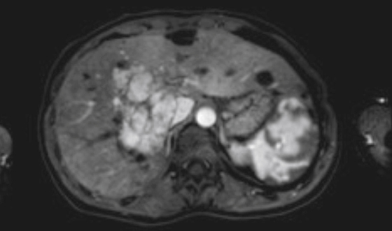
Because of the functional reticuloendothelial cells, FNH may be detectable on sulfur colloid scanning as an area of increased uptake. On MRI, FNH is only slightly hyperintense on T2-weighted images, but the central scar may be markedly hyperintense (see figures).


