CASE 92
1. Which arteries are depicted in the two figures? (Choose all that apply.)
A. Aorta
2. Which artery has a thickened, enhancing wall?
A. Aorta
3. What is the most likely diagnosis in a young patient?
4. What is the most commonly involved artery in this disease?
A. Aorta
ANSWERS
References
Gotway MB, Araoz PA, Macedo TA, et al. Imaging findings in Takayasu’s arteritis. AJR Am J Roentgenol. 2005;184(6):1945–1950.
Reddy GP, Gunn M, Mitsumori LM, et al. Multislice CT and MRI of the thoracic aorta. In: Webb WR, Higgins CB, eds. Thoracic imaging: pulmonary and cardiovascular radiology. ed 2 Philadelphia: Lippincott Williams & Wilkins; 2010.
Cross-Reference
Cardiac Imaging: The REQUISITES, ed 3, pp 393–394.
Comment
Clinical Features
Takayasu arteritis is an idiopathic disease that is characterized by wall thickening of the aorta or arch vessels, or both, and stenosis of the aorta and its branches. Other arteries, such as the pulmonary arteries, can be involved.
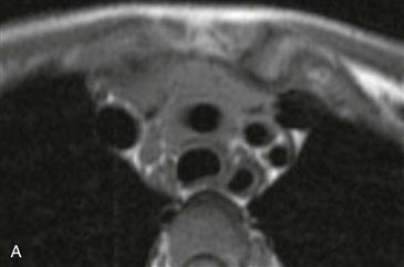
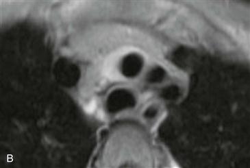
Imaging
In Takayasu arteritis, MRI and CT can show stenosis (Figs. A–B), occlusion, or dilation of the aorta and its branches or a combination of all three. In patients in the active phase of arteritis, gadolinium-enhanced MRI demonstrates wall thickening and enhancement of the involved vessels (Fig. B). Noninvasive imaging with MRI or CT may be particularly valuable in patients with severe stenosis or occlusion of the arch vessels or of the abdominal aorta because it may be especially difficult to pass an angiography catheter into the thoracic aorta.


