CASE 91
History: A patient presents with cough.
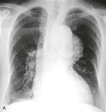
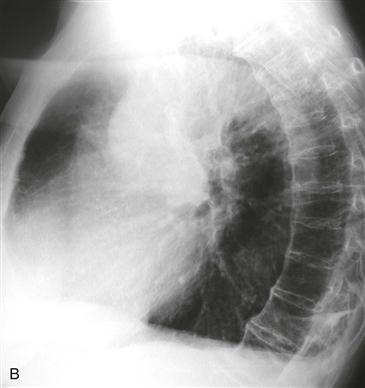
1. What should be included in the differential diagnosis based on Figs. A and B? (Choose all that apply.)
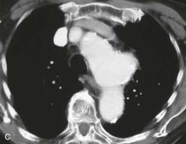
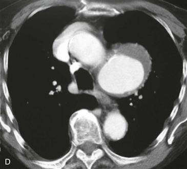
2. What is the most likely diagnosis based on Figs. C and D?
3. What is the most common cause of aortic aneurysm?
B. Syphilis
4. What is an advantage of MRI over CT for evaluation of the thoracic aorta?
ANSWERS
Reference
Reddy GP, Gunn M, Mitsumori LM, et al. Multislice CT and MRI of the thoracic aorta. In: Webb WR, Higgins CB, eds. Thoracic imaging: pulmonary and cardiovascular radiology. ed 2 Philadelphia: Lippincott Williams & Wilkins; 2010.
Cross-Reference
Cardiac Imaging: The REQUISITES, ed 3, pp 377–379.
Comment
Clinical Features
The thoracic aorta is considered to be enlarged when its diameter exceeds 4 cm. Thoracic aortic enlargement is termed an aneurysm when the diameter is more than 5 cm. The maximum diameter of the aorta is an important determinant of the risk of rupture. If the diameter is 6 cm or greater, the risk of rupture in the short term is greater than 30%. Atherosclerosis is the most common cause of thoracic aortic aneurysm. Aneurysms most commonly occur in the descending aorta and aortic arch.
True and False Aneurysm
In a true aneurysm of the aorta, all three layers of the aortic wall are intact. In contrast, a false aneurysm results from a focal disruption of one or more layers of the aortic wall and may be contained by the adventitia and surrounding fibrous tissue.
Imaging
CT and MRI are the best imaging methods for evaluation of a thoracic aortic aneurysm (Figs. A–D). On CT and MRI, true aneurysms have wide necks (as in the patient in this case), and false aneurysms have narrow necks (<50% of the aneurysm diameter). True aneurysms most commonly are secondary to atherosclerosis. Other etiologies include infection, connective tissue disorders such as Marfan syndrome, aortitis, idiopathic cystic medial necrosis, complications of aortic valve disease, and aneurysm of the ductal remnant. In the imaging assessment of a thoracic aortic aneurysm, the demonstration of mural thrombus is important in patients with peripheral embolization.


