CASE 90
1. What should be included in the differential diagnosis? (Choose all that apply.)
A. Metastasis
B. Thrombus
C. Myxoma
D. Angiosarcoma
2. What is the most common mass in the heart?
A. Thrombus
B. Myxoma
C. Metastasis
D. Angiosarcoma
A. Left atrium
C. Right atrium
4. What is the most appropriate management of the mass?
B. Chemotherapy
ANSWERS
Reference
Restrepo CS, Largoza A, Lemos DF, et al. CT and MR imaging findings of malignant cardiac tumors. Curr Probl Diagn Radiol. 2005;34(1):12–21.
Cross-Reference
Cardiac Imaging: The REQUISITES, ed 3, pp 280–281.
Comment
Clinical Features and Treatment
Myxoma is usually located in the left atrium and is one of the most common benign tumors of the heart. Patients with myxoma can be asymptomatic or develop symptoms of mitral stenosis or constitutional symptoms, including fever, anemia, and elevated erythrocyte sedimentation rate. Embolization of a myxoma to the systemic circulation can occur and can cause transient ischemic attacks, stroke, or other systemic organ disease. Other complications include occlusion of the mitral valve and malignant degeneration. This tumor is usually resected even though it is benign.
Imaging
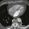
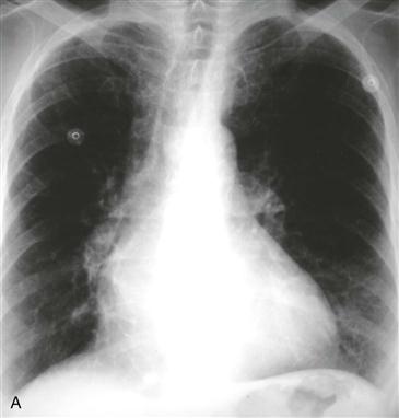
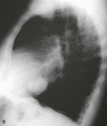
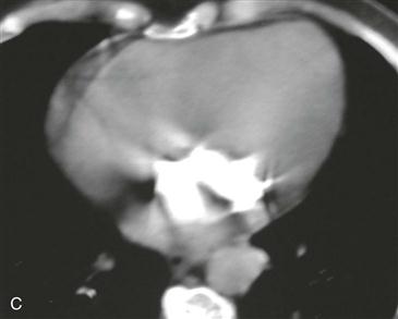
Myxoma is well circumscribed and often has a narrow pedicle that is attached to the fossa ovalis of the atrial septum. A broad attachment to the atrial wall is less common. Radiographs can show a mass or show nonspecific findings (Figs. A and B). Myxomas can calcify (Fig. C), although dense calcification is uncommon. On black blood MRI, a myxoma typically enhances with administration of a contrast agent. Because of fibrosis, calcification, or iron deposition, myxomas can be dark on steady-state free precession MRI, mimicking the appearance of a thrombus. It is important to look for contrast enhancement on black blood images to differentiate myxoma from a thrombus, which does not enhance.