CHAPTER 9. PREPARING AND SETTING UP INTRAVENOUS MEDICATIONS
INTRODUCTION
Intravenous fluids or medication for administration are usually prepared by nursing staff. However, junior doctors should be able to perform this simple task.
INDICATIONS
• Administration of intravenous fluids.
• Administration of intravenous medications.
• Intravenous replacement of electrolytes.
CONTRAINDICATIONS
• Cellulitis at the cannula site – replace the cannula elsewhere if suspected (see Chapter 8).
EQUIPMENT
PRACTICAL PROCEDURE
• Wash your hands and wear gloves.
• Open the intravenous giving set from its sterile packaging.
• Place the intravenous bag of fluid on fluid stand.
• Check the cannula site to ensure it is free from local infection and the use-by date on the bag of fluid.
• Twist off the plastic end of the access port on the fluid bag.
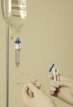
• Carefully push the sharp plastic bevel of the giving set into the access port of the bag of fluid, taking care not to touch the bevel and thus maintain sterility (Fig. 9.1). Allow fluid to run into the giving set chamber by opening the roller clamp (Fig. 9.2).
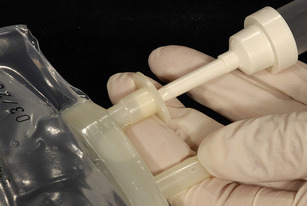 |
| Fig. 9.1 |
• Fluid should be allowed to run from the giving set chamber through the administration set tubing until air bubbles are flushed out. The roller clamp can then be closed to halt further flow.
• Remove the bung from the already sited intravenous cannula and flush the cannula with 10 mL of sterile normal saline to ensure patency and intravenous placement.
• Attach the end of the giving set to the cannula and open the giving set up again.
• Observe the giving set chamber. A steady flow of drips confirms free flow of fluid through the giving set, which can be adjusted by manipulating the roller mechanism on the giving set.


• Ensure that the bag of fluid is kept hanging above the patient to avoid retrograde flow of blood into the giving set.
• If the fluid fails to run through, check the giving set for links. If this fails to work, disconnect the giving set from the cannula and manipulate the position of the intravenous cannula. Try flushing the cannula with saline to confirm patency and intravenous position, then reconnect to the giving set.
COMPLICATIONS
• Air embolism. Air bubbles in the plastic giving set tubing may be inadvertently flushed intravenously. Patients with patent intracardiac foramina may be at risk of paradoxical arterial embolic events. This problem can be avoided by opening the giving set to allow free flow of fluid until all bubbles are flushed out. Alternatively tap the giving set tubing until air bubbles rise through the tubing and dissipate within the giving set chamber.
• Poor fluid flow associated with new pain at the cannula insertion site usually means subcutaneous extravasated fluid with the cannula replacing the no longer in situ. Consider cannula if in situ repositioning does not resolve the problem.