CASE 89

History: A 73-year-old woman presents with abdominal distention and constipation.
1. Which of the following should be included in the differential diagnosis of the imaging finding shown in the figures? (Choose all that apply.)
2. What is the most common cause of ascites?
A. Cirrhosis and severe liver disease
3. In the supine patient, what is the lowest, most dependent part of the abdomen?
A. Rectouterine pouch (pouch of Douglas)
B. Hepatorenal recess (Morison’s pouch)
4. What is the dog ears imaging sign of ascites?
B. Medial displacement of the lateral liver edge from the thoracoabdominal wall
C. Small bowel loops lined up in a polycyclic or arcuate arrangement
D. Diaphragmatic crus displaced laterally on CT
ANSWERS
CASE 89
Mass Effect Displacing Bowel
1. B, C, D, and E
2. A
3. A
4. A
References
Gore RM, Gore MD. Ascites and peritoneal fluid collections. In: Gore RM, Levine MS, eds. Textbook of Gastrointestinal Radiology. 2nd ed Philadelphia: WB Saunders; 2000:1969–1979.
Thoeni RF. The role of imaging in patients with ascites. Am J Roentgenol. 1995;165:16–18.
Cross-Reference
Gastrointestinal Imaging: THE REQUISITES, 3rd ed, p 132.
Comment
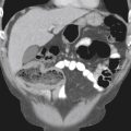
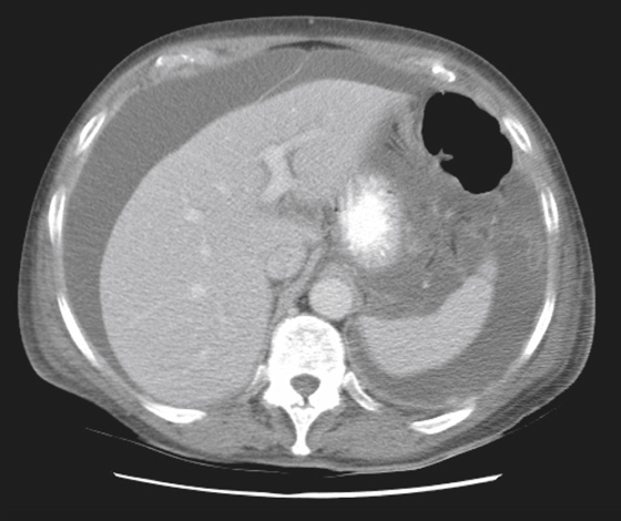
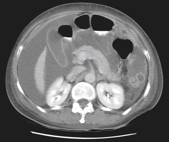
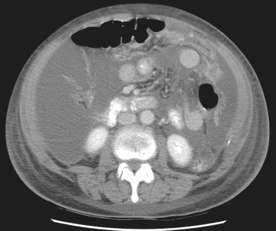
Bowel can be displaced by a number of processes in the abdomen. Fluid in the peritoneal cavity, ascites, and blood can displace bowel. Large amounts cause air-filled loops of bowel to rise as the fluid level increases (see figures). Because the abdomen is dome-shaped in most of us, when the bowel reaches the top of the dome, it will appear as if the bowel (with air in it) has collected in a circle in the middle of the abdomen. This is called centralization of bowel loops and is a wonderful sign for a large amount of fluid in the abdomen on plain films of the abdomen. Lesser degrees of fluid in the abdomen may be suspected when there is separation (more than 2 cm) of the properitoneal fat stripe from air in the ascending or descending colon. A less-specific sign is loss of the inferior margin of the liver. This is usually a fat–soft tissue interface. However, when fluid occupies the Morrison pouch it becomes a soft tissue–soft tissue interface, which is an invisible interface on plain film. This sign does require the patient to have some intraperitoneal fat, and the extremely thin patient may be an exception. Another sign of fluid in the abdomen is fluid in the lateral pelvic recesses, giving the dog ears sign. This is also a soft sign and can be reproduced by fluid in bowel and even loops of collapsed bowel. Mass effect owing to massive enlargement of viscera (e.g., hepatosplenomegaly) or large tumors can also displace bowel loops.