CASE 89
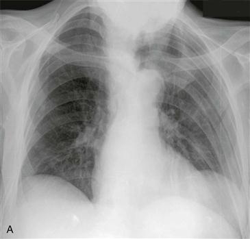
1. What should be included in the differential diagnosis based on Fig. A? (Choose all that apply.)
A. Lung cancer
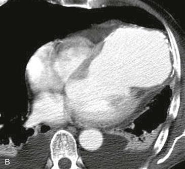
2. What is the most likely diagnosis based on Fig. B?
A. Lung cancer
3. What characteristic is most specific for the CT diagnosis of a true aneurysm?
B. Calcification along the ventricular wall
C. Wide ostium
D. Large size
4. This aneurysm puts the patient at high risk for which of the following complications?
B. Sudden death
ANSWERS
Reference
White RD. MR and CT assessment for ischemic cardiac disease. J Magn Reson Imaging. 2004;19(6):659–675.
Cross-Reference
Cardiac Imaging: The REQUISITES, ed 3, pp 235–237.
Comment
Pathology, Etiology, and Treatment
Left ventricular aneurysms result from transmural myocardial infarction. True aneurysms have focal wall thinning and akinesis, with bulging during systole. Most true aneurysms are located in the anteroapical region of the left ventricle and have wide necks. A false aneurysm represents a contained rupture. Most false aneurysms are inferoposterior in location and are connected to the left ventricle via a narrow ostium. True aneurysms are managed medically, but they can be resected if they cause dysfunction, such as heart failure, arrhythmia, or peripheral embolization. False aneurysms are typically managed surgically.
Imaging Findings and Diagnostic Criteria
Radiographs may show a contour abnormality along the left ventricle (Fig. A). Calcification or thrombus may be present (Fig. B). With MRI or CT, true and false aneurysms can be differentiated on the basis of their ostia. The size of the ostium (>50% of the aneurysm diameter) is the most specific feature of a true aneurysm. A true aneurysm typically has an ostium that is greater than 50% of the aneurysm diameter (Fig. B), whereas a false aneurysm typically has an ostium that is less than 50% of the aneurysm diameter.


