CASE 88

History: A patient presents with cyanosis.
1. What findings are seen? (Choose all that apply.)
A. Right ventricular hypertrophy
C. Pulmonary infundibular stenosis
2. What is the most likely diagnosis?
A. Atrioventricular septal defect
C. Transposition of the great arteries
D. Pulmonary atresia with ventricular septal defect
3. What is a common complication of surgery for tetralogy of Fallot?
4. What artifact is indicated by the arrow in Fig. C?
B. Aliasing
D. Phase wrap
ANSWERS
Reference
Reddy GP, Higgins CB. Magnetic resonance imaging of congenital heart disease: evaluation of morphology and function. Semin Roentgenol. 2003;38(4):342–351.
Cross-Reference
Cardiac Imaging: The REQUISITES, ed 3, pp 359–367.
Comment
Anomalies of Tetralogy of Fallot
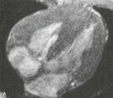
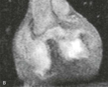
Tetralogy of Fallot is the most common cyanotic congenital heart disease. The four primary lesions of tetralogy of Fallot are an overriding aorta, ventricular septal defect, pulmonary infundibular stenosis, and right ventricular hypertrophy. Pulmonary stenosis can be present at multiple levels, including infundibular (most common), valvular, supravalvular, and peripheral. Approximately 25% of patients with tetralogy of Fallot have a right aortic arch, usually a mirror-image arch.
MRI
MRI can be performed for comprehensive evaluation of tetralogy of Fallot (Figs. A–C). Contrast-enhanced magnetic resonance angiography (MRA) can show the pulmonary artery sizes and can identify peripheral pulmonary arterial stenoses. Velocity-encoded cine phase contrast images can be obtained to measure differential right and left pulmonary flow and to quantify regurgitation in the postoperative setting. Cine MRI is employed for the quantitative appraisal of right ventricular function after surgery.


