CHAPTER 87
Hallux Rigidus
David Wexler, MD, FRCS (Tr & Orth); Dawn M. Grosser, MD; Todd A. Kile, MD
Definition
Degenerative joint disease or loss of articular cartilage from the first metatarsophalangeal (MTP) joint leading to painful restriction of motion is called hallux rigidus. The normal range of motion of the first MTP joint is 30 to 45 degrees of plantar flexion to almost 90 degrees of dorsiflexion. The limited range of motion and pain with hallux rigidus are exacerbated by overgrowth of bone (osteophytes or “bone spurs”) on the dorsal aspects of the base of the proximal phalanx and the head of the metatarsal, which impinge on one another as the great toe dorsiflexes [2]. Hallux rigidus is the second most common problem in the first MTP joint, after hallux valgus; 1 in 40 people older than 50 years will develop hallux rigidus [3].
In general, the cause is unknown, although it is associated with generalized osteoarthritis of other joints and repeated microtrauma (e.g., in soccer players). Sustaining repetitive turf toe–type injuries may lead to this form of early joint degeneration [4]. As the plantar capsuloligamentous complex of the first MTP joint is injured by hyperflexion of the great toe, it may acutely compress the articular surfaces of the joint, causing articular damage, or become chronically unstable, predisposing the MTP joint to degeneration and hallux rigidus [5].
Symptoms
Patients typically report pain, either intermittent or constant, that occurs with walking and is relieved by rest. It is insidious in onset and may be associated with stiffness, swelling, and sometimes inflammation. On occasion, there can be locking due to a cartilaginous loose body. Patients may notice that they are walking on the outside of the foot to avoid pushing off with the great toe during the terminal stance and toe-off phases of the gait cycle. As degeneration increases, the pain may intensify and result in an alteration of gait.
Physical Examination
On inspection, there will usually be swelling around the MTP joint with tenderness of the joint line. Dorsal osteophytes may be palpable and may cause irritation of overlying skin with shoe wear abrasion. Pain is reproduced with forcible dorsiflexion of the great toe, which is also restricted in range of movement. Plantar flexion may also be affected. Patients may have an antalgic (painful) gait, and single-stance heel raise may be difficult secondary to a painful MTP joint, as opposed to posterior tibial tendon deficiency. Findings of the neurologic examination, including strength, sensation, and reflexes, are typically normal.
Functional Limitations
Functional limitations include walking long distances, running any distance, and ascending stairs. As the severity increases, walking even short distances, daily errands, and standing for long periods may be difficult. Flexible shoes as well as shoes with a tight toe box may prove to be uncomfortable. This may lead to pressure areas dorsally over the osteophytes.
Diagnostic Studies
Plain anteroposterior and lateral standing radiographs will usually suffice in confirming the diagnosis (Fig. 87.1). The signs are consistent with degenerative joint disease, namely, loss of joint space and congruency, large dorsal osteophytes (bone spurs), sclerosis (increased density of bone), and subchondral cysts. There may be evidence of a loose body. This disease process has been divided into three grades on the basis of the severity of radiographic and clinical findings, which help guide surgical treatment.
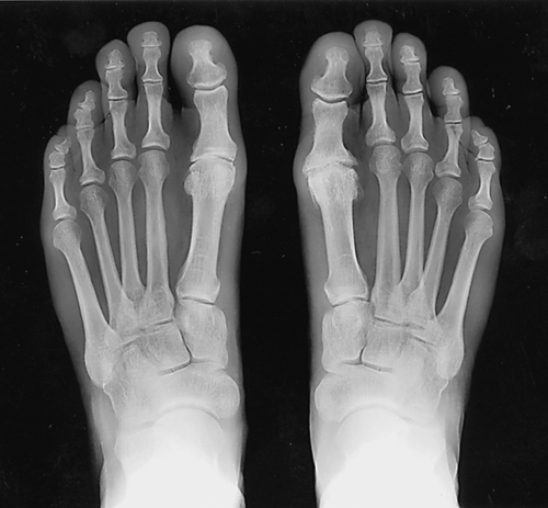
Grade I demonstrates small dorsal osteophytes with preservation of the MTP joint space on radiographic examination and typically intermittent pain with ambulation. Grade II demonstrates moderate dorsal osteophyte formation and asymmetric joint space narrowing radiographically and often constant pain with ambulation. Grade III has extensive osteophytes and severe dorsal and plantar joint space narrowing, often with noticeable loose bodies; clinically, patients will have constant pain with ambulation and significant limitation of motion [6].
Treatment
Initial
Nonsteroidal anti-inflammatory drugs may provide symptomatic relief. Footwear modifications and orthoses to limit stresses at the MTP joint (carbon fiber inserts or Morton’s extension orthotic devices) as well as avoidance of high heels or shoes with very flexible soles may be useful conservative treatment options [7].
Rehabilitation
More advanced shoe modifications can be made by a certified pedorthist. These include a steel shank and possibly a rocker-bottom. These may be applied to the soles of many different types of shoes, including athletic shoes. Anecdotally, many patients prefer first to try a steel shank because there is no cosmetic change to the shoe. With a rocker-bottom, the sole is altered, and this is sometimes less cosmetically acceptable to patients.
If there is evidence of other foot deformities, such as pes planus (flatfoot), orthotic inserts may provide correction and help with gait biomechanics.
Physical therapy is not generally necessary but may include basic mobilization and distraction techniques as well as strengthening exercises of the flexor and extensor hallucis muscles to enhance joint stability. Modalities such as contrast baths and ice may help with pain control.
Procedures
Intra-articular x-ray–guided or ultrasound-guided injection of the MTP joint with local anesthetic and steroid may provide short-term relief.
Surgery
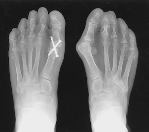
The principal indications for surgery are continuing pain and failed nonoperative management. Depending on the severity of the degeneration, there are two broad approaches to surgery. For grade I and grade II, the appropriate treatment is joint preserving; the impinging dorsal osteophytes are excised and the joint is debulked, thus improving dorsiflexion [8–10]. A phalangeal osteotomy (i.e., Moberg procedure) may also be used to improve dorsiflexion. The second approach is joint sacrificing for grade III disease. These procedures range from resection arthroplasty, resulting in a floppy or flail shortened toe, to arthrodesis (Fig. 87.2), resulting in a stiff, rigidly fixed toe [6,11,12]. A number of manufacturers have tried to produce artificial great toe joints, made of Silastic, metal, and polyethylene or ceramic [13,14], but the long-term results of these have not lived up to expectations. A randomized controlled trial comparing arthrodesis to arthroplasty found better improvement in pain, satisfaction of patients, and cost ratio with arthrodesis. Patients receiving arthroplasty had minimal improvement in range of motion, had continued altered gait mechanics, and required removal secondary to loosening in a significant number [15]. Interposition arthroplasty (with use of autologous tissue attached between the two joint surfaces) has also been advocated as a reasonable option instead of fusing the joint for grade III disease [16]. Arthroscopic surgery has also been attempted.
Potential Disease Complications
Hallux rigidus may produce intractable pain and reduced mobility.
Potential Treatment Complications
Analgesics and nonsteroidal anti-inflammatory drugs have well-known side effects that most commonly affect the gastric, hepatic, and renal systems. Steroid injection can rarely introduce infection.
Complications of surgery can range from failure of improvement with insufficient osteophyte resection to toe shortening with subsequent transfer metatarsalgia (pain under the metatarsal heads of the lesser toes). Arthroplasty complications include implant failure, silicone wear and subsequent debris production, foreign body reaction, and osteolysis. Arthrodesis complications include malunion and nonunion.


