CHAPTER 85
Chronic Ankle Instability
Michael D. Osborne, MD; Stephan M. Esser, MD
Definition
Chronic ankle instability is a condition characterized by a constellation of symptoms, typically including pain, weakness, and a feeling that the ankle episodically gives way, that persist after an acute lateral ankle sprain. Although chronic ankle instability may occur after a single ankle sprain, it is more commonly a sequela of repeated sprains. It has been reported to occur in up to 40% of individuals with a history of ankle sprain and as late as 61⁄2 years after an initial injury [1]. Anatomic lateral ankle ligament laxity and mechanical instability, peroneal muscle weakness, and ankle proprioceptive deficits are three primary factors thought to cause and to perpetuate symptoms. Arthrogenic muscle inhibition of the peroneal and soleus muscles has also been implicated as a possible contributing factor [2]. These causative factors may coexist with other pathologic processes of the ankle (such as those listed in the differential diagnosis section), which may serve to amplify and to perpetuate symptoms of functional instability. The establishment of additional diagnoses does not preclude a diagnosis of chronic ankle instability.
Symptoms
Usual symptoms are ankle pain, swelling around the lateral malleolus, weakness of the ankle evertors, and a feeling that the ankle is episodically unstable. The term functional instability describes the subjective sensation of “giving way” that often persists after ankle sprains [3]. Functional instability may occur in the absence of true mechanical ligament laxity and vice versa. Symptoms can continue for months or years after the original injury, range from mild to severe, and often are manifested as recurrent acute lateral ankle sprains.
Physical Examination
Objective findings are variable and can often be minimal. Potential examination findings in patients with chronic ankle instability may include reduced passive or active ankle range of motion, lateral ankle swelling, ecchymosis, lateral ankle tenderness (typically over the lateral ligament complex or peroneal tendons), weakness of the peroneal muscles, proprioceptive deficits (manifested by decreased ability to perform a single-leg stance), and mechanical laxity (demonstrated by increased motion on anterior drawer or talar tilt test compared with the contralateral ankle) [4]. Abnormal alignment, such as calcaneal varus, calcaneal valgus, or pes planus, may be evident. A limp may also be observed. Examination of the affected ankle should always be compared with the contralateral unaffected ankle.
Findings of the neurologic examination, including sensation and deep tendon reflexes, are commonly normal. Results of manual muscle testing should also be normal, with the exception of muscles surrounding the ankle that may exhibit weakness from disuse or because of pain. Balance testing commonly demonstrates deficits in the affected limb but may also reveal impairments in the contralateral ankle, making it unclear whether these findings represent a preexisting risk for injury or are the result of previous inversion injuries [5].
Functional Limitations
Affected persons may have difficulty participating in sports, particularly high-demand sports that require quick starts and stops, cutting, and jumping (such as soccer, football, and basketball) as well as sports that involve a lot of lateral movement (such as tennis). When symptoms are severe, limitations can include difficulty with climbing steps, ambulation, and activities that require prolonged standing. It is estimated that functional instability prevents 6% of patients from returning to their occupation; 5% to 15% remain occupationally disabled 9 months to 61⁄2 years later, whereas 36% to 85% of patients report full recovery within a period of 3 years [1,2,6,7].
Diagnostic Studies
The diagnosis is made by confirming a history of prior sprain with subsequent development of typical symptoms of functional instability in conjunction with consistent examination findings. Adjunctive diagnostic testing can be helpful in establishing the diagnosis, particularly by identifying pathologic changes and conditions that may produce similar symptoms. Testing that may be useful when the diagnosis of chronic ankle instability is being considered includes routine radiography, stress radiography, computed tomography, bone scan, magnetic resonance imaging, ankle arthrography, and magnetic resonance arthrography.
Routine radiographs are useful to rule out old or chronic fractures (most commonly of the fibula, tibia, talus, and fifth metatarsal), to assess the integrity of the ankle mortise, and to assess for ankle arthritis. A routine radiographic series should include anteroposterior, lateral, and mortise views. Widening of the ankle mortise may indicate a syndesmotic disruption or significant deltoid ligament tear. Radiographs should be obtained in all cases with a history of significant trauma at initial injury.
Stress radiographs may be helpful in determining the presence of chronic mechanical instability. Although the routine use of stress radiographs remains controversial, a finding of more than 5 mm of anterior displacement of the talus during anterior drawer testing is considered to be abnormal [8]. Inversion stress radiographs are considered abnormal with a finding of more than 5 degrees of side-to-side difference in tibiotalar tilt [7]. However, a review found the published data regarding stress radiographs too variable to determine accepted normal values for acute and chronic sprains [9]. The sensitivity of stress radiographs in diagnosis of chronic lateral ligament tears (surgically confirmed) is low, although specificity is high [8].
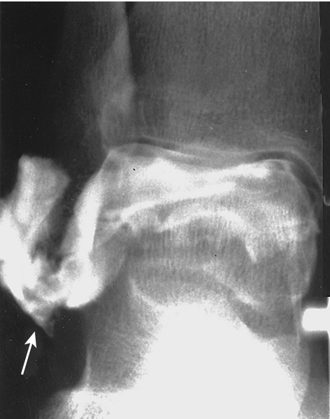
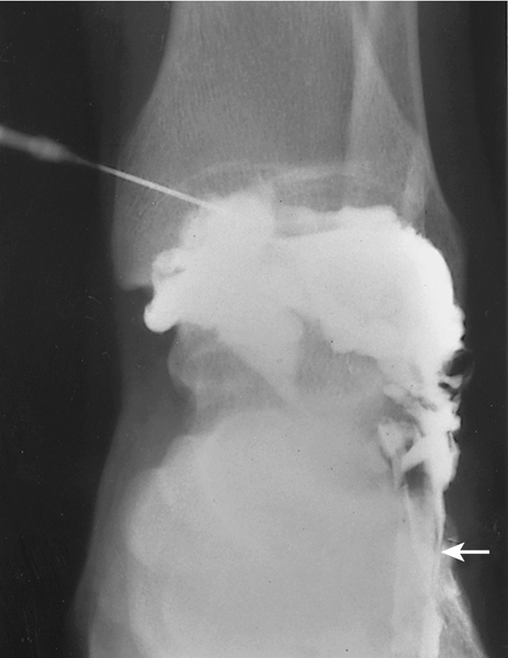
Computed tomography can identify subtle talus fractures and other bone disease, such as tumors. Bone scans are particularly helpful in identifying stress fractures and can be a useful screening tool to evaluate for ongoing ankle disease, such as significant arthritis, infection, tumors, and reflex sympathetic dystrophy. Magnetic resonance imaging and magnetic resonance arthrography generally give the most information about soft tissue injury, although they also can be helpful in identifying fractures (such as osteochondral fractures), tumors, and chronic infections. Magnetic resonance imaging and magnetic resonance arthrography both have high specificity for identification of chronic ligament tears, but magnetic resonance arthrography has higher sensitivity [10]. The appropriate timing of advanced imaging is variable and governed by the clinical suspicion of further injury or pathologic change not evident on routine radiographs or persistent symptoms despite appropriate treatment. Figures 85.1 and 85.2 demonstrate anterior talofibular and calcaneofibular ligament tears as observed with ankle arthrography.
Treatment
Initial
The initial treatment regimen depends, in part, on symptom acuity and whether a recent sprain has occurred. Initial treatment options include ice massage, compression, elevation, taping or bracing, and nonsteroidal anti-inflammatory drugs or analgesics. The goal with bracing at this juncture is to prevent recurrent sprains and further tissue trauma. Comprehensive literature reviews indeed validate the use of ankle supports to prevent reinjury [11–13]. Many patients can successfully be weaned from external supports after rehabilitation. However, high-demand athletes may choose to brace or to tape during athletic participation indefinitely. A thin lateral heel wedge will put the ankle in slight valgus alignment and potentially diminish symptoms of instability and the tendency for spontaneous ankle inversion with activity. This can be considered in patients with normal alignment for short-term symptom management. Eventually, restoration of neutral foot and ankle alignment should be pursued through orthotic prescription. A three-quarter-length rigid medial longitudinal arch support is recommended in patients with pes planus. In patients with varus or valgus alignment of the ankle, wearing of a shoe that has a firm heel counter is recommended.
Rehabilitation
Rehabilitation starts by normalizing ankle range of motion, with primary emphasis on restoring ankle dorsiflexion and eversion. This typically includes Achilles tendon stretching with the knee straight (to stretch the gastrocnemius) and flexed 30 degrees (to stretch the soleus) as well as eversion (posterior tibialis) stretching. Care must be taken to avoid recurrent inversion stress to the ankle, which can perpetuate lateral capsuloligamentous laxity.
As symptoms allow, the patient begins an ankle muscle group strengthening program with an emphasis on ankle evertor strengthening. Resistance exercises can begin when there is no pain through the available range of motion, with full weight bearing [14]. The rehabilitation program may start with low-level strengthening, such as submaximal static exercises, and progress in a pain-free fashion to dynamic and isokinetic strengthening. Typically, a combination of open and closed kinetic chain strengthening is employed in the rehabilitation process [15]. Open kinetic chain exercises include the use of ankle weights and resistance tubing. Closed kinetic chain exercises are more functionally based; the foot is planted on the ground, and the patient engages in an activity that requires the activation of antagonistic muscles that stabilize the ankle. Because eccentric muscle contractions place the greatest strain on the muscle, this mode of strengthening should be reserved for the final stages of the rehabilitation program.
Balance challenge and proprioceptive exercises are an important part of chronic ankle instability rehabilitation and have been found to decrease symptoms of functional instability as well as to reduce the rate of reinjury [16–18]. Ankle disks or wobble boards are devices that facilitate proprioceptive training. These exercises can be started without specialized equipment by having the patient perform a single-leg stance on the affected ankle; the skill level is then increased by having the patient close the eyes or stand on a pillow.
Functional exercises and sport-specific drills can begin when the patient has full range of motion, no pain, and at least 85% peroneal strength compared with the contralateral ankle [19]. These exercises add progressively difficult challenges and facilitate the attainment of dynamic strength and balance. Examples of these exercises are jogging, running, double-leg jumping, single-leg hopping, skipping rope, figure-eight drills, lateral cutting drills, and plyometrics. Patients should start at a low level of intensity and progress with increased intensity and difficulty only if they remain pain free while performing the exercise and have no pain or swelling after the training session.
Adjunctive modalities may be helpful throughout the rehabilitation process. These may include regular ice application (ice massage, ice pack, ankle Cryo/Cuff) after therapy sessions or heat application (superficial heat or ultrasound) to facilitate range of motion of a stiff joint. Electrical stimulation may be helpful for pain and edema control.
Procedures
Corticosteroid injections around the ankle ligaments are not advised and may further weaken the ligaments, accelerating mechanical instability. Prolotherapy, the injection of an irritant solution around the capsuloligamentous structures of the ankle, can be considered. The injectate (typically a low-concentration dextrose solution or a combination of phenol, glycerin, and glucose) acts as a local tissue irritant and activates the inflammatory cascade [20]. Given its mechanism of action, it can sometimes be a painful treatment. The purported result of this treatment is ligament and capsular hypertrophy causing reduction in laxity and thereby decreased pain [20]. At this time, no randomized controlled or large case series exists for the use of prolotherapy as a treatment of chronic ankle instability, but there is some suggestion of benefit in the treatment of knee laxity and pain due to osteoarthritis [21,22]. Further research is required.
Surgery
Surgery should be considered for patients who sustain recurrent lateral ankle sprains or exhibit significant symptoms of functional instability despite appropriate rehabilitation interventions. The goal of surgery is to restore mechanical stability to the ankle and thereby significantly reduce or eliminate chronic symptoms of instability. Late ankle reconstruction for chronic lateral instability is successful in approximately 85% of patients, regardless of the type of surgical procedure performed [23]. Intra-articular lesions (especially syndesmotic widening), osteochondral lesions of the talus, and ossicles are predictors of unsatisfactory results of ligament reconstruction [24]. Primary anatomic repair of the anterior talofibular and calcaneofibular ligaments is the preferred method of ankle ligament reconstruction [23]. However, in cases in which there is excessive joint laxity or insufficient capsular tissue is available, reconstructions such as the Chrisman-Snook procedure with use of the split peroneal brevis tendon, the semitendinosus, or an allograft may be preferred [25,26]. If the patient has significant varus predisposing to ankle instability, a Dwyer calcaneal closing wedge or lateral calcaneal slide may be appropriate.
Potential Disease Complications
Potential long-term sequelae of chronic ankle instability include the development of ankle impingement syndrome; chronic peroneal tendinopathy or subluxation; tibiotalar osteochondral injury; degenerative arthritis; superficial peroneal neuropathy; and chronic pain syndromes, such as complex regional pain syndrome type I (reflex sympathetic dystrophy).
Potential Treatment Complications
Potential treatment complications include frostbite from overly aggressive use of ice; exacerbation of edema from inappropriate ankle taping, wrapping, or bracing; and pain exacerbation or reinjury during physical therapy. Analgesics and nonsteroidal anti-inflammatory drugs have well-known side effects that most commonly affect the gastric, hepatic, and renal systems. Surgical complications include failed repair, wound or bone infection, loss of ankle range of motion from an aggressive reconstruction, and persistent ankle pain despite appropriate rehabilitation or surgical repair or reconstruction.


